RAPIDLY PROGRESSIVE INTERSTITIAL LUNG DISEASE IN DERMATOMYOSITIS A


- Slides: 22
RAPIDLY PROGRESSIVE INTERSTITIAL LUNG DISEASE IN DERMATOMYOSITIS A POTENTIAL USE FOR JAK/STAT Brian Abe MD Ph. D INHIBITORS ? Division of Immunology & Rheumatology Stanford University Hospital & Clinics
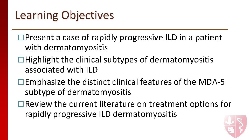
Learning Objectives �Present a case of rapidly progressive ILD in a patient with dermatomyositis �Highlight the clinical subtypes of dermatomyositis associated with ILD �Emphasize the distinct clinical features of the MDA-5 subtype of dermatomyositis �Review the current literature on treatment options for rapidly progressive ILD dermatomyositis
Case Presentation 48 yo M with no PMH presents to the emergency department with rash, painful tongue ulcers, and shortness of breath
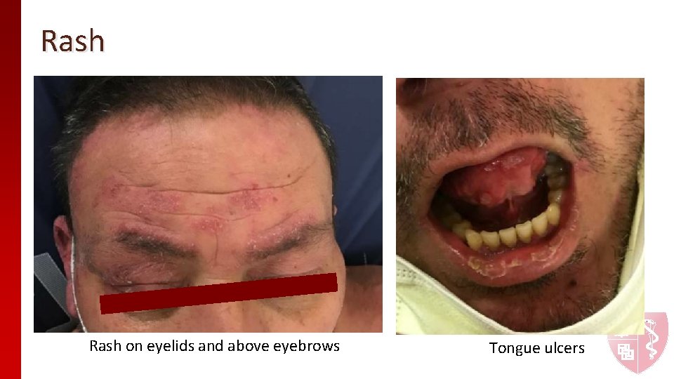
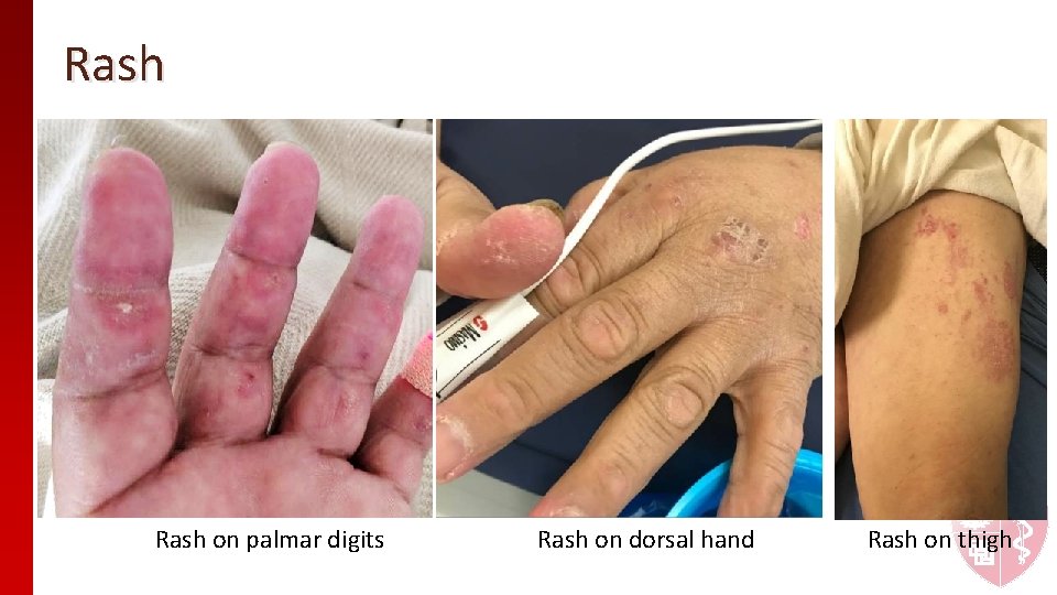
Case Presentation � Developed rash on hands and back. Not painful, not pruritic. Occurred after carrying “wet lumber” with his brother 4 months prior to admission. � Rash spread to his eyes, scalp, thighs. � One month later, he developed painful ulcers on his tongue, making eating painful. He denied difficulty swallowing. � His rash progressed, and he presented to a local ED and was diagnosed with contact dermatitis. He was treated with IM solumedrol 125 mg x 1 and 40 mg prednisone tapered over 2 weeks. He noted mild improvements in his skin but no improvement on his tongue. � He developed progressive shortness of breath.
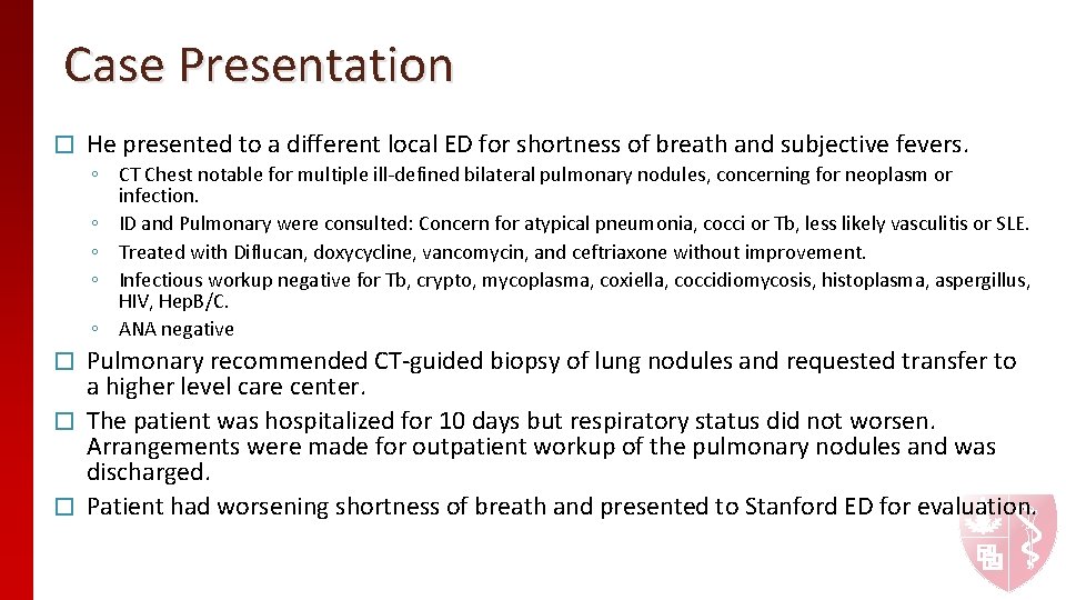
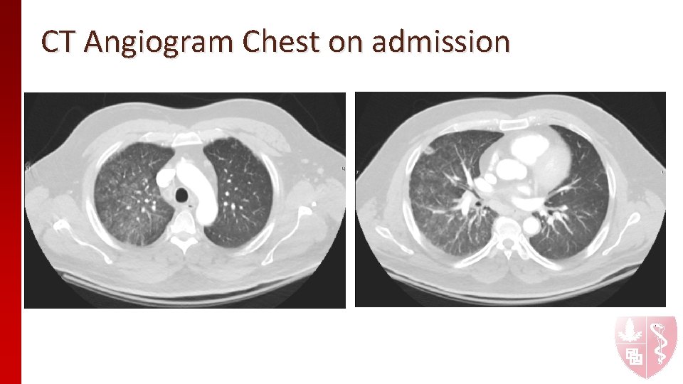
Case Presentation � He presented to a different local ED for shortness of breath and subjective fevers. ◦ CT Chest notable for multiple ill-defined bilateral pulmonary nodules, concerning for neoplasm or infection. ◦ ID and Pulmonary were consulted: Concern for atypical pneumonia, cocci or Tb, less likely vasculitis or SLE. ◦ Treated with Diflucan, doxycycline, vancomycin, and ceftriaxone without improvement. ◦ Infectious workup negative for Tb, crypto, mycoplasma, coxiella, coccidiomycosis, histoplasma, aspergillus, HIV, Hep. B/C. ◦ ANA negative Pulmonary recommended CT-guided biopsy of lung nodules and requested transfer to a higher level care center. � The patient was hospitalized for 10 days but respiratory status did not worsen. Arrangements were made for outpatient workup of the pulmonary nodules and was discharged. � Patient had worsening shortness of breath and presented to Stanford ED for evaluation. �
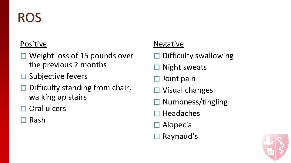
ROS Positive � Weight loss of 15 pounds over the previous 2 months � Subjective fevers � Difficulty standing from chair, walking up stairs � Oral ulcers � Rash Negative � Difficulty swallowing � Night sweats � Joint pain � Visual changes � Numbness/tingling � Headaches � Alopecia � Raynaud’s
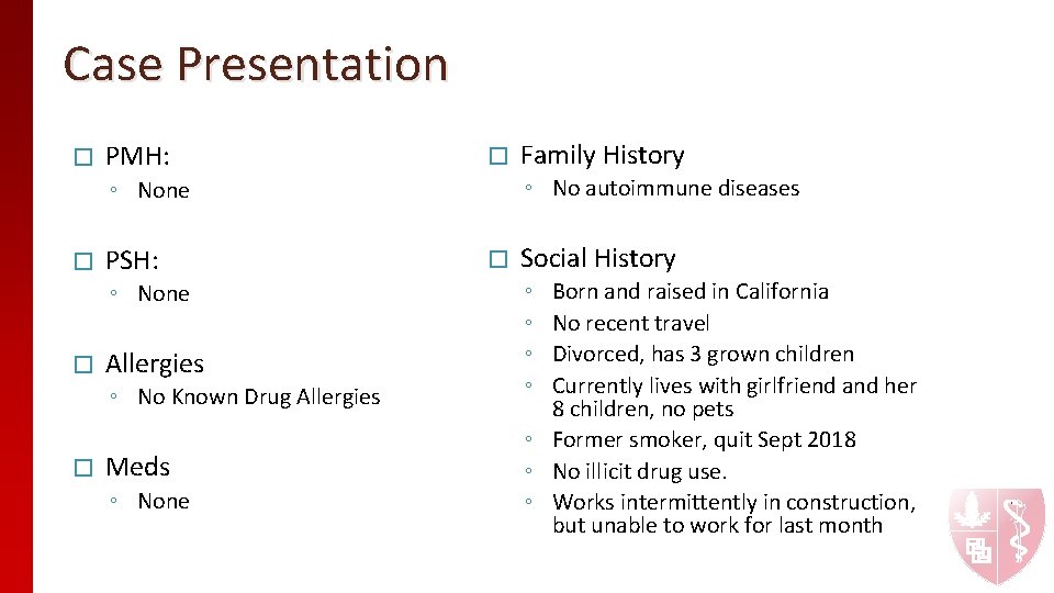
Case Presentation � PMH: � PSH: � ◦ No autoimmune diseases ◦ None � Allergies ◦ No Known Drug Allergies � Meds ◦ None Family History Social History ◦ ◦ Born and raised in California No recent travel Divorced, has 3 grown children Currently lives with girlfriend and her 8 children, no pets ◦ Former smoker, quit Sept 2018 ◦ No illicit drug use. ◦ Works intermittently in construction, but unable to work for last month
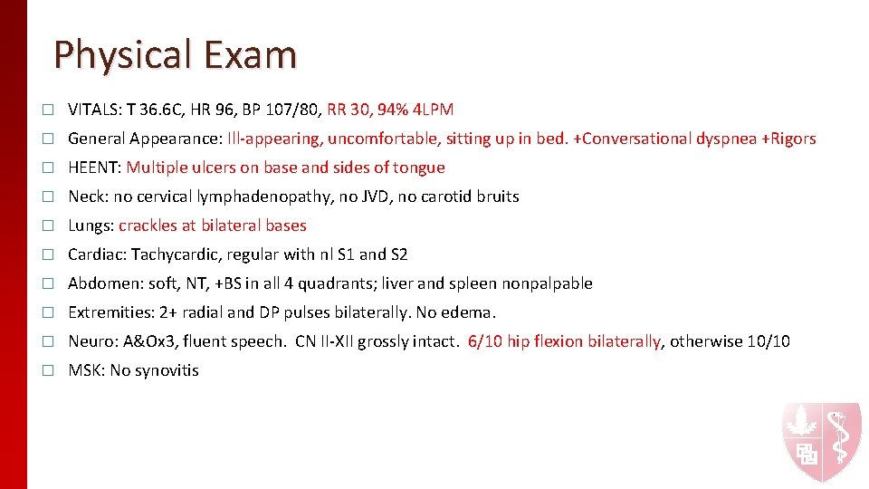
Physical Exam � VITALS: T 36. 6 C, HR 96, BP 107/80, RR 30, 94% 4 LPM � General Appearance: Ill-appearing, uncomfortable, sitting up in bed. +Conversational dyspnea +Rigors � HEENT: Multiple ulcers on base and sides of tongue � Neck: no cervical lymphadenopathy, no JVD, no carotid bruits � Lungs: crackles at bilateral bases � Cardiac: Tachycardic, regular with nl S 1 and S 2 � Abdomen: soft, NT, +BS in all 4 quadrants; liver and spleen nonpalpable � Extremities: 2+ radial and DP pulses bilaterally. No edema. � Neuro: A&Ox 3, fluent speech. CN II-XII grossly intact. 6/10 hip flexion bilaterally, otherwise 10/10 � MSK: No synovitis
Rash on eyelids and above eyebrows Tongue ulcers
Rash on palmar digits Rash on dorsal hand Rash on thigh
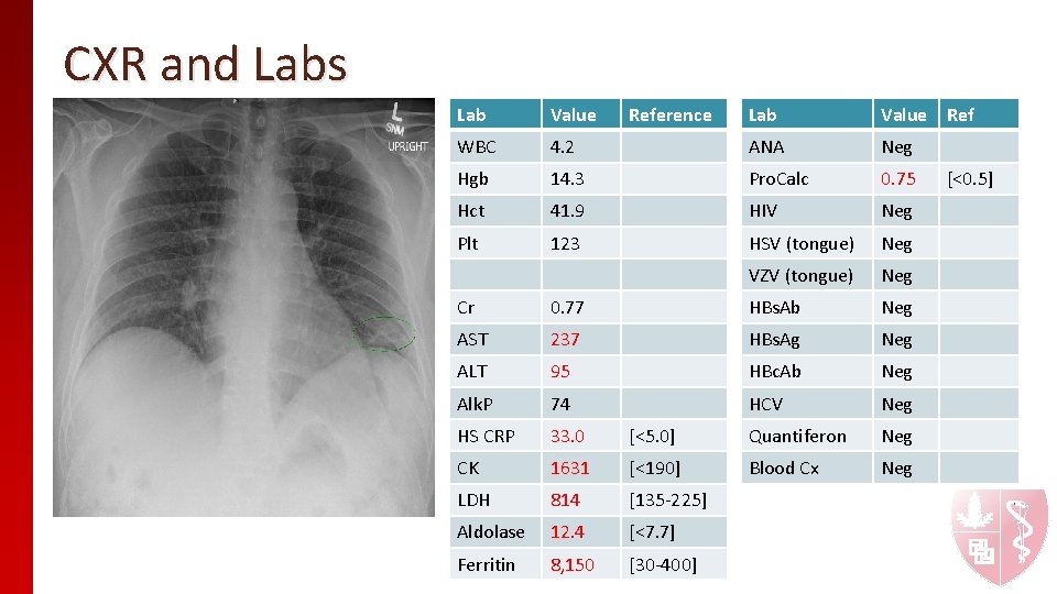
CXR and Labs Lab Value Ref 4. 2 ANA Neg Hgb 14. 3 Pro. Calc 0. 75 Hct 41. 9 HIV Neg Plt 123 HSV (tongue) Neg VZV (tongue) Neg Lab Value WBC Reference Cr 0. 77 HBs. Ab Neg AST 237 HBs. Ag Neg ALT 95 HBc. Ab Neg Alk. P 74 HCV Neg HS CRP 33. 0 [<5. 0] Quantiferon Neg CK 1631 [<190] Blood Cx Neg LDH 814 [135 -225] Aldolase 12. 4 [<7. 7] Ferritin 8, 150 [30 -400] [<0. 5]
CT Angiogram Chest on admission
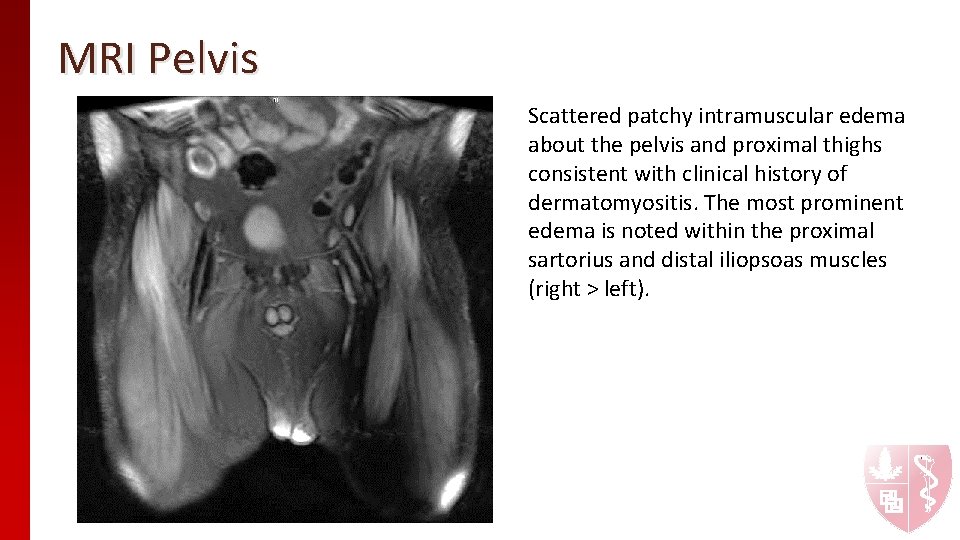
MRI Pelvis Scattered patchy intramuscular edema about the pelvis and proximal thighs consistent with clinical history of dermatomyositis. The most prominent edema is noted within the proximal sartorius and distal iliopsoas muscles (right > left).
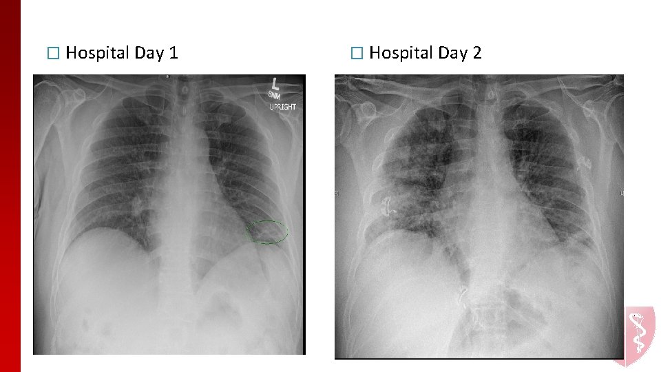
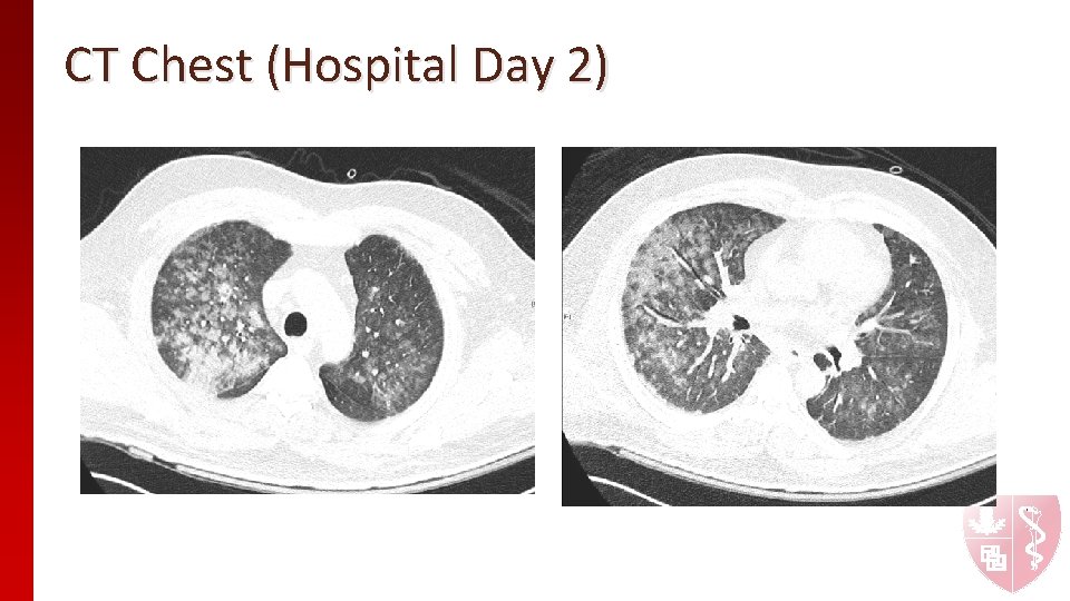
Clinical Course � Within 24 hours of admission patient has rapid respiratory decline, now on 12 L HFNC � On Zosyn, doxycycline, valacyclovir � ID does not think this is an infection � Pulmonary is concerned for rapidly progressing ILD � Skin biopsy shows vacuolar interface with vascular ectasia, papillary dermal edema and dermal mucin.
� Hospital Day 1 � Hospital Day 2
CT Chest (Hospital Day 2)
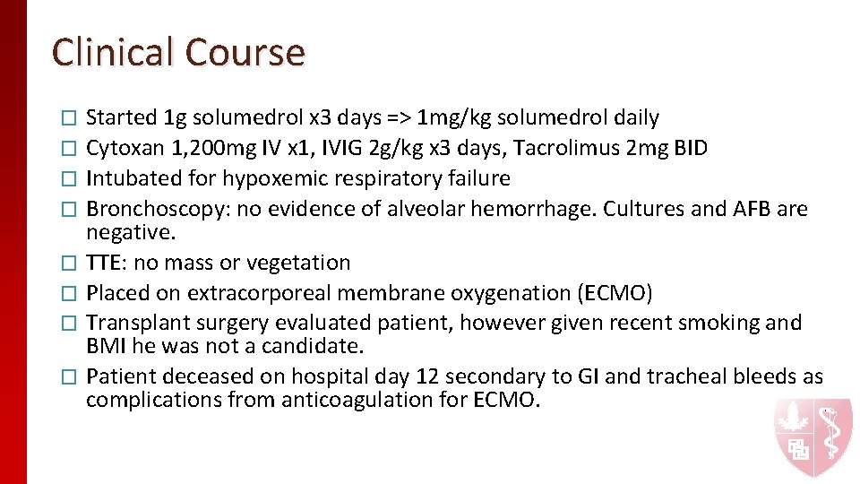
Clinical Course Started 1 g solumedrol x 3 days => 1 mg/kg solumedrol daily � Cytoxan 1, 200 mg IV x 1, IVIG 2 g/kg x 3 days, Tacrolimus 2 mg BID � Intubated for hypoxemic respiratory failure � Bronchoscopy: no evidence of alveolar hemorrhage. Cultures and AFB are negative. � TTE: no mass or vegetation � Placed on extracorporeal membrane oxygenation (ECMO) � Transplant surgery evaluated patient, however given recent smoking and BMI he was not a candidate. � Patient deceased on hospital day 12 secondary to GI and tracheal bleeds as complications from anticoagulation for ECMO. �
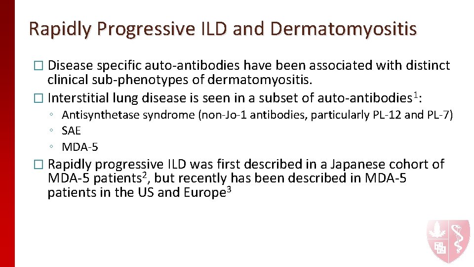
Rapidly Progressive ILD and Dermatomyositis � Disease specific auto-antibodies have been associated with distinct clinical sub-phenotypes of dermatomyositis. � Interstitial lung disease is seen in a subset of auto-antibodies 1: ◦ Antisynthetase syndrome (non-Jo-1 antibodies, particularly PL-12 and PL-7) ◦ SAE ◦ MDA-5 � Rapidly progressive ILD was first described in a Japanese cohort of MDA-5 patients 2, but recently has been described in MDA-5 patients in the US and Europe 3
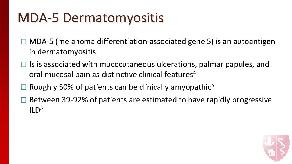
MDA-5 Dermatomyositis � MDA-5 (melanoma differentiation-associated gene 5) is an autoantigen in dermatomyositis � Is is associated with mucocutaneous ulcerations, palmar papules, and oral mucosal pain as distinctive clinical features 4 � Roughly 50% of patients can be clinically amyopathic 5 � Between 39 -92% of patients are estimated to have rapidly progressive ILD 5
Management of RP-ILD DM Early recognition of the disease and early intervention can reduce disease related morbidity and improve survival � In addition to high dose corticosteroids, aggressive immunosuppression includes 6, 7: � ◦ ◦ ◦ Cyclophosphamide Mycophenolate mofetil Calcinuerin inhibitors Intravenous immunoglobulin Rituximab Plasmapharesis Extracorporeal membrane oxygenation has been suggested to avoid the risk of pneumomediastinum that can be associated with mechanical ventilation 8 � Lung transplant should be considered in RP-ILD DM as they have favorable outcomes compared to patients with idiopathic pulmonary fibrosis 9 � Recent case reports are suggesting tofacinib as an emerging therapeutic option in patients with refractory RP-ILD 10 -13 �
Summary � � � � MDA-5 dermatomyositis is a clinical phenotype with mucocutaneous ulcers and palmar papules MDA-5 dermatomyositis can present as amyopathic Early recognition of MDA-5 dermatomyositis is vital as there is a high prevalence of rapidly progressive ILD There are no established guidelines on treating MDA-5 dermatomyositis Treatment often includes high dose corticosteroids, cyclophosphamide, IVIG, and calcineurin inhibitors Additional therapeutic considerations include plasmapheresis, extracorporeal membrane oxygenation, and lung transplant Recent case reports suggest Jak/Stat inhibitors may be effective for this clinical phenotype of dermatomyositis
References 1. Fujimoto, M. , Watanabe, R. , Ishitsuka, Y. & Okiyama, N. Recent advances in dermatomyositis-specific autoantibodies. Curr. Opin. Rheumatol. 28, 636– 644 (2016). 2. Sato, S. et al. Autoantibodies to a 140 -kd polypeptide, CADM-140, in Japanese patients with clinically amyopathic dermatomyositis. Arthritis Rheum. 52, 1571– 1576 (2005). 3. Moghadam-Kia, S. , Oddis, C. V. & Aggarwal, R. Anti-MDA 5 Antibody Spectrum in Western World. Curr. Rheumatol. Rep. 20, 78 (2018). 4. Fiorentino, D. , Chung, L. , Zwerner, J. , Rosen, A. & Casciola-Rosen, L. The mucocutaneous and systemic phenotype of dermatomyositis patients with antibodies to MDA 5 (CADM-140): A retrospective study. J. Am. Acad. Dermatol. 65, 25– 34 (2011). 5. Kurtzman, D. J. B. & Vleugels, R. A. Anti-melanoma differentiation–associated gene 5 (MDA 5) dermatomyositis: A concise review with an emphasis on distinctive clinical features. J. Am. Acad. Dermatol. 78, 776– 785 (2018). 6. González-Moreno, J. et al. Rapidly progressive interstitial lung disease due to anti-MDA 5 antibodies without skin involvement: a case report and literature review. Rheumatol. Int. 38, 1293– 1296 (2018). 7. Moghadam-Kia, S. , Oddis, C. V. & Aggarwal, R. Modern Therapies for Idiopathic Inflammatory Myopathies (IIMs): Role of Biologics. Clin. Rev. Allergy Immunol. 52, 81– 87 (2017). 8. Truong, L. , Youseef, F. , Ajaz, U. & Desai, S. Extracorporeal Membrane Oxygenation Used in the Treatment of Rapidly Progressive Interstitial Pneumonia in a Patient With Amyopathic Dermatomyositis and Positive Melanoma Differentiation-Associated Gene 5. J. Clin. Rheumatol. 29, 1053– 1054 (2018). 9. Ameye, H. , Ruttens, D. , Benveniste, O. , Verleden, G. M. & Wuyts, W. A. Is lung transplantation a valuable therapeutic option for patients with pulmonary polymyositis? Experiences from the leuven transplant cohort. Transplant. Proc. 46, 3147– 3153 (2014). 10. Hornig, J. et al. Ansprechen einer Dermatomyositis mit Lungenbeteiligung auf eine Januskinase-Inhibitor-Therapie. Z. Rheumatol. 952– 957 (2018). doi: 10. 1007/s 00393 -018 - 0565 -8 11. Kurasawa, K. et al. Tofacitinib for refractory interstitial lung diseases in anti-melanoma differentiation-associated 5 gene antibody-positive dermatomyositis. Rheumatology 57, 2114– 2119 (2018). 12. Kato, M. et al. Successful Treatment for Refractory Interstitial Lung Disease and Pneumomediastinum With Multidisciplinary Therapy Including Tofacitinib in a Patient With Anti-MDA 5 Antibody-Positive Dermatomyositis. J. Clin. Rheumatol. 29, 1053– 1054 (2019). 13. Moghadam-Kia, S. , Charlton, D. , Aggarwal, R. & Oddis, C. V. Management of refractory cutaneous dermatomyositis: potential role of Janus kinase inhibition with tofacitinib. Rheumatology (2019). doi: 10. 1093/rheumatology/key 366