Radiological Category Musculoskeletal Principal Modality 1 General Principal
: General Principal Modality (2): CT Case Report #0218")


- Slides: 12
Radiological Category: Musculoskeletal Principal Modality (1): General Principal Modality (2): CT Case Report #0218 Submitted by: Scott Reabe, M. D. Faculty reviewer: Ronald Bilow, M. D Date accepted: 01 April 2005
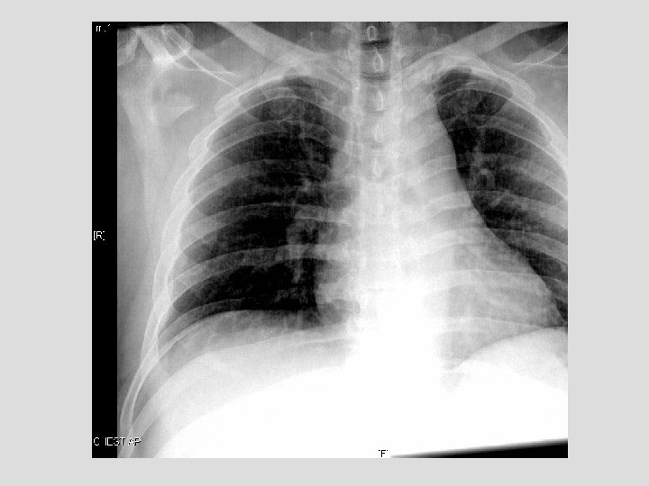
Case History 47 yo male presents to the ER s/p seizure, now with right shoulder pain.
Test Your Diagnosis Which one of the following is your choice for the appropriate diagnosis? After your selection, go to next page. • Normal Chest • Scapular Fracture • Coracoid Process Fracture • Bankart Fracture
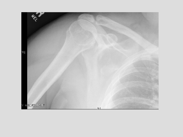
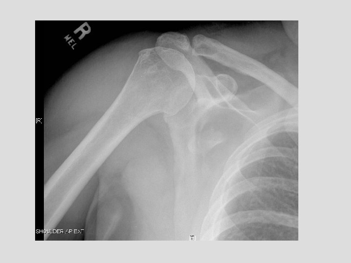
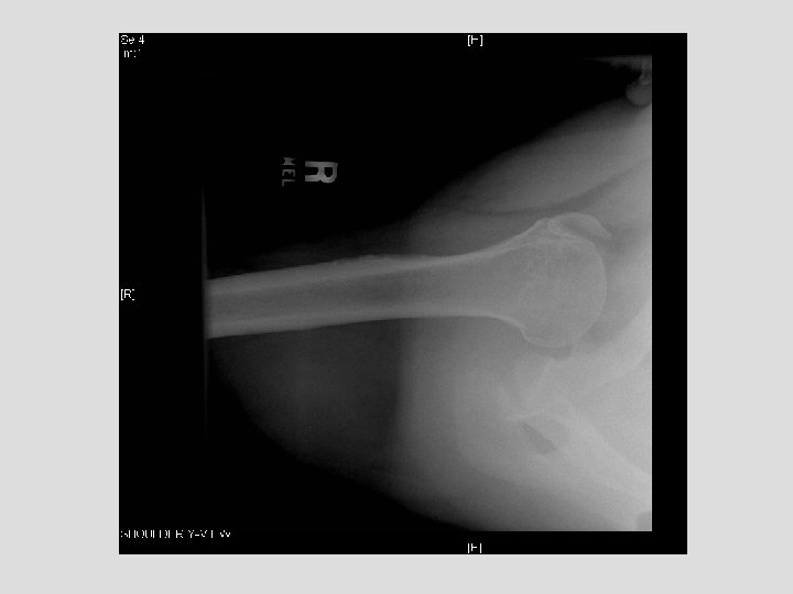
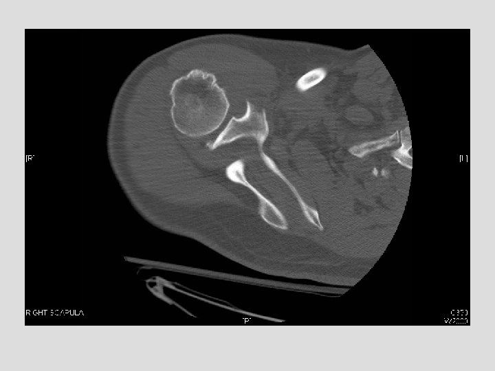
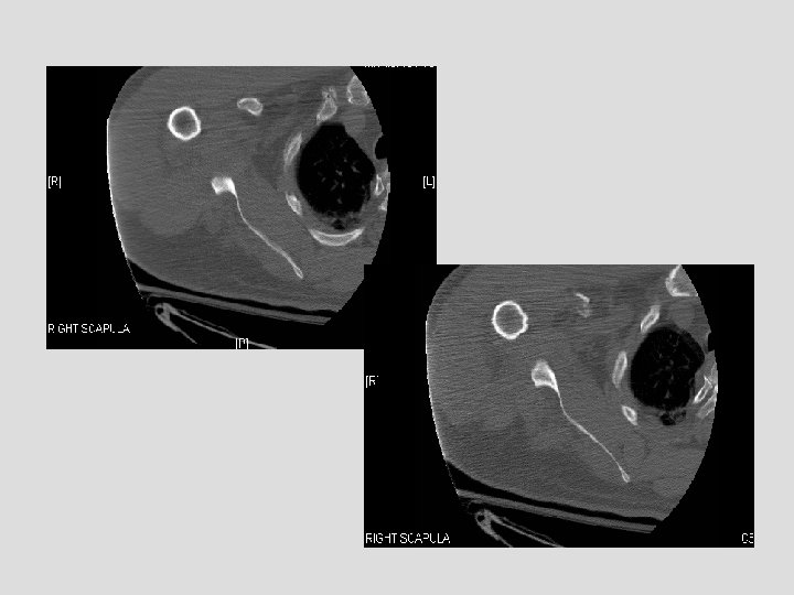
Findings and Differentials Findings: On the CXR, there is a piece of bone overlying the right scapula, which could easily be missed. The follow-up shoulder series and CT more clearly demonstrate a fracture. The CT clearly shows the donor site to be the coracoid process. Differentials: • Coracoid avulsion fracture
Discussion Patient’s that have seizures are prone to shoulder dislocations and put abnormal stress on other muscles. This case is not the typical pattern of injury in a seizure patient, but probably resulted from abnormal muscle contraction; the pectoralis major and minor forcefully contracted, and in this case the minor stressed its attachment to the coracoid such that it resulted in this injury. Ogawa et al. proposed a classification of coracoid fractures based on extent and relation to coracoclavicular ligamentous attachment. Type I is more common and involves the base of the coracoid and is associated with more extensive shoulder injuries and Type II involves the coracoid process itself (this case) and may be an islolated injury. Brant, W. , Helms, C. Fundamentals of Diagnostic Radiology, 2 nd Ed. 1999. Lippincott, Williams and Wilkins. Ppgs. 1001 -1015. Harris. The Radiology of Emergency Medicine, 4 rth Ed. 2000. Lippincott, Williams and Wilkins. Ppgs. 331 -335.
Diagnosis Coracoid avulsion fracture.