Primary somatosensory somesthetic cortex SI Somatosensory cortex a
 cortex (SI)")

")


 § Aδ mechanical nociceptors §")


- Slides: 41
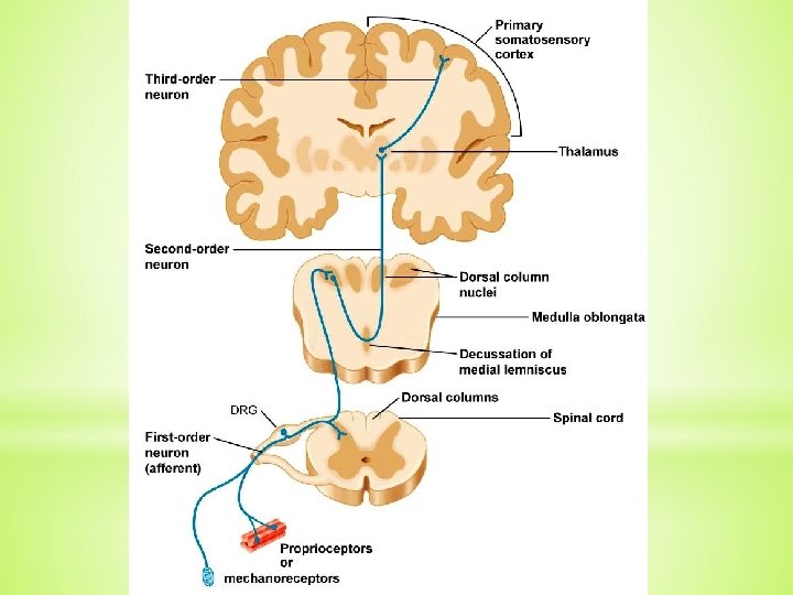
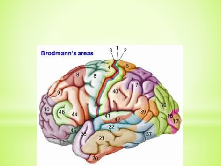
Primary somatosensory (somesthetic) cortex (SI)
Somatosensory cortex – a. 3, 1, 2
Secondary somatic sensory area (SII)
PAIN q pain is a protective modality q International Association for the Study of Pain (IASP): „Pain is an unpleasant sensory and emotional experience associated with actual or potential tissue damage, or described in terms of such damage“. q pain threshold: individual and social influences
Nociceptors/nocisensors q nocer – to injure or to hurt in Latin q are activated by noxious mechanical, thermal or chemical stimuli q detect signals from damaged tissue or the threat of damage q free nerve endings found in the skin, muscles, joints, bones and viscera
Nociceptors q nociceptors of Aδ fibres (5 -40 meters/sec) § Aδ mechanical nociceptors § Aδ thermal nociceptors q nociceptors of C fibres (0. 5 -2. 0 meters/sec) § C polymodal nociceptors – react to thermal, mechanical and chemical stimuli q silent nociceptors (MIA = mechanically insensitive afferents) - responsive after inflammation and tissue injury
Factors that activate nociceptors
q Peripheral sensitization q Central sensitization = activity- or use-dependent neuronal plasticity in the spinal cord § hyperalgesia - exaggerated or prolonged response to noxious inputs § allodynia - pain induced by normally innocuous inputs
Nociceptive afferents q Aδ fibres – thin myelinated fibres § sharp, localized pain q C non-myelinated fibres § dull, non-localized pain Spinoreticular tract
Pain pathways from trunk and limbs PALEOSPINOTHALAMIC PATHWAY NEOSPINOTHALAMIC PATHWAY q tr. spino-reticulo-thalamicus § diffuse, non-localized pain § autonomic and reflexive responses to pain stimuli § emotional and affective reactions to pain § intralaminar nuclei of the thalamus § postcentral gyrus, insula and cingulate gyrus q tr. spino-thalamicus lat. § sharp, localized pain § ventral posterolateral (VPL) and posterior nucleus of the thalamus § postcentral gyrus
Cortex Thalamus Paleospinothalamic pathway Neospinothalamic pathway Spinal cord
Pain pathways from head TRACTUS TRIGEMINO-RETICULOTHALAMICUS q diffuse, non-localized pain TRACTUS TRIGEMINOTHALAMICUS ANTERIOR q sharp, localized pain
Cortex Thalamus Medulla
The phylogenetically old, paleospinothalamic and trigeminoreticulothalamic, pathways through the RF are concerned with the arousal and affective (emotional) aspects of somatic sensory stimuli. In contrast, the direct, neospinothalamic and anterior trigeminothalamic, pathways are analytic, encoding information about modality, intensity, and location.
Referred pain
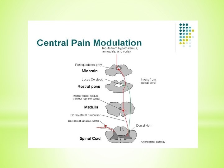
MODULATING SYSTEMS OF NOCICEPTIVE PATHWAYS q level of modulation of nociceptive pathways § spinal cord (“gate control theory”) § RF of brain stem § periaquaeductal gray matter (PAG)
“Gate control theory”
Stress-induced analgesia
Some necessary components of proper motor control q Volition q Coordination of signals to many muscle groups q Proprioception q Postural adjustments q Sensory feedback q Compensation for the physical characteristics of the body and muscles q Unconscious processing q Adaptability
Levels of movement regulation q spinal cord q brain stem q cortex q cerebellum q basal ganglia
Lower motor neurons – spinal cord q α motoneurons q γ motoneurons q Somatotopic organization § medial column – axial muscles § lateral column – limb muscles § anteriorly – extensors § posteriorly - flexors
q Motor neurons have highly branched, elaborate dendritic trees, enabling them to integrate the inputs from large numbers of other neurons and to calculate proper outputs. q The combination of an individual motor neuron and all of the muscle fibers that it innervates is called a motor unit.
q size of motor unit § small motor unit – 1 motoneuron innervates several muscle fibers (extraocular muscles, muscles of hand) § large motor unit – 1 motoneuron innervates 500 – 1000 muscle fibers (back muscles)
Lower motor neurons – brain stem q Somatomotor nuclei CN III, IV, VI, XII q Branchiomotor nuclei CN V, VII, IX, X, XI
Supraspinal system of movement control q Medial system § bilateral § terminates on the interneurons or the medial column of lower motor neurons § controls maintenance of balance and postural movements q Lateral system § mostly cross the midline and descend contralaterally § terminates on the interneurons or the lateral column of lower motor neurons § controls fine manipulative movements of the hand fingers q The “third” motor system § aminergic pathways of the brain stem
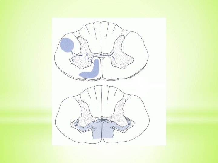
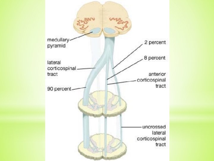
Medial system q Subcortical pathways § Medial and lateral vestibulospinal tracts - control of balance and postural movements § Tectospinal tract (sup. colliculus) – coordination of movements of the head and eyes during watching § Medial (pontine) and lateral (medullary) reticulospinal tracts - control of postural movements q Cortical pathways § Anterior corticospinal tract - bilateral, medial column of lower motor neurons
Lateral system q Subcortical pathways § Rubrospinal tract - contralaterally descends to the lateral column q Cortical pathways § Lateral corticospinal tract
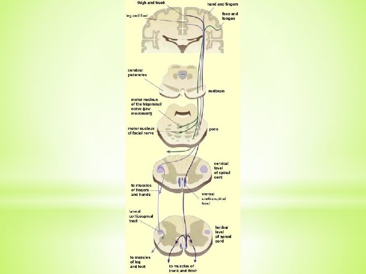
Corticospinal and corticonuclear tracts
The third system q the oldest one q nuclei of RF – raphespinal and coeruleospinal tracts q control of involuntary emotional movements
Motor cortex
Planning of movements
Illustrations were copied from: Neuroscience Online, the Open-Access Neuroscience Electronic Textbook Department of Neurobiology and Anatomy University of Texas Medical School at Houston