Physiology of Vision Photochemistry of Vision Dr Sumera


 and Scotopsin separate")


- Slides: 37
Physiology of Vision Photochemistry of Vision Dr. Sumera Gul
Learning Objectives • At the end of the lecture the students should be able to: • Discuss Light and Dark adaptation • Explain the mechanism of signal transmission in retinal neurons • Discuss the neurotransmitters released by retinal neurons • Explain the functions of various cells in retinal layers
Review of Previous Lecture
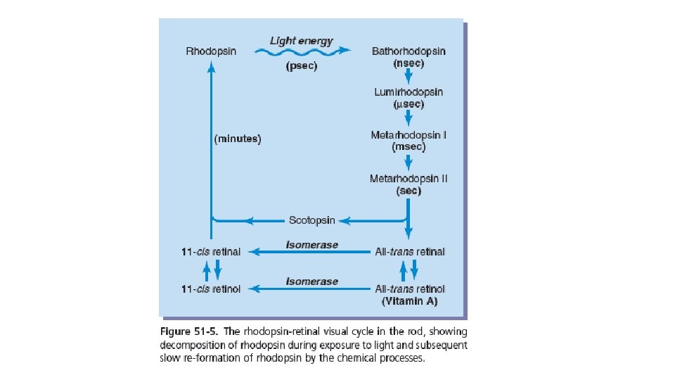
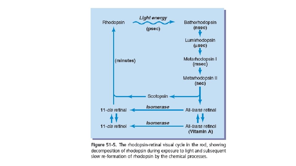
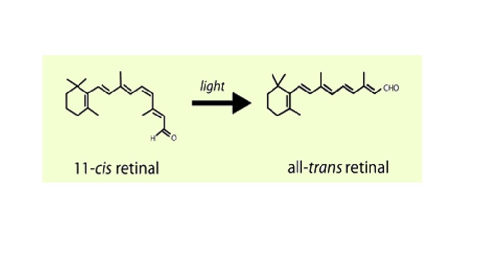
Rhodopsin-Retinal Visual Cycle • After exposure to light • Retinal (retinene) and Scotopsin separate
Color Vision • Blue sensitive pigment • Red sensitive pigment • Green sensitive pigment • Only one pigment is present in a cone
Color Blindness • Protanope • Deuteranope • Red-green color blindness X-linked recessive • 8% of all women are carriers
Light and Dark Adaptation
Light Adaptation • What will happen if a person has been in bright light for hours?
Light Adaptation • Large portions of the photochemicals in both the rods and the cones will have been reduced to retinal and opsins. • Furthermore, much of the retinal of both the rods and the cones will have been converted into vitamin A. • Low concentrations of the photosensitive chemicals in the rods and cones • sensitivity of the eye to light is correspondingly reduced.
Dark Adaptation • What will happen if a person remains in darkness for a long time?
Dark Adaptation • The retinal and opsins in the rods and cones are converted back into the light-sensitive pigments. • Vitamin A is converted back into retinal to increase light-sensitive pigments.
Dark Adaptation • After remaining is bright light when a person is exposed to complete darkness • Sensitivity if retina is very low initially • After 1 min increases 10 fold
Dark Adaptation • Dark Adaptation Curve • Early portion is due to adaptation of cones (4 times rapid) • Rods are slowly adapting • More sensitive • 100 or more rods converge on a single ganglion
Light Dark Adaptation • Change in pupillary size • Neural Adaptation
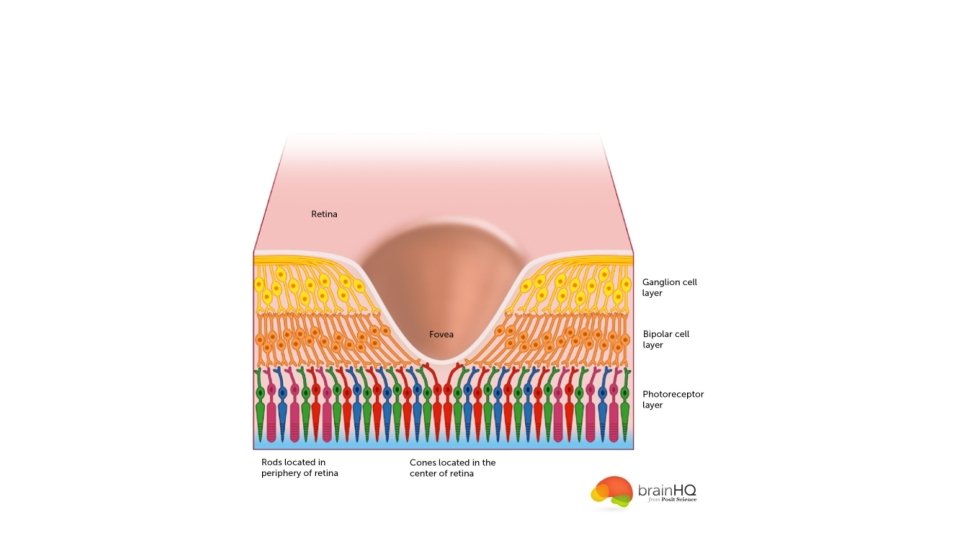
Neural Functions of Retina
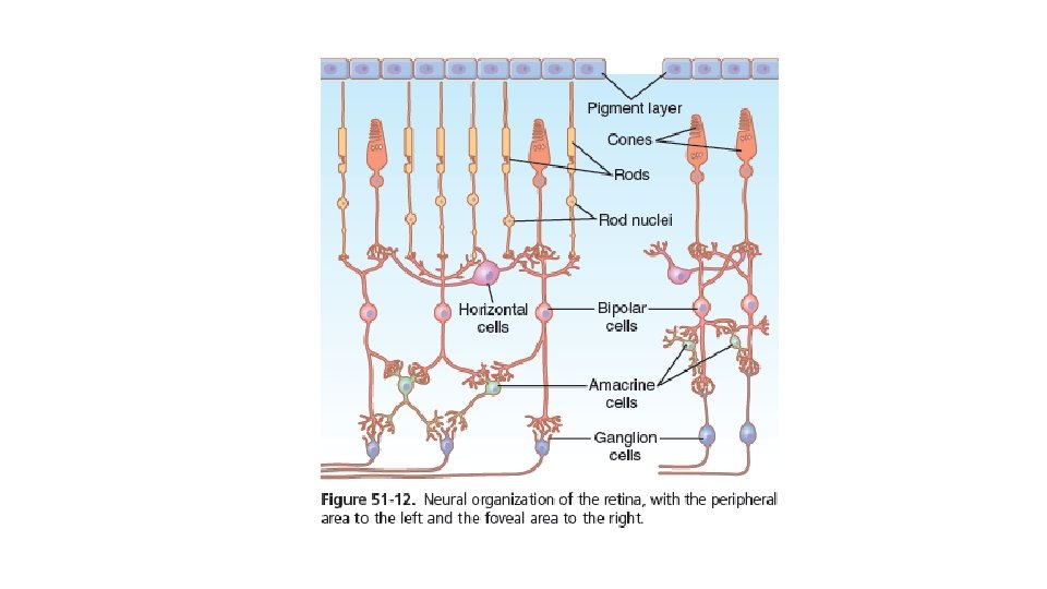
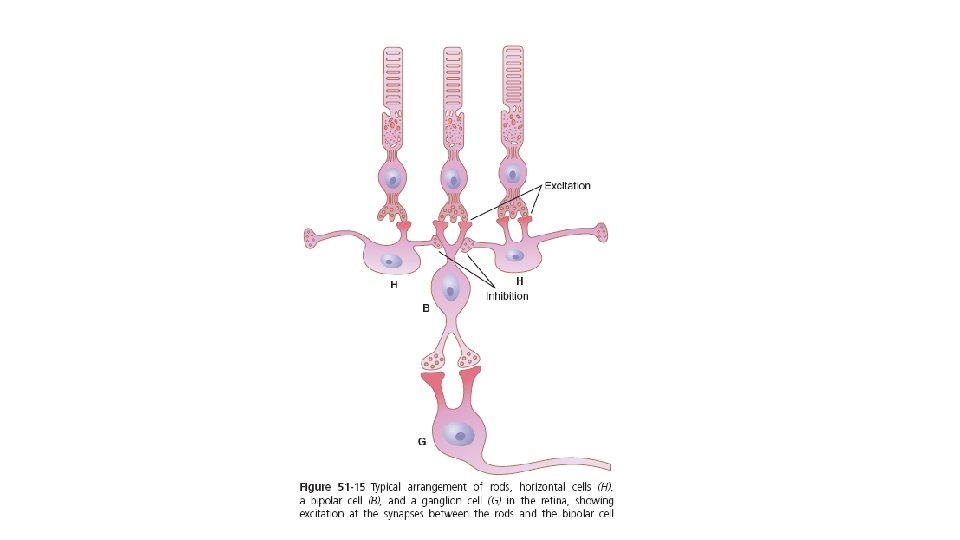
Neurotransmitters of Retina • Glutamate is released from Rods and Cones at their synapse with bipolar cells • Amacrine cells release at least 8 neurotransmitters including • GABA, glycine, Ach, Dopamine, Indolamine
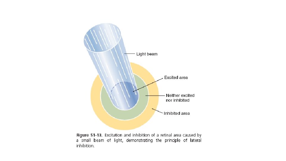
Horizontal Cells • Horizontal cells are inhibitory • Responsible for lateral inhibition • It enhances the visual contrast and allows high visual accuracy
Bipolar Cells • Can be depolarizing or hyperpolarizing cells • Can be due to different types of cells altogether • Or due to direct stimulation by Rods/Cones or via horizontal cells
Amacrine Cells • 30 different types Rods vision Onset and offset of visual signals Light turned on or off Respond to movement of spot
Ganglion Cells • 1. 6 million ganglion cells in each retina • 100 million rods • 3 million cones
Ganglion Cells • Peripheral retina is more sensitive to light • As there are more rods there which are 30 -300 times more sensitive to light. • And around 200 rods converge on a single optic nerve fibre.
W type Ganglion Cells Transmit signals at lower velocity mostly from rods Sensitive to detect directional movement in peripheral retina Important for vision in dark
X type Ganglion Cells • Small fields • Dendrites do not spread widely • Final and discrete visual image • Probably responsible for color vision
Y type Ganglion Cells • Largest of all cells • Fast • Broad dendritic fields • Respond rapidly to changes in visual image • Less discrete
P and M cells • Magnocellular cells • Parvocellular cells
Magnocellular Cells • Alpha cells • Not generally sensitive to color • Parasol cells • More sensitive to contrast black and white • To the magnocellular layer of lateral geniculate body • Sensitive to rapid movements • Larger receptive field • Faster
Parvocellular cells • Beta cells • Sustained response to color • Midget ganglion cells • Fine details • Parvocellular layer of LGN • Small receptive field • Slower
Questions?