The Special Senses Vision 2 Professor A M


 • Stimulus: •")


 Light")


 Distribution of photoreceptors")


 • Same in")
: Activation of rhodopsin: • In the dark:")


")
- Slides: 57
The Special Senses Vision - 2 Professor A. M. A Abdel Gader MD, Ph. D, FRCP (London & Edinburgh), FRSH (London) Professor of Physiology, College of Medicine & King Khalid University Hospital Riyadh, Saudi Arabia
The Physiology of Vision Objectives: At the end of this lecture the student should be able to: • Understand the optical bases of image formation on the retina • Understand explain the optical bases of common refractive errors • Understand the electrical bases of the photoreceptor function • Understand the nature and function visual pigments Understand color vision
The Physiology of Vision Objectives: At the end of this lecture the student should be able to: • Understand the optical bases of image formation on the retina • Understand explain the optical bases of common refractive errors • Understand the electrical bases of the photoreceptor function • Understand the nature and function visual pigments Understand color vision
Physiology of Vision Light Receptor: Retina (Photoreceptors) • Stimulus: •
Light • Definition: ‘elctromagnetic’ radiation that is capable of exciting the human eye’ • Extremely fast
Which travels faster: light or sound?
Electromagnetic spectrum & The visible light spectrum
The Electromagnetic Spectrum
Visible light & Duplicity Theory of vision Visible light Spectrum • Extends from 397 to 723 nm • Eye functions under two 2 conditions of illumination: – Bright light (Photopic vision)…Cones – Dim light (Scotopic vision). . Rods Duplicity theory of vision
Duplicity theory • Photopic visibilty curve peaks at 505 nm • Scotopic “” ” “ “ 550 nm
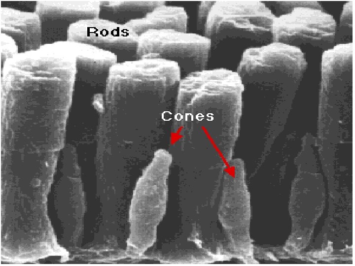
Photoreceptors Rods & Cones Morphology & Distribution
Retina Back of retina, pigment epithelium (Choroid) Light
Rods and Cones Figure 17. 13
Photoreceptors Figure 16. 11
Retina: distribution photoreceptors
Receptor density (cells x 103 / mm 2) Distribution of photoreceptors
Normal Fundus Photoreceptors are not distributed uniformly across the retina Optic disc Macula 5000 um 650, 000 cones Fovea 1500 um 100, 000 cones Foveola 350 um 25, 000 cones
Human foveal pit INL Light ONL Foveola
Low Convergence Cone-Fed Circuits Retinal ganglion cell Bipolar cell Cone High Convergence Rod-Fed Circuits Retina ganglion cell Bipolar cell Rod Convergence rod/cone cells
Retina: photoreceptors • 100, 000 rods • 5, 000 cones Cones Fovea High light levels Color Good acuity Rods Periphery Low light levels Monochromatic Poor acuity
Electrophysiology of Vision Genesis of electrical responses
Retinal photoreceptors mechanism Light Absorption by photosensitive substances Structural change in photosensitive substances Phototransduction Action potential in the optic nerve
Action Potential Propagated and “All-or-None” Receptor Potential Local & Graded
Retina: Neural Circuitry Light hits photoreceptors, sends signal to the bipolar cells Bipolar cells send signal to ganglion cells Ganglion cells send signal to the brain
In Darkness
Photoreception-cont.
Retina Light
Electrophysiology of Vision Electric recording in Retinal cells: • Rods & Cones: Hyperpolarization • Bipolar cells: Hyper- & Depolarization • Horizental cells: Hyperpolarization • Amacrine cells: Depolarizing potential • Ganglion cells: Depolarizing potential
outer segment Disk membrane Intracellular disk Intracellular space Disk membrane Extracellular space Visual pigment Extracellular space Intracellular space Visual pigment Plasma membrane Connecting cilium ROD CELL CONE CELL Rods and Cones
Rods Light Environment Dim light - scotopic Bright light - photopic Spectral sensitivity 1 pigment 3 pigments Color discrimination No Yes Absolute sensitivity High Low Speed of response Slow Fast Rate of dark adaptation Fast Slow Starlight Moonlight No color vision Poor acuity Scotopic Absolute threshold Cones Indoor lighting Good color vision Best acuity Mesopic Cone threshold Sunlight Photopic Rod Saturation begins Best acuity Indirect Ophthalmoscope Damage Possible Comparison Scotopic and Photopic systems
Photoreceptor pigments
Photoreceptor pigments • Composition: – Retinine 1 (Aldehyde of vitamin A) • Same in all pigments – Opsin (protein) • Different amino acid sequence in different pigments Rhodopsin (Rod pigment): Retinine + scotopsin
Photoreceptor compounds -cont Rhodopsin (visual purple, scotopsin): Activation of rhodopsin: • In the dark: retinine 1 in the 11 -cis configuration Light All-trans isomer Metarhodopsin II Closure of Na channels
Visual cycle Rhodopsin Light Prelumirhdopsin Inermediates including Metarhodopsin II Vitamin A + Scotopsin Retinine & Scotopsin
Light Change in photopigment Metarhodopsin II Activation of transducin Activation of phophodiesterase Decrease IC cyclic GMP Closure of Na channels Hyperpolarization of receptor Decrease release of synaptic tramitter Action potential in optic nerve fibres
From light reception to receptor potential
Retina: Neural Circuitry Light hits photoreceptor s, sends signal to the bipolar cells Bipolar cells send signal to ganglion cells Ganglion cells send signal to the brain
Photoreception
Photoreception- cont.
Retina • 100, 000 rods • 5, 000 cones • 1, 000 ganglion cells Convergence
Convergence Cones • Photoreceptors • Ganglion cells Rods
Convergence and Ganglion Cell Function Figure 17. 18
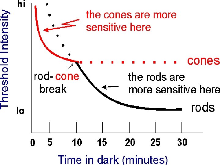
Dark adaptation
Dark adaptation: Increased sensitivity of the photoreceptors when vision shifts from bright to dim light
Dark adaptation • Reaches max in 20 minutes • First 5 minutes …… threshold of cones • 5 to 20 mins ……. Sensitvity of rods Mechanism of dark adaptation: Regeneration of rhodopsin
Dark adaptation-cont. In vitamin A deficiency What happens to Dark adaptation? Night blindness (Nyctalopia)