PERIODONTAL CASE STUDY PROJECT DENTAL HYGIENE CLINICAL PRACTICE


- Slides: 19

PERIODONTAL CASE STUDY PROJECT DENTAL HYGIENE CLINICAL PRACTICE II DIEM LE

PATIENT PROFILE • 33 year old Asian male • Health history reveals: • a current heavy smoker, been a smoker for 15 years • Family history of Diabetes • School related stress • No medications • Minor Dental Anxiety • Vitals WNL • ASA Class II • Dental history reveals: • Brushes with soft toothbrush 2 x daily • Flosses 3 x daily • TMJ pain and click • No night guard • Grinds and clenches his teeth • Last dental visit was 1 year ago

EXTRA ORAL AND INTRA ORAL FINDINGS • TMJ: • Bilateral crepitus • pain in cold seasons • Generalized attrition • Hypocalcification mesial of #7 -#10, and cervical 1/3 on #9 • Angles classification of occlusion: Tendency to class II on molar right, canine right, and canine left. Class I occlusion on molar left. • 75% overbite • 4 mm over jet • Slight crowding on lower anteriors which lead to torsoversion on mandibular anteriors • Decalcification on cervical 1/3 on #29 • Short Lingual Frenum • Moderately coated tongue • Tonsils slightly enlarged, nicotine stomatitis on hard palate

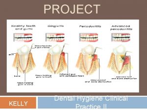
GINGIVAL DESCRIPTION Generalized moderate redness, shiny, spongy, enlarged, rounded slight edematous tissue with rolled margins and bulbous papillae

INTRA ORAL PHOTOS -Photo was taken on 01/28/14 -Green Arrow- generalized mod attrition

INTRA ORAL PHOTOS Photos were taken on 01/28/14 Notes: Green Arrow- generalized mod attrition Red Arrow- Generalized mod tobacco stain on linguals Black arrow- Fracture and tobacco stain on tooth #29 Yellow arrow- Generalized slight marginal redness White arrow- Torsoversion on mandibular anteriors due to crowding

DENTAL CHART NOTE: -Green arrows-Carious lesions on occlusal of Teeth #14, #30, #31 -Red arrows- fractured tooth, needs restorations

PERIODONTAL CHARTING

ASSESSMENT FINDINGS • • • No furcations No mobility or mucogingival involvement BOP was observed on all teeth Generalized slight marginal redness Generalized heavy ledges of supragingival calculus on mandibular anteriors Generalized heavy ledges of subgingival calculus Generalized heavy biofilm on the cervical 1/3 of the teeth & interproximally Plaque Control Record was 48% Generalized tobacco stain Carious lesions on occlusal of Teeth #14, #30, #31 Average CAL were 2

PERIODONTAL EVALUATION Note. General average CAL is 2, with localized CAL of 3 and one CAL of 4 on tooth #18 Distal. No noticeable recession

FACTORS • Periodontal Risk Factors • -Smoking • -Stress • Contributory Factors • -Calculus, malocclusion, OH care

PERIODONTAL DIAGNOSIS • Generalized slight active Chronic Periodontitis with moderate active chronic Periodontitis on teeth #3, 15, 18, 29 • AAP II

RADIOGRAPHS • • • Generalized slight vertical bone loss -Crestal Irregularities on teeth #11, 12, 14, 15, 18, 19 -Green arrows indicate calculus subgingivally on mesial and distal of tooth #3 *Radiographs were taken at patient dentist office on 02/27/14. These were the only available radiographs since patient’s dentist did not approve to retake another FMX at MCC due to ALARA principles.

RADIOGRAPHS • Generalized slight bone loss with localized moderate bone loss on teeth # 20, 28, 29, 30 • Green arrows indicate impacted wisdom teeth on teeth #17, 32. • *Radiographs were taken at patient dentist office on 02/27/14. These were the only available radiographs since patient’s dentist did not approve to retake another FMX at MCC due to ALARA principles.

Treatment Plan This is the patient treatment plan. I diagnosed that he had high oral cancer risk due to smoking habits and active moderate perio due to smoking and infrequent recalls. I recommended the modified Stillman method due to his interproximal calculus and slight localized recession on teeth #910. He has moderate tobacco stain so I recommended motor polishing and Sodium Fluoride Tray.

PROCEDURES • First Second, and Third visit completed assessments – Took intra-oral photos on second visit-01/28/14 • Fourth visit • Medical History, EOE, IOE, Vital Signs • Plaque index & home care • Review Brushing technique- focus on the cervical 1/3 of the tooth w/ a modified Stillman method • Local anesthesia administer by Professor Ligor, 5% lidocaine Topical applied to all injection sites, Right PSA and Right MSA Lidocaine, 2% with Epinephrine 1: 100, 000, 1 cartridge, (36 mg Lido, . 018 mg Epi) • Debridement on on teeth #2 -3 using magnetostrictive power inserts and hand scaling • Fifth visit • Medical History, Vital Signs, EOE, IOE • Plaque index, Re-assess upper right • Local anesthesia administer by Dr. Terkoski, 5% lidocaine Topical applied to all injection sites, Right MSA and Right ASA Lidocaine, 2% with Epinephrine 1: 100, 000, 1 cartridge, (36 mg Lido, . 018 mg Epi) • Debridement on on teeth #4 -9 using magnetostrictive power inserts and hand scaling

Procedures • Sixth visit -Medical History, Vital Signs, EOE, IOE -Plaque index, Re-assess upper right -Local anesthesia administer by Professor Fernandez, 5% lidocaine Topical applied to all injection sites, -Right IA and Right Buccal Lidocaine, 2% with Epinephrine 1: 100, 000, 1 cartridge, (36 mg Lido, . 018 mg Epi) -Debridement on on teeth #25 -31 using magnetostrictive power inserts and hand scaling • Seventh visit -Medical History, Vital Signs, EOE, IOE -Plaque index, Re-assess lower right -Local anesthesia administer by Professor Ligor, 5% lidocaine Topical applied to all injection sites, -Left PSA and Left MSA, Left ASA, l Lidocaine, 2% with Epinephrine 1: 100, 000, 1 cartridge, (36 mg Lido, . 018 mg Epi) -Debridement on on teeth #9 -15 using magnetostrictive power inserts and hand scaling

PROCEDURES • • Eighth visit Medical History, Vital Signs, EOE, IOE Plaque index, Re-assess upper left Local anesthesia administer by Dr. Terkoski, 5% lidocaine Topical applied to all injection sites, -Left IA and Left Buccal Lidocaine, 2% with Epinephrine 1: 100, 000, 1 cartridge, (36 mg Lido, . 018 mg Epi) • Debridement on on teeth #25 -31 using magnetostrictive power inserts and hand scaling • Motor polishing • Ninth visit • Medical History, Vital Signs, EOE, IOE • Plaque index, Re-assess all teeth • Motor polishing • Fluoride Tray Treatment with Sodium Fluoride 1. 23%, 4 minutes • Handed patient dental hygiene report • Patient survey

SUMMARY Even though this was my first patient, I am glad I got the toughest periodontal case as my first patient. I was able to learn how to perform a thorough periodontal assessment and practice my debridement skills. When reviewing the photos, I realize that one would not have guessed that the patient’s periodontitis was not that bad due to his smoking which masks the effects on his gingival margins. I was only to really determine that he had generalized slight bone loss due to the x-rays sent from his dentist. I think his bone loss is worse now since those x-rays were from almost 2 years ago. My goal was to reduce his calculus and plaque index by 50% and to have him gradually quit smoking. He stated that he does want to quit smoking eventually as well but did not give me a start date. I realized that after 2 weeks of not seeing my patient due to spring break, calculus built up on areas that I had already debrided because he smokes more when he is working or stressed. I continue to encourage him to have a start date or short term goals to cut down on the number of cigarettes per week. I was able to determine that my patient had slight active chronic periodontitis with active moderate chronic periodontitis on tooth #15, 18. This patient has been smoking heavily for 15 years which was a big risk factor contributing to his bone loss as well as malocclusions and lack of professional dental hygiene care. I hope that he continues a 3 -month re-care, improve his oral hygiene care, and set a start date to quit smoking. There is no date for re-evaluation because patient will be busy working and have no time to come back for a re-evaluation.
 Gingival description
 Periodontal case study
 Unfolding clinical reasoning case study
 Pacific university dental hygiene
 Pen grasp
 Adpie dental hygiene
 Maine dental hygiene association
 Ethical dilemma examples in dental hygiene
 Dental hygiene schedule template
 Cscc hesi
 Maryland state board dental examiners
 Vermont dental hygiene association
 Ouhsc dental hygiene
 Best case worst case average case
 Private equity lbo case study
 Project failure case study
 Business analyst case study
 Favela bairro project case study
 Edinburgh tram project case study
 Project scope management case study
