Neuroscience II Pathology Ghadeer Hayel MD Central nervous


- Slides: 34

Neuroscience II Pathology Ghadeer Hayel, MD

Central nervous system Characteristic Features of Cellular Pathology in CNS 2

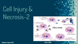
Neurons – Acute neuronal injury ▪ ▪ Within 12 -24 hours of an irreversible hypoxic-ischemic insult, neuronal injury becomes evident microscopically Shrinkage of the cell body, pyknosis of the nucleus, disappearance of the nucleolus, loss of Nissl substance, and intense eosinophilia of the cytoplasm “red neurons” 3

Neurons – Axonal injury/reaction ▪ ▪ A change observed in the cell body of the neurons during regeneration of the axon (sprouting). Cell body enlargement and rounding, peripheral displacement of the nucleus, enlargement of the nucleolus, and peripheral dispersion of Nissl substance (central chromatolysis) 4

Astrocyte Injury and Repair ▪ Astrocytes repair & scar formation in CNS, (gliosis) ▪ After injury they undergo hypertrophy and hyperplasia. The nucleus enlarges (more vesicular) & the nucleolus becomes prominent. The cytoplasm expands with bright pink hue & extends multiple processes (gemistocytic astrocyte). ▪ 5

Astrocyte Injury and Repair ▪ Unlike elsewhere in the body, fibroblasts participate in healing after brain injury to a limited extent except in specific settings (penetrating brain trauma or around abscesses). 6

Astrocyte Injury and Repair ▪ ▪ In long-standing gliosis, the cytoplasm of reactive astrocytes shrinks in size, & cellular processes become tightly interwoven (fibrillary astrocytes). Rosenthal fibers: thick, elongated, brightly eosinophilic protein aggregates in astrocytic processes in chronic gliosis & in some low-grade gliomas. (pilocytic astrocytoma) 7

Microglial cells Long-lived resident phagocytes in CNS, derived from embryonic yolk sac. ▪ Activated by tissue injury, infection, or trauma. ▪ May develop elongated nuclei - rod cells (infections). ▪ Aggregates around necrosis microglial nodules. ▪ Or around a dying neuron Neuronophagia. ▪ 8

9

Demyelinating diseases of CNS Ghadeer Hayel, MD

Myelin. . ▪ ▪ ▪ Axons in CNS are tightly ensheathed by myelin. It is an electrical insulator allows rapid propagation of neural impulses. Consists of multiple layers of highly specialized, closely apposed plasma membranes. Assembled by oligodendrocytes. Dominant component in the white matter, so most diseases of myelin are primarily white matter disorders. 11

Differences b/w CNS & PNS Myelin 1) PNS myelin is made by Schwann cells, CNS myelin is made by oligodendrocytes. In PNS each Schwann cells provides myelin for only one internode, while in the CNS, many internodes are created by processes coming from a single oligodendrocyte. 3) The specialized proteins and lipids are also different. 4) Most diseases of CNS myelin do not involve the PNS to any significant extent, and vice versa. 2) 12

Diseases of myelin are separated to two groups: I. Demyelinating diseases +acquired conditions. +damage to previously normal myelin. +causes: (1)immune mediated, (2)oligodendrocytes viral infection (progressive multifocal leukoencephalopathy JC virus, a polyomavirus), or (3) injury caused by drugs or other toxic agents. 13

diseases of myelin are separated to two groups: II. Leukodystrophy or dysmyelinating diseases: +Myelin is not formed properly or has abnormal kinetics +Caused by mutations that disrupt the function of proteins required for the formation of normal myelin sheaths. 14

Multiple Sclerosis (MS) ▪ The most common demyelinating disease. ▪ Episodes of disease activity, separated in time which ▪ ▪ ▪ produce white matter lesions, separated in space. M: F 1: 2, rare in childhood & after the age of 50. The lesions of are caused by an autoimmune response directed against components of the myelin sheath. Course is variable, commonly multiple relapses followed by episodes of remission; typically, recovery during remissions is not complete. 15

Multiple Sclerosis (MS) • So over time there is usually a gradual accumulation of neurologic deficits. • Unilateral visual impairment due to optic nerve involvement is a frequent initial manifestation. • Brainstem involvement produces cranial nerve signs; ataxia & nystagmus, while spinal cord lesion give rise to motor & sensory impairment. • The CSF in patients shows a mildly elevated protein level, moderate pleocytosis, & increased immunoglobulin(Ig) with oligoclonal bands. 16

A white matter disease. Lesions plaques: discrete, slightly depressed, glassyappearing, and gray in color, and commonly near the ventricles. 17

lesions are sharply defined microscopically: + Active plaques (ongoing myelin breakdown): contain abundant macrophages stuffed with myelin debris (lipid), also perivascular cuffs of Lymphocytes. +Inactive plaques (quiescent): inflammation mostly disappears, leaving little to no myelin, & gliosis. 18

NEURODEGENERATIVE DISEASES • Progressive loss of neurons, affecting groups of neurons with functional interconnections. (even if not immediately adjacent. ) • Caused by the accumulation of protein aggregates, often are resistant to degradation by normal cellular proteases • The clinical phenotype is determined more by the distribution of the aggregates than by the nature of the aggregating protein. • Many of the protein aggregates are capable of spreading to healthy neurons. (like prions). 19

20

Alzheimer Disease (AD) 21

Alzheimer Disease (AD) • The most common cause of dementia in older adults. • Rare before 50, incidence increases with age (1% 60 to 64, reaching 47% in 85 and older). • Manifests with the insidious onset of impaired higher intellectual function, memory impairment, & altered mood and behavior. • Aβ (amyloid β) and tau proteins accumulation is the fundamental abnormality. • AD is an eventual feature of the cognitive impairment in trisomy 21 individuals (Down syndrome). 22

Dementia is a general term for loss of memory and other mental abilities severe enough to interfere with daily life of a conscious patient, is not a specific disease it’s an umbrella term. 23

24

AD – Pathogenesis § Aβ generation is the critical initiating event to develop AD, it’s derived from a membrane protein amyloid precursor protein (APP). § APP processed in 2 ways pathways: (1)Starts with α-secretase (non-amyloidogenic), no Aβ generation. (2)Starts with β-secretase (amyloidoigenic), Aβ generation. § APP gene located on chromosome 21 (Down syndrome). § Aβ is highly prone to aggregation and causing neural dysfunction, & elicits a local inflammatory response that can result in further cell injury. 25

26

27

A variable degree of cortical atrophy, resulting in a widening of the cerebral sulci that is most pronounced in the frontal, temporal, and parietal lobes. 28

The atrophy produces a compensatory ventricular enlargement (hydrocephalus ex vacuo) 29

• Microscopy: Amyloid plaques (extracellular - accumulation of Aβ amyloid) and neurofibrillary tangles (intracellular -Tau accumulation). 30

Neuritic plaques are focal, spherical collections of dilated, tortuous, processes of dystrophic neurites around a central amyloid (Aβ) core. Aβ deposition without neurites termed diffuse plaques. 31

Neurofibrillary tangles: Tau containing bundles of filaments in neurons cytoplasm (encircle the nucleus), <flame shapes> Where ? cortical neurons (entorhinal cortex), & the pyramidal cells of hippocampus, amygdala, basal forebrain, the raphe nuclei. 32

Clinically: Insidious onset of impaired higher intellectual function & memory, & altered mood & behavior. Over time, disorientation & aphasia. In final stages they are disabled, mute & immobile. Death intercurrent pneumonia or other infections. 33

“can the arts ever illuminate a condition that by its very nature resists all understanding? ” William Utermohlen’s selfportraits, the first, made in 1967, the rest from 1996 the year following his diagnosis of Alzheimer’s disease, to 2000, charting his decline. 34
 Ghadeer khum
 Which neuron is rare
 Nervous
 Nerve cell processes
 Building vocabulary activity: the central nervous system
 Central nervous system for kids
 Central nervous system amusement park
 Hydra neurons
 Central nervous sysytem
 Bser aer
 Cerebellum central nervous system
 Neuroglia
 Division of nervous system
 Neuroscience example
 Neuroscience psychology
 Via indiretta gangli della base
 An introduction to model-based cognitive neuroscience
 Ucf psychology major requirements
 Cornell neuroscience major
 Matlab neuroscience tutorial
 Australian neuroscience nurses association
 Porter neuroscience research center
 Collaborative neuroscience research llc
 Nature neuroscience
 Halle berry neuron
 Systems neuroscience textbook
 Federation of european neuroscience societies
 Uw madison neuroscience
 Bg loop
 Neuroscience example
 Unsolved problems in neuroscience
 Dynamical systems neuroscience
 Usc neuroscience
 Jelle bruineberg
 Why is chess addictive

