Muscular Tissue Muscular contractile tissue is composed of
 tissue is composed of cells called muscle fibers. •")


- Slides: 17
Muscular Tissue • Muscular (contractile) tissue is composed of cells called muscle fibers. • Muscle fibers contain actin and myosin filaments; interactions result in animal movement. • Three types of vertebrate muscle tissue are: • Skeletal • Cardiac • Smooth
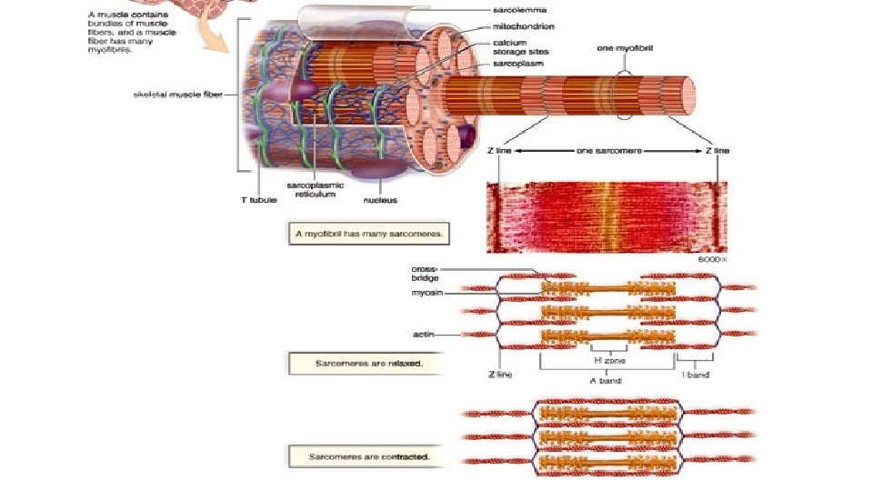
Microscopic Anatomy & Physiology • Myofibrils are contractile portions of fibers that lie parallel and run length of fiber. • Protein filaments: • thick (made of myosin) • thin (made of actin) • Sarcomere has repeating bands of actin and myosin that occur between two Z lines in myofibril. • I band contains only actin filaments. • H zone contains only myosin filaments. • Anatomy of skeletal muscle 2. ram
Sliding Filament Model • As a muscle fiber contracts, sarcomeres within myofibrils shorten. • As sarcomere shortens, actin filaments slide past myosin; I band shortens and H zone disappears. • Sliding filament theory: • actin filaments slide past myosin filaments because myosin filaments have crossbridges that pull actin filaments inward, toward their Z line.
Sliding Filament Model • Contraction process involves sarcomere shortening, filaments themselves remain same length. • ATP supplies energy for muscle contraction. • Myosin filaments break down ATP to form crossbridges that attach to and pull actin filament.
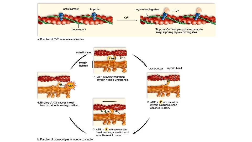
Muscular Contraction • Ca 2+ ions bind to troponin, which causes tropomyosin threads to shift position. • Change in structure of tropomyosin exposes myosin heads with ATP binding sites. • The myosin heads function as ATPase enzymes, splitting ATP into ADP + P • After attaching to actin filaments, myosin cross-bridges bend forward, actin filament is pulled along.
http: //highered. mheducation. com/sites/0072495855/student_view 0/c hapter 10/animation__sarcomere_contraction. html
Fast twitch & slow twitch muscles • Slow twitch muscle fibers • contract slowly, but keep going for a long time • more mitochondria for aerobic respiration • less SR Ca 2+ remains in cytosol longer • long distance runner • “dark” meat = more blood vessels • Fast twitch muscle fibers • contract quickly, but get tired rapidly • store more glycogen for anaerobic respiration • sprinter • “white” meat
Diseases of Muscle tissue • ALS • amyotrophic lateral sclerosis • Lou Gehrig’s disease • motor neurons degenerate • Myasthenia gravis • auto-immune • antibodies to acetylcholine receptors Stephen Hawking
Botox • Bacteria Clostridium botulinum toxin • blocks release of acetylcholine • botulism can be fatal muscle
Rigor mortis no life, no breathing u no breathing, no O 2 u no O 2, no aerobic respiration u no aerobic respiration, no ATP u no ATP, no Ca 2+ pumps u Ca 2+ stays in muscle cytoplasm u muscle fibers continually contract u § tetany or rigor mortis u eventually tissues breakdown & relax § measure of time of death
Signal Transduction Pathway Fig. 49. 2 Copyright © 2002 Pearson Education, Inc. , publishing as Benjamin Cummings
• Sensory reception begins with the detection of stimuli by sensory receptors. • Exteroreceptors detect stimuli originating outside the body. • Interoreceptors detect stimuli originating inside the body. Copyright © 2002 Pearson Education, Inc. , publishing as Benjamin Cummings
Sensory Processing • Transduction. • The conversion of stimulus energy into a change in membrane potential. • Amplification. • The strengthening of stimulus energy that is can be detected by the nervous system. • Transmission. • The conduction of sensory impulses to the CNS. • Integration. • The processing of sensory information. Copyright © 2002 Pearson Education, Inc. , publishing as Benjamin Cummings
Sensory Receptors • Mechanoreceptors respond to mechanical energy. • Pain receptors = nocioceptors. • Different types of pain receptors respond to different types of pain. • Thermoreceptors respond to heat or cold. • Respond to both surface and body core temperature. • Chemoreceptors respond to chemical stimuli. • Electromagnetic receptors respond to electromagnetic energy. • Photoreceptors respond to the radiation we know as visible light. Copyright © 2002 Pearson Education, Inc. , publishing as Benjamin Cummings
• http: //www. mindbites. com/series/423 -biology-the-nerve-impulse