Midwall left ventricular systolic function in cardiac hypertrophy
- Slides: 21
Midwall left ventricular systolic function in cardiac hypertrophy J Mayet, B Wasan, B Ariff, N Chapman, M Shahi, NR Poulter, PS Sever, RA Foale, SAMc. G Thom Peart-Rose Clinic and Department of Cardiology St Mary’s Hospital, Imperial College, London
Is LV systolic function preserved in LVH? • Discrepancy between experimental and human studies – Former suggest myocardial function depressed – Latter indicate it is preserved • Experimental and human studies not strictly compatible – Experimental studies measure myocardial or myofibril function – Human studies have assessed whole heart function by utilising endocardial measurements
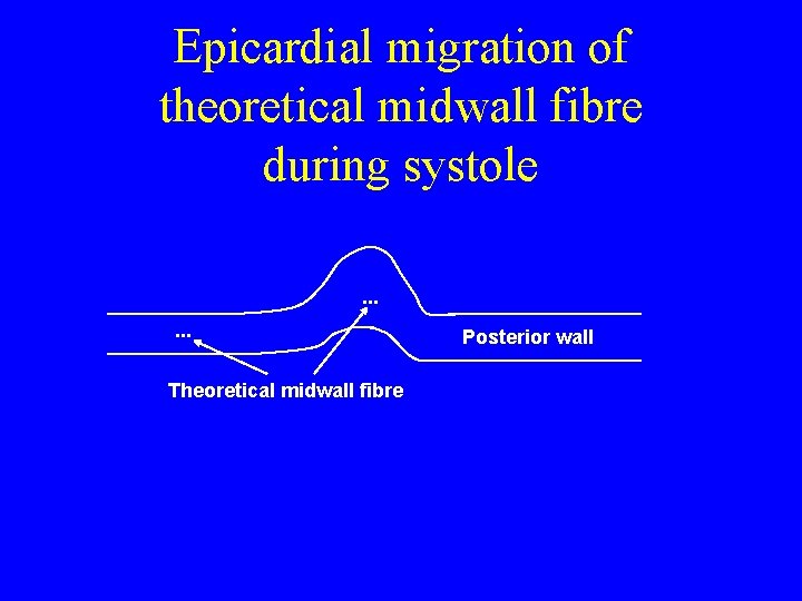
Midwall LV shortening • It is often assumed that the inner and outer parts of the LV wall thicken equally in systole • Myocardial shortening in subendocardial is greater than subepicardial layers • Therefore a theoretical mid-point in the LV wall shows relative migration toward the epicardium throughout contraction
Epicardial migration of theoretical midwall fibre during systole. . . Theoretical midwall fibre Posterior wall
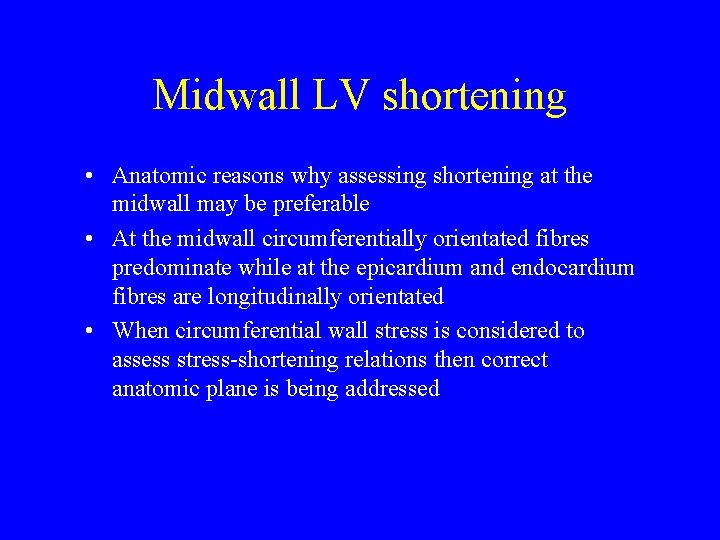
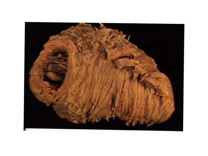
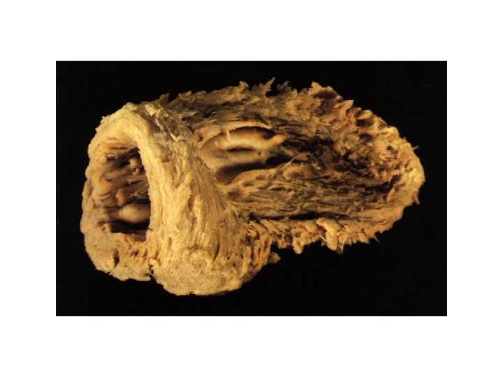
Midwall LV shortening • Anatomic reasons why assessing shortening at the midwall may be preferable • At the midwall circumferentially orientated fibres predominate while at the epicardium and endocardium fibres are longitudinally orientated • When circumferential wall stress is considered to assess stress-shortening relations then correct anatomic plane is being addressed
What is the relationship between endocardial and midwall shortening in normal subjects and those with LVH? • 38 previously untreated hypertensives with LVH compared with normotensive controls (healthy volunteers) • No patient had significant co-morbidity • Each underwent full two-dimensional echocardiographic examination
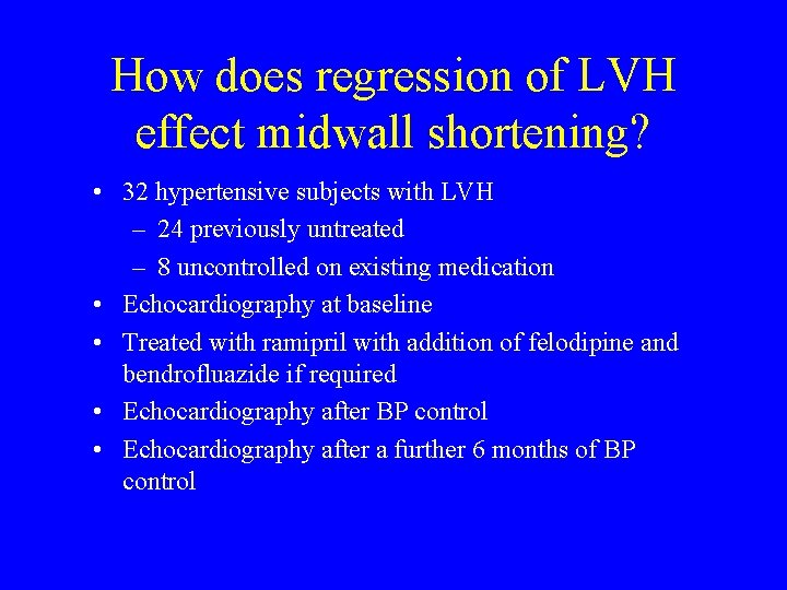
How does regression of LVH effect midwall shortening? • 32 hypertensive subjects with LVH – 24 previously untreated – 8 uncontrolled on existing medication • Echocardiography at baseline • Treated with ramipril with addition of felodipine and bendrofluazide if required • Echocardiography after BP control • Echocardiography after a further 6 months of BP control
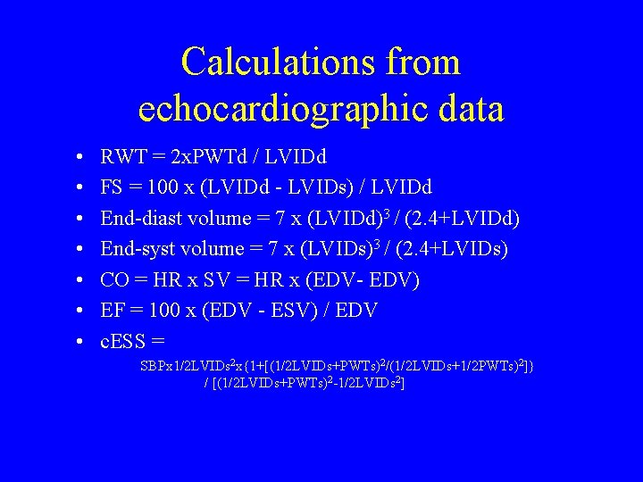
Calculations from echocardiographic data • • RWT = 2 x. PWTd / LVIDd FS = 100 x (LVIDd - LVIDs) / LVIDd End-diast volume = 7 x (LVIDd)3 / (2. 4+LVIDd) End-syst volume = 7 x (LVIDs)3 / (2. 4+LVIDs) CO = HR x SV = HR x (EDV- EDV) EF = 100 x (EDV - ESV) / EDV c. ESS = SBPx 1/2 LVIDs 2 x{1+[(1/2 LVIDs+PWTs)2/(1/2 LVIDs+1/2 PWTs)2]} / [(1/2 LVIDs+PWTs)2 -1/2 LVIDs 2]
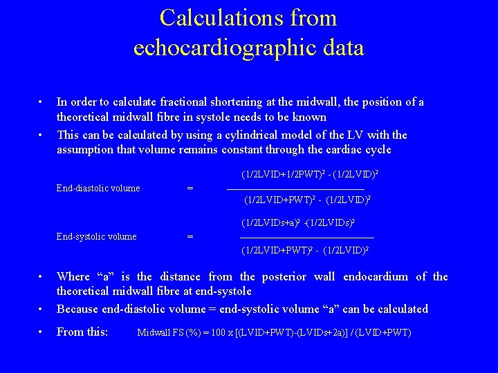
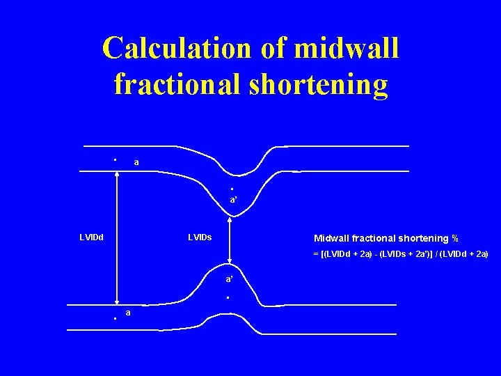
Calculations from echocardiographic data • • In order to calculate fractional shortening at the midwall, the position of a theoretical midwall fibre in systole needs to be known This can be calculated by using a cylindrical model of the LV with the assumption that volume remains constant through the cardiac cycle (1/2 LVID+1/2 PWT)2 - (1/2 LVID)2 End-diastolic volume = ______________________ (1/2 LVID+PWT)2 - (1/2 LVID)2 (1/2 LVIDs+a)2 -(1/2 LVIDs)2 End-systolic volume = _____________________ (1/2 LVID+PWT)2 - (1/2 LVID)2 • • Where “a” is the distance from the posterior wall endocardium of theoretical midwall fibre at end-systole Because end-diastolic volume = end-systolic volume “a” can be calculated • From this: Midwall FS (%) = 100 x [(LVID+PWT)-(LVIDs+2 a)] / (LVID+PWT)
Calculation of midwall fractional shortening. a . a’ LVIDd LVIDs Midwall fractional shortening % = [(LVIDd + 2 a) - (LVIDs + 2 a’)] / (LVIDd + 2 a) a’ . . a
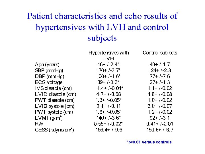
Patient characteristics and echo results of hypertensives with LVH and control subjects *p<0. 01 versus controls
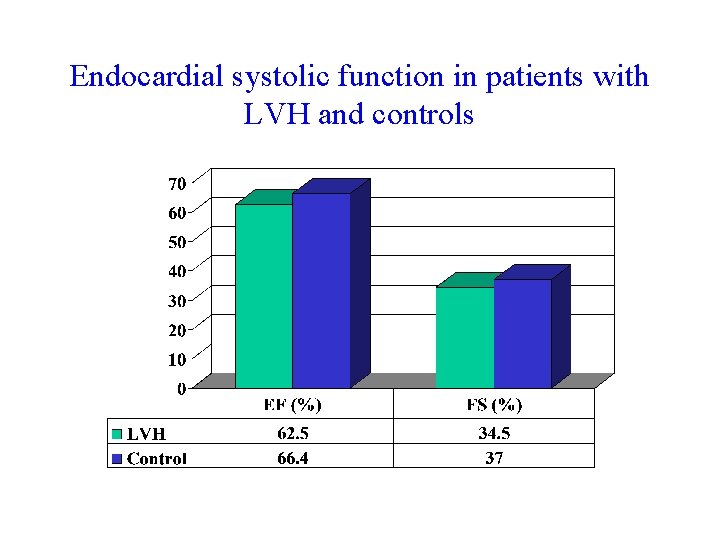
Endocardial systolic function in patients with LVH and controls
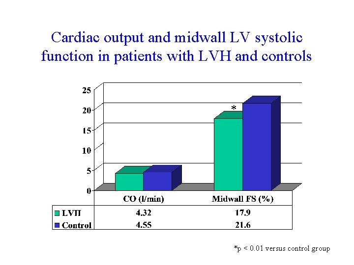
Cardiac output and midwall LV systolic function in patients with LVH and controls * *p < 0. 01 versus control group
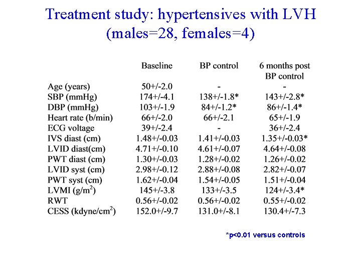
Treatment study: hypertensives with LVH (males=28, females=4) *p<0. 01 versus controls
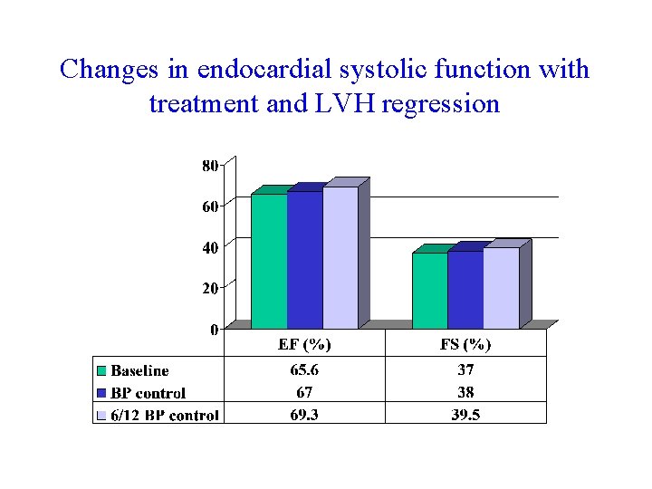
Changes in endocardial systolic function with treatment and LVH regression
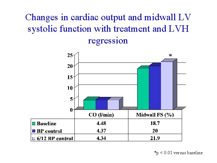
Changes in cardiac output and midwall LV systolic function with treatment and LVH regression * *p < 0. 01 versus baseline
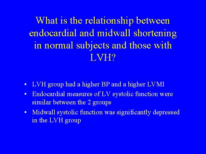
What is the relationship between endocardial and midwall shortening in normal subjects and those with LVH? • LVH group had a higher BP and a higher LVMI • Endocardial measures of LV systolic function were similar between the 2 groups • Midwall systolic function was significantly depressed in the LVH group
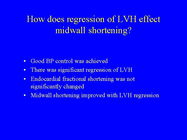
How does regression of LVH effect midwall shortening? • Good BP control was achieved • There was significant regression of LVH • Endocardial fractional shortening was not significantly changed • Midwall shortening improved with LVH regression
Discussion • In spite of similar endocardial systolic function, midwall systolic function is significantly depressed in subjects with LVH secondary to hypertension • Midwall shortening is improved with regression of LVH using an ACE inhibitor and calcium antagonist based treatment regime • A reduced midwall shortening has been found to be associated with a lower exercise performance • Depressed midwall shortening has been shown to be an independent predictor of an adverse outcome in hypertensive subjects, particularly in those subjects with additional LVH