Lameness in Racehorses Paul R Earl Facultad de


 often referred")

 and thickening and lameness.")


- Slides: 28
Lameness in Racehorses Paul R Earl Facultad de Ciencias Biológicas Universidad Autónoma de Nuevo León San Nicolás, NL 66451, Mexico
Millions and millions of dollars are involved in horse lameness. It is caused mainly by overexercise on hard surfaces. The first point is that 70 % of horses on dirt tracks have front cannon bone problems, whereas 4. 1 times less (17%) on turf tracks have. The cannon bone is the main fused leg bone. The second point is that maturity is reached at 4 -5 years of age, and young bones are soft. This is an appeal for all racetracks to switch from dirt to grass.
Racehorses run farther as they get older, staring at ¼ or 3/8 th of a mile (3 furlongs) going to 2 miles usually. Nonetheless, it is speed not distance that causes the damage. Still, the owner of Man o’ War at 3 years old winning the Preakness at 1 ¼ and the Belmont Stakes at 1 ½ miles thought the Kentuky Derby at a 1 1/8 miles was too long for him! There is more to it than if stake winners only. Some owners only want to win stake races. Then those horses that cannot reach a “Gallant Fox” triple crown standard will be sold or crippled or both.
The main sign of trouble ahead is heat in the legs. Infrared thermogram. What is your diagnosis?
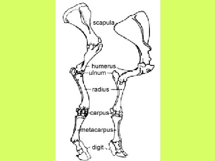
The anatomy of the legs
Some Problems with the Hooves Subsolar Infections are the commonest hoof infections, and common causes of acute lameness. A penetrating wound in the sole, frog or white line generally caused by the horse stepping on a sharp object like a nail causing lacerations. Bruised Soles are one of the most common causes of lameness in both shod and unshod horses. Injury to the sole may cause damage to the sensitive structures underneath and results in bruising. The result can be damage to the many tiny blood vessels underneath the sole and consequent hemorrhage.
Normal and contracted heels
Splints that were parts of the toes millions of years ago can make a horse from 2 to 3 years lame. The ligament between each side bone-splint--and the cannon bone fuse possibly causing a temporary soreness. Bucked shins is a very common condition possibly caused by microfractures and the like caused in turn by concussion and stress. The surface, the periosteum, may separate from the front of the cannon bone in the process of laying down more bone. Many trainers force the horse to buck as inevitable. This is done by fast works, not galloping long distances. This scale is: walk, jog or trot, gallop, breeze, work or race.
Desmitis of Supporting Ligaments like the with impar ligament and the proximal suspensory ligament desmitis is a separate diagnosis from navicular disease. Will some of these lame horses develop inflammation and degeneration of the navicular bone? Such horses are separated from those with abnormal fluid in the navicular bone, although a few horses have both fluid in the bone and desmitis of the supporting ligaments. These horses are diagnosed with navicular disease. The treatment is the same, mainly depending on rest. .
Thrush is an infective condition of the frog and its sulci which results in degeneration of the horn and the production of foul smelling grey/black discharge. In severe or neglected cases the infection can spread to involve the underlying corium. The degeneration of the horn is due to infection with bacteria and especially fungi. Wet unhealthy stable conditions, poor routine foot care, prolonged confinement, overgrown ragged frogs and long or high heels which produce deep sulci. Most of these conditions can be corrected by a blacksmith.
Nail Prick occurs at shoeing when a nail penetrates the live structures of the foot. It happens most commonly if the horse is restless or fractious, the hoof wall is particularly thin or broken, the shoe is too small or the hoof has been dumped. Dumping means Dumping is excessive rasping of the outside of the walll usually at the toe, but it can extend all the way around the wall. Nail Bind occurs when the nail of the shoe is driven close to, but not into the sensitive structures of the foot resulting in pressure and inflamation.
Tendonitis It is inflammation of the superficial deep digital flexor tendons (DDFT) often referred to as a bowed tendon due to the bowed appearance of the tendons on the back of the cannon bone. The “strap” that crosses the cannon bone in front is the extender ligament Tendonitis of the DDFT is fairly rare. The damage can range from mild tearing of tendon fibers to actual rupture of the entire tendon and the bone-attached ligaments. Tears in tendons are best diagnosed by ultrasound. Tendonitis is predominately a forelimb problem.
The 2 primary causes of superficial DDFT are either acute trauma due to overload, or a cumulative damage due to repeated damage to the tendons. Aside from poor conditioning and misapplied racing bandages, high racing speed can bow a horse. Hyperextension of the distal interphalangeal joint as a result of allowing the to grow and thus rise may cause chronic or acute tearing of the DDF tendon fibers. Normally, tendons are elastic so that they can stretch with routine overloading. This is referred to as elastic deformation.
Clinical signs of superficial DDFT include heat, tendon swelling (edema) and thickening and lameness. X-ray images of lame horses likely are less effective than ultrasound. Inflammation may be best demonstrated by IR photographs. Note that in many late cases of inflammation, antiinflammatary drugs are contraindicated, as inflammation speeds up the healing process. After tendon fibers are torn, hemorrhage results in blood clots likely along with swelling and inflammation. As the inflammation and the blood clots subside, scar tissue forms in the place of the displaced fibers.
Typical bowed tendon. This horse might be checked for bone chips with X rays.
A low bow in the left leg
Laminitis—inflammation of the laminas of the foot--is a common, painful and sometimes disastrous condition of the horse hoof that has always been known. Laminitis occurs when the coffin bone (distal phalanx) and the inner aspect of the hoof wall are separated. Nevertheless, a blacksmith can likely cure a given problem with well managed racehorses. Laminitis is—essentially—a problem of neglected horses.
Bring the hoof and the coffin bone back into their original positions through trimming. Optimize blood flow through the foot by a little walking or roping. Removing the cause of laminitis: overload of the coffin bone.
There are 2 kinds of laminas. Dermal laminae grow outwards from the laminar dermis attached to the coffin bone. They are made of living connective tissues that fold into primary finger-like structures; each of these in turn is folded into hundreds of feather-like secondary laminas. These dermal laminas interdigitate with corresponding epidermal laminae, each again of primary and secondary generations, that project inward from the inner horny surface of the wall, anchoring the coffin bone to the hoof wall. The amount of folding in the laminas increases the surface area of bonding.
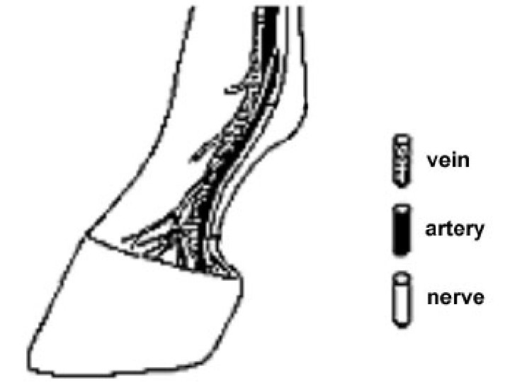
The blood supply to the equine foot is unusual. All arteries have receptors in their walls that can bind specific chemical messengers circulating throughout the body. The binding of these chemicals some like bradykinin causes the dilation or constriction of the arteries changing the amount of blood reaching the tissues. Blood rich in the oxygen and nutrients necessary to sustain living tissues is carried in arteries from the heart to the hoof. It then passes through a capillary bed in the dermis where oxygen and nutrients can diffuse to the cells.
Acute laminitis occurs anywhere from 24 to 72 hours after the initial damage to the basement membrane and is heralded by the clinical signs of pain. Signs of acute laminitis include: 1/ severe lameness, reluctance to move or even to recumbency 2/ typical stance to try to get weight off the toes: front feet out in front if only the forelimbs are involved; all 4 legs in under the body if all limbs are involved; pointing with a leg if only one limb is involved 3/ increased digital pulses are almost always present 4/ continued heat in the foot
Disposing Factors in Laminitis: Numerous predisposing factors have been identified. They can be divided into 6 major groups: 1/ High heels 2/ Carbohydrate overload: excess grain, lush pasture, sudden feed change 3/ Endotoxemia, sepsis, shock: colic, intestinal obstruction, pleuritis, retained placenta, metritis, abortion 4/ Excessive unilateral weight bearing: postfracture repair, severe lameness 5/ management: unconditioned animals worked on a hard surface, cold water ingestion by overheated horse, overweight animal
Therapy: The treatment of horses that develop acute laminitis can be considered an emergency. A delay of even a few hours can make the difference between a successful outcome and a failure. If any of the known risk factors for laminitis have occurred, therapy should be started before the clinical signs become visible. The treatment should be focused on restoring the circulation of the hoof and of course the reattachment of the laminas to the coffin bone. It is at least 8 weeks before prognosis. In chronic cases, 4 months to a year are required before future performance abilities can be estimated.
Navicular disease Finding excessive fluid in the navicular bone, especially diffuse fluid in the medullary cavity, indicates navicular disease. The diagnosis is considerably refined for about 10 years via the use of magnetic resonance for soft tissues. Ultrasound produces poor images in boney tissue. X ray is often used. Again, the basic cause is not at all always overwork pulling on bones, ligaments and muscles, although the basic treatment is always rest. The main cause is high heels and sometimes bar shoes.
A low bow with navicular disease
What have we learnt ? We should know 2 facts about lameness: 1/ racing on turf rather than dirt will vastly reduce injuries, and 2/ velocity over pulse gives the horse’s capability at a given moment. Running on grass will GREATLY reduce owner costs, but raise racetrack costs. Training horses as a single set not as individuals is the universal rule. As very many are quite beyond their capacities, lameness is the common result and has the common factor of uneconomic rest.