JCM OSCE Questions and Answers 6 th May


 • It contains amatoxin. – Cytotoxic")
 • This is the mushroom eaten by this")


 What are the possible complications? • # scaphoid: – – AVN")


 What is the likely diagnosis? Dextrocardia (c) How would you confirm")

 Biliary colic, acute cholecystitis, acute cholangitis, hepatitis, liver abscess, RLL pneumonia")
 What is the most likely diagnosis? Liver abscess (d) Give 2")

 What is the name of this condition? Purple urine bag syndrome")
 Give 3 common risk factors for this condition. – Constipation –")
 and indirubin (red) or")

 Briefly describe your management. Change urinary catheter +/- antibiotics")


 Comment on the blood gas and calculate the anion gap. Anion")

- Slides: 39
JCM OSCE Questions and Answers 6 th May 2015 Queen Mary Hospital
Question 1 a A family of 2 members developed abdominal pain, nausea and vomiting one hour after eating mushroom. It was picked at the hillside and wellcooked. In addition to the usual steps to manage as food poisoning and to trace the culprit, what single most important question should you ask to assess the risk and severity of mushroom poisoning?
Answer 1 a • Time and onset of symptoms. • The most toxic mushrooms are cytotoxic. Time is needed for the uptake of toxins into cells and deprivation of end organ reserve. • Delayed onset (> 6 hours) usually implies potentially fatal mushroom ingestion.
Question 1 b • It was lucky that their relatives got some uncooked specimen for your identification. Who should you consult? How should you prepare and store the specimen?
Answer 1 b • Hong Kong Poison Information Centre – Amatoxin/ phallotoxin analysis by TRL – Friendly collaboration with Prof. S. W. Chiu (mycologist) for recognition of mushroom species
Answer 1 b • Preparation of specimens: – Fresh intact specimens are the best – Save more samples if possible, together with original package – Wrap in aluminium foil or wax paper and then put into paper bag – Store in refrigerator but do not freeze – Cooked specimen, gastric content and stool may contain mushroom remnants that may be useful if fresh specimen is not available
Question 1 c • Here is the most deadly mushroom in the world. Do you know its name and action? (Pictures 1 and 2)
Picture 1 Picture 2
Question 1 c • Amanita Phalloides (Death Cap) • It contains amatoxin. – Cytotoxic – GI symptoms in 6 – 10 hours – Fulminant liver failure and renal impairment after 2 – 3 days
Question 1 d (Distinction Q. ) • This is the mushroom eaten by this family (Picture 3). Do you know its name? Picture 3
Answer 1 d • Macrolepiota • It causes self-limiting GI symptoms.
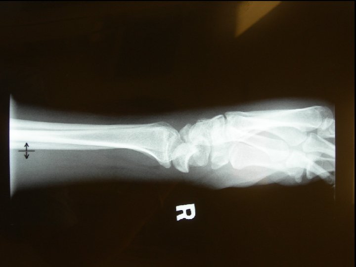
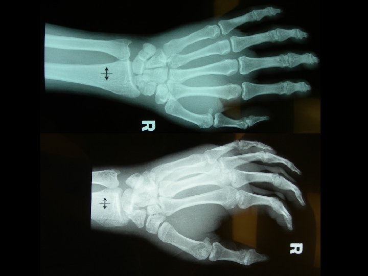
Question 2 • A 37 -year-old man attended A&E because of pain over his right wrist for one week after a fall from ladder. (a) What are the X-rays abnormalities?
Answer 2 a • Transcaphoid perilunate dislocation • Common in young adults • Fall on oustretched hand • Mechanism – bending with dorsal compression and tension on palmar surface owing to forced dorsiflexion
Perilunate Dislocation 60% with scaphoid fracture Radiolunate articulation is preserved Capitate dislocates almost always dorsal to the lunate capitolunate joint is disrupted, and the lunate projects through the space of Poirier • Proximal pole of scaphoid retains its articulation with the lunate, whereas the distal pole retains its relationship with the distal carpal row • • Image courtesy: http: //www. pt. ntu. edu. tw/hmchai/Kines 04/KINupper/Wrist. htm
Space of Poirier • Space between “greater arc” and “lesser arc” • Greater arc – Radiocapitate (yellow) and ulnocapitate (purple) ligaments interdigitate, forming arcuate ligament • Lesser arc – Scapholunate and lunotriquetral ligaments Image courtesy: https: //indianclinicalknowledgedotnet 1. wordpress. com/2012/10/24/space-of-poirier/
Question 2 (b) What are the possible complications? • # scaphoid: – – AVN Non-union Secondary OA Sudeck atrophy • Perilunate dislocation: – – Median nerve palsy (carpal tunnel syndrome) Sudeck atrophy AVN Long-term carpal instability: Dorsal/Volar Intercalated Segment Instability (DISI/VISI) (c) What is your treatment? – Prompt ORIF
Question 3 a • A 20 years old man had a routine preemployment check in a health care centre. He was referred to A&E because his ECG was commented “abnormal” by the ECG machine. He was asymptomatic. Another ECG performed in A&E showed no serial changes. • Describe the ECG.
1. RAD 2. R in a. VR 3. R wave regression over chest leads
Question 3 (b) What is the likely diagnosis? Dextrocardia (c) How would you confirm the diagnosis by simple means in A&E? Physical examination, CXR
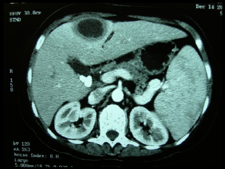
Question 4 • A 34 -year-old woman complained right upper quadrant pain and fever for five days. Mild localised tenderness was elicited at the right upper quadrant. She was not pregnant. BP was 109/61 mm. Hg and pulse rate was 102/min. (a) Suggest 4 differential diagnoses. (b) List 3 abnormalities of the CT film.
Answer 4 (a) Biliary colic, acute cholecystitis, acute cholangitis, hepatitis, liver abscess, RLL pneumonia (b) CT: multiple gallstones; complicated cystic lesion (infected cyst or abscess), hepatosplenomegaly
Question 4 (c) What is the most likely diagnosis? Liver abscess (d) Give 2 possible causative organisms of this disease in (i) Developed: E. Coli (most common), K. pneumoniae, Bacteroides, Enterococci, Anaerobeic Streptococci, and Microaerophilic Streptococci (ii) Developing: Entamoeba histolytica
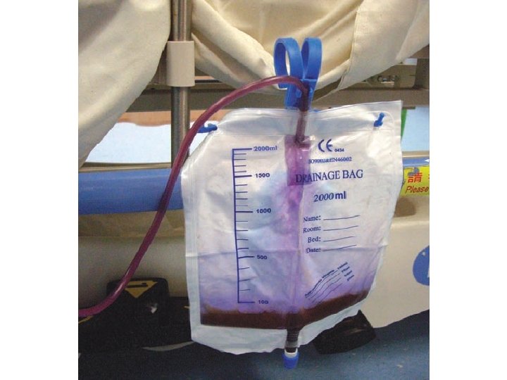
Question 5 • An elderly was brought to the Accident and Emergency Department because of colour changes of his urine in urinary catheter and bedside bag. He was bed-ridden and needed to use long term urinary catheterisation. • (Photo)
Question 5 (a) What is the name of this condition? Purple urine bag syndrome
Question 5 (b) Give 3 common risk factors for this condition. – Constipation – DM – Advanced age – Women – Chronically catheterized – Bacterial urinary infections that produce sulphatase/ phosphatase
Question 5 • The etiology is due to indigo (blue) and indirubin (red) or to their mixture that becomes purple. • ***Escherichia coli, Klebsiella pneumoniae, Enterobacter agglomerans, Pseudomonas aeruginosa, Proteus species, Providencia species, Enterococcus species and Streptococcus faecalis
www. medscape. com
Question 5 (c) Briefly describe your management. Change urinary catheter +/- antibiotics
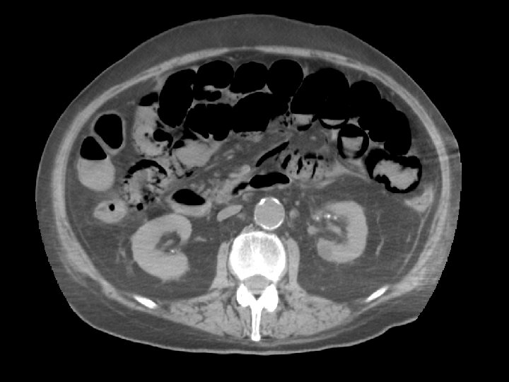
Question 6 • A 64 -year-old man presented to Emergency room because of abdominal pain, vomiting and shortness of breath. The abdomen was distended with no pulsatile mass. His ECG showed atrial fibrillation.
Question 6 • The result of arterial blood gas was as follows: • p. H 7. 029, p. CO 2 1. 93 k. Pa, p. O 2 23. 1 k. Pa, BE – 27, HCO 3 3. 8 mmol/L, Sa. O 2 99% • Na 136 mmol/L, K 4. 0 mmol/L, Cl 95 mmol/L • H’Stix 11. 7 mmol/L • Urgent CT abdomen was performed.
Question 6 (a) Comment on the blood gas and calculate the anion gap. Anion gap metabolic acidosis: Na (+ K) – Cl - HCO 3 = 136 (+ 4) – 95 - 3. 8 = 41. 2 (b) Describe the features of CT abdomen. Pneumatosis intestinalis, porto-venous gas, pneumoperitoneum (c) What is the most likely diagnosis? Ischaemic bowel
END