JCM OSCE AED UCH 592012 Case 1 11M


")
")
")
")


 location of")


- Slides: 47
JCM OSCE AED UCH 5/9/2012
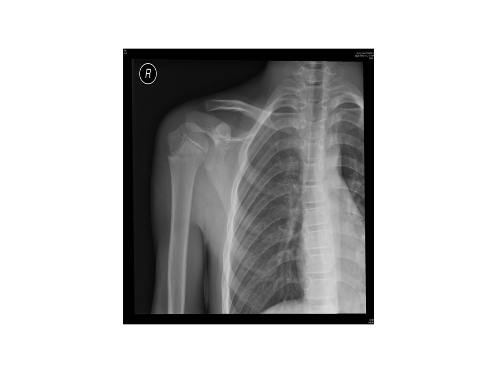
Case 1 • 11/M • Fought with classmate • Right arm pain with tenderness
1. What is the X-ray finding? 2. Name three differential diagnoses from the X -ray finding. 3. What will be your management?
1. What is the X-ray finding? • Rediolucent transverse line at neck of right humerus 2. Name three differential diagnoses from the X -ray finding. 3. What will be your management?
1. What is the X-ray finding? • Rediolucent transverse line at neck of right humerus 2. Name three differential diagnoses from the X-ray finding. • Normal variant of epiphyseal line • Little League’s syndrome (overuse syndrome) • Post-traumatic fracture of NOH 3. What will be your management?
1. What is the X-ray finding? • 2. Rediolucent transverse line at neck of right humerus Name three differential diagnoses from the X-ray finding. • Normal variant of epiphyseal line • Little League’s syndrome (overuse syndrome) • Post-traumatic fracture of NOH 3. What will be your management? • Immobilization (arm sling) and FU
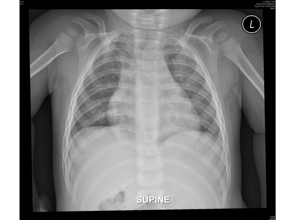
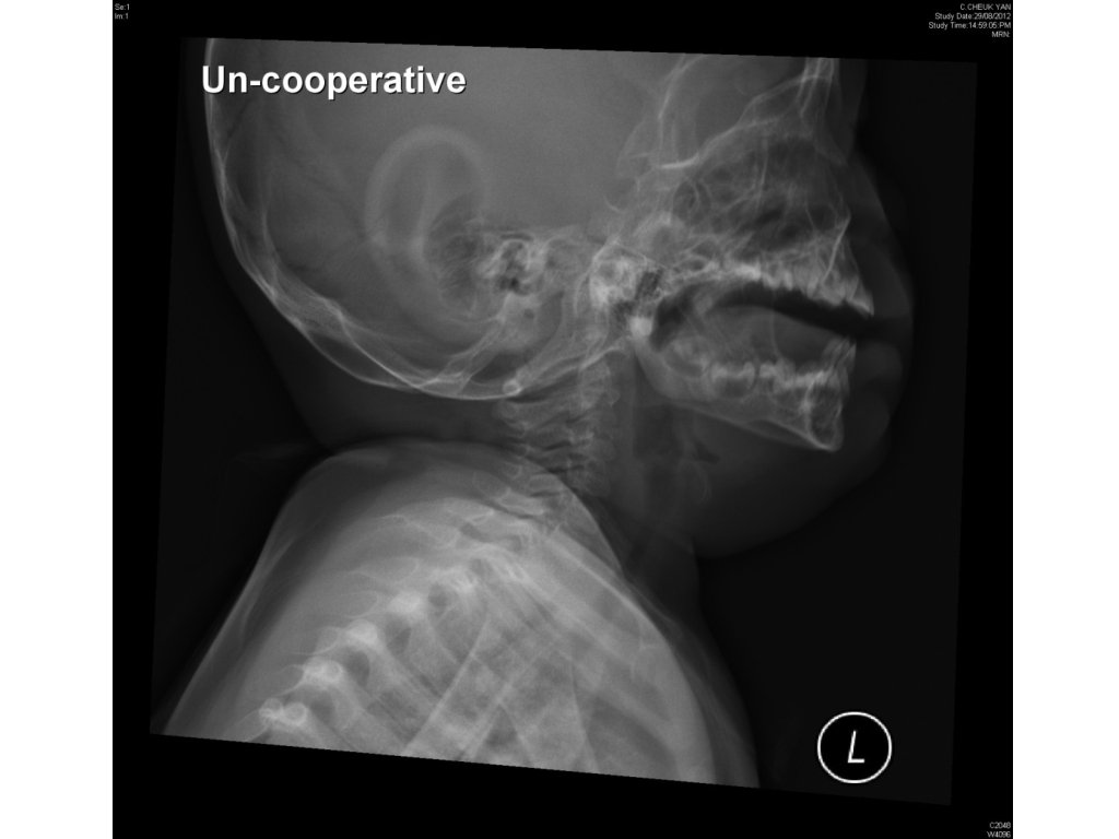
Case 2 • 13 months/M • Cough for 3 days • Noisy breathing for a day
1. Name 2 CXR findings. Are they pathological? 2. Name 3 neck X-ray findings. 3. What is the diagnosis? 4. Name 2 medications for the disease.
1. Name 2 CXR findings. Are they pathological? • Steeple sign (subglottic tracheal narrowing) - pathological. • Right deviation of trachea – normal variant. 2. Name 3 neck X-ray findings. 3. What is the diagnosis? 4. Name 2 medications for the disease.
1. Name 2 CXR findings. Are they pathological? • Steeple sign (subglottic tracheal narrowing) - pathological. • Right deviation of trachea – normal variant. 2. Name 3 neck X-ray findings. • Prevertebral soft tissue swelling, subglottic tracheal narrowing, ballooning of hypopharynx 3. What is the diagnosis? 4. Name 2 medications for the disease.
1. Name 2 CXR findings. Are they pathological? • Steeple sign (subglottic tracheal narrowing) - pathological. • Right deviation of trachea – normal variant. 2. Name 3 neck X-ray findings. • 3. What is the diagnosis? • 4. Prevertebral soft tissue swelling, subglottic tracheal narrowing, ballooning of hypopharynx Croup Name 2 medications for the disease.
1. Name 2 CXR findings. Are they pathological? • Steeple sign (subglottic tracheal narrowing) - pathological. • Right deviation of trachea – normal variant. 2. Name 3 neck X-ray findings. • 3. Prevertebral soft tissue swelling, subglottic tracheal narrowing, ballooning of hypopharynx What is the diagnosis? • 4. Croup Name 2 medications for the disease. • Dexamethasone (po/IM/IV) • Nebulized Adrenaline
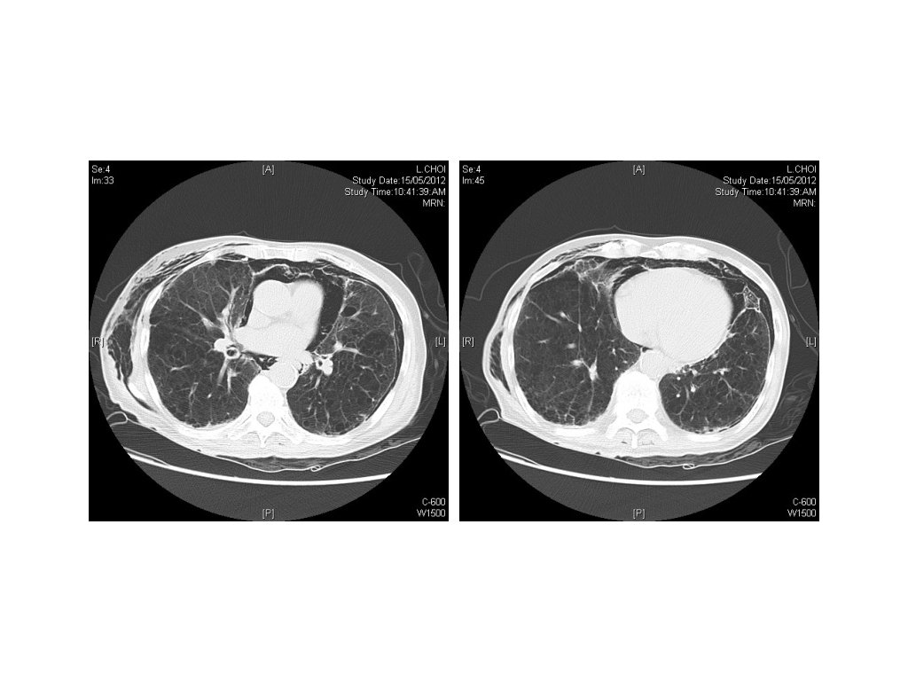
Case 3 • 90/M • Known COAD • Chest pain for a day
1. Besides the emphysematous and fibrotic changes of the lung, what are the X-ray findings? 2. What are the two differential diagnoses? 3. What further imaging is helpful? 4. If the patient deteriorates with SOB, what will be your management?
1. Besides the emphysematous and fibrotic changes of the lung, what are the X-ray findings? • Pneumomediastinum, pneumothorax, and subcutaneous emphysema 2. What are the two differential diagnoses? 3. What further imaging is helpful? 4. If the patient deteriorates with SOB, what will be your management?
1. Besides the emphysematous and fibrotic changes of the lung, what are the X-ray findings? • Pneumomediastinum, pneumothorax, and subcutaneous emphysema 2. What are the two differential diagnoses? • Spontaneous pneumomediastinum and pneumothorax • Ruptured oesophagus 3. What further imaging is helpful? 4. If the patient deteriorates with SOB, what will be your management?
1. Besides the emphysematous and fibrotic changes of the lung, what are the X-ray findings? • 2. What are the two differential diagnoses? • Spontaneous Pneumomediastinum and pneumothorax • Ruptured oesophagus 3. What further imaging is helpful? • 4. Pneumomediastinum, pneumothorax, and subcutaneous emphysema Water soluble contrast study If the patient deteriorates with SOB, what will be your management?
1. Besides the emphysematous and fibrotic changes of the lung, what are the X-ray findings? • 2. Pneumomediastinum, pneumothorax, and subcutaneous emphysema What are the two differential diagnoses? • Spontaneous Pneumomediastinum and pneumothorax • Ruptured oesophagus 3. What further imaging is helpful? • 4. Water soluble contrast study If the patient deteriorates with SOB, what will be your management? • Chest drain insertion
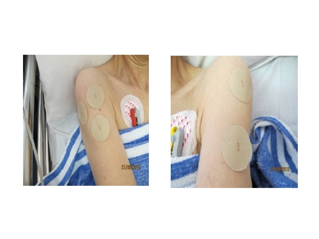
Case 4 • A 59 years old lady was recently prescribed transdermal patch for treatment of her Alzheimer’s disease because of poor oral drug compliance. • The patient was found to have vomiting, diarrhoea, dizziness and generalized limb weakness the day after starting the patch.
• The patient was fully conscious, with blood pressure 137/73 mm. Hg and pulse rate of 58 bpm. • Her pupil size were 2 mm, chest clear on auscultation and bowel sounds were normal.
1. What is the most likely ingredient in the patches? 2. Which drug class does the drug belong to? 3. Which enzyme is inhibited by the drug? 4. What is the most important step in the management of this patient? 5. Name the antidote(s) for overdose of this drug.
1. What is the most likely ingredient in the patches? • Answer: Rivastigmine 2. Which drug class does the drug belong to? 3. Which enzyme is inhibited by the drug? 4. What is the most important step in the management of this patient? 5. Name the antidote(s) for overdose of this drug.
1. What is the most likely ingredient in the patches? • 2. Answer: Rivastigmine Which drug class does the drug belong to? • Answer: Carbamate (1 mark) / Cholinesterase inhibitor (0. 5 mark) 3. Which enzyme is inhibited by the drug? 4. What is the most important step in the management of this patient? 5. Name the antidote(s) for overdose of this drug.
1. What is the most likely ingredient in the patches? • 2. Answer: Rivastigmine Which drug class does the drug belong to? • 3. Answer: Carbamate (1 mark) / Cholinesterase inhibitor (0. 5 mark) Which enzyme is inhibited by the drug? • Answer: Acetylcholinesterase 4. What is the most important step in the management of this patient? 5. Name the antidote(s) for overdose of this drug.
1. What is the most likely ingredient in the patches? • 2. Which drug class does the drug belong to? • 3. Answer: Carbamate (1 mark) / Cholinesterase inhibitor (0. 5 mark) Which enzyme is inhibited by the drug? • 4. Answer: Acetylcholinesterase What is the most important step in the management of this patient? • 5. Answer: Rivastigmine Answer: Search and remove all the patches (N. B. 2 more patches in the axilla of this patient) Name the antidote(s) for overdose of this drug.
1. What is the most likely ingredient in the patches? • 2. Answer: Rivastigmine Which drug class does the drug belong to? • 3. Answer: Carbamate (1 mark) / Cholinesterase inhibitor (0. 5 mark) Which enzyme is inhibited by the drug? • 4. Answer: Acetylcholinesterase What is the most important step in the management of this patient? • 5. Answer: Search and remove all the patches (N. B. 2 more patches in the axilla of this patient) Name the antidote(s) for overdose of this drug. • Answer: Atropine (Pralidoxime usually not indicated in carbamate poisoning)
• Note: • Clinical presentation in overdose: CNS depression, vomiting, diarrhoea, sweating, bradycardia, miosis, muscle weakness, muscle fasciculations (DUMBELS + nicotinic over-stimulation) etc • A patient died from pre-renal failure from excessive diarrhoea and vomiting • Patch resembles colour of skin so may be missed if not searched carefully • Pseudocholinesterase level may be low but not predictive of clinical severity
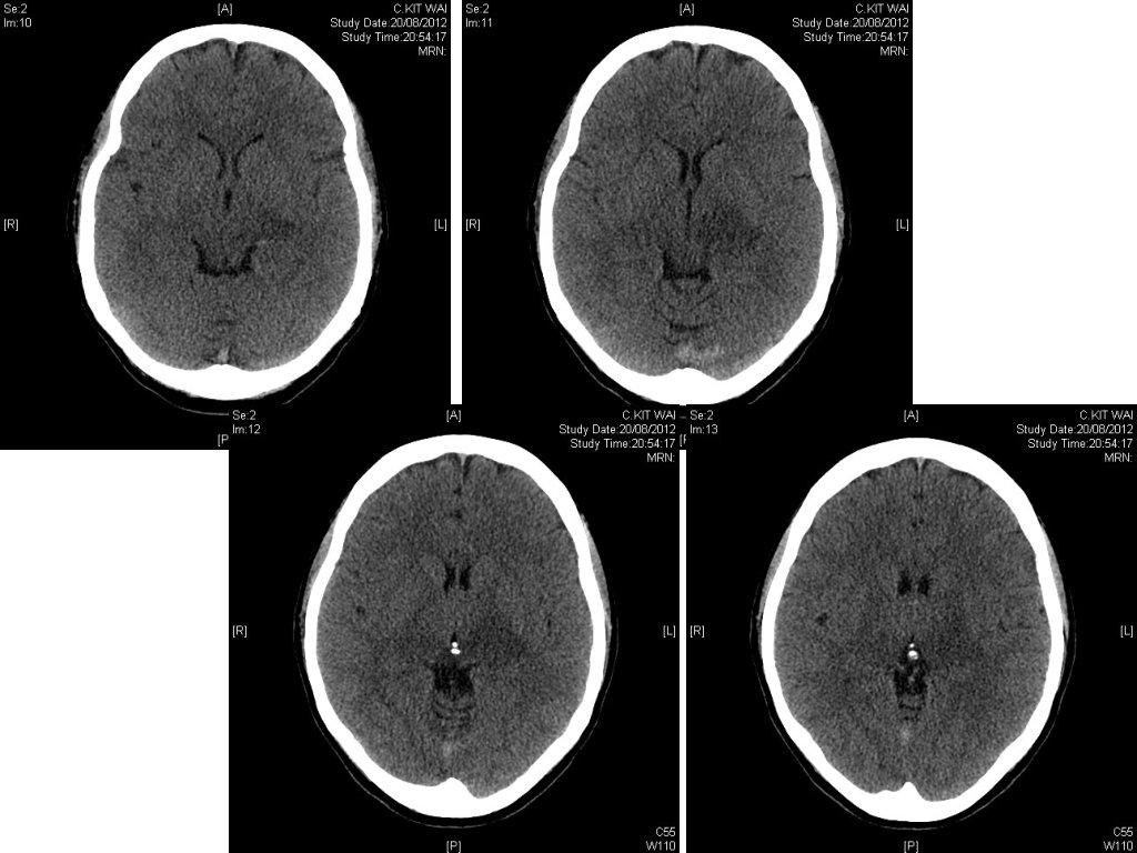
Case 5 • 47/F • Hx of Antithrombin III deficiency • Presented with • Headache for a few days • Right side weakness for a day
• BP 130/80, p 90/min • Afebrile • GCS 13/15; E 3 V 4 M 6 • PERL • Right hemi-paresis with power grade 4/5
1. What is the CT finding? 2. What is the diagnosis? 3. Any laboratory test can help to make the diagnosis? 4. Any further imaging can help to confirm the diagnosis? 5. Name the mainstay of treatment.
1. What is the CT finding? • Hyperdensity at sagittal and transverse sinuses 2. What is the diagnosis? 3. Any laboratory test can help to make the diagnosis? 4. Any further imaging can help to confirm the diagnosis? 5. Name the mainstay of treatment.
1. What is the CT finding? • 2. Hyperdensity at sagittal and transverse sinuses What is the diagnosis? • Cerebral sinus thrombosis 3. Any laboratory test can help to make the diagnosis? 4. Any further imaging can help to confirm the diagnosis? 5. Name the mainstay of treatment.
1. What is the CT finding? • 2. Hyperdensity at sagittal and transverse sinuses What is the diagnosis? • 3. Cerebral sinus thrombosis Any laboratory test can help to make the diagnosis? • D-dimer 4. Any further imaging can help to confirm the diagnosis? 5. Name the mainstay of treatment.
1. What is the CT finding? • 2. What is the diagnosis? • 3. Cerebral sinus thrombosis Any laboratory test can help to make the diagnosis? • 4. D-dimer Any further imaging can help to confirm the diagnosis? • 5. Hyperdensity at sagittal and transverse sinuses MRI +/-MR venogram, or CT venogram Name the mainstay of treatment.
Magnetic resonance venogram showing the cerebral venous system and most frequent (%) location of cerebral venous and sinus thrombosis, as reported in the International Study on Cerebral Venous and Dural Sinuses Thrombosis (n=624). 44. Saposnik G et al. Stroke 2011; 42: 1158 -1192 Copyright © American Heart Association
CT venogram of our patient
1. What is the CT finding? • 2. What is the diagnosis? • 3. Cerebral sinus thrombosis Any laboratory test can help to make the diagnosis? • 4. D-dimer Any further imaging can help to confirm the diagnosis? • 5. Hyperdensity at sagittal and transverse sinuses MRI +/-MR venogram, or CT venogram Name the mainstay of treatment.
1. What is the CT finding? • 2. Hyperdensity at sagittal and transverse sinuses What is the diagnosis? • 3. Cerebral sinus thrombosis Any laboratory test can help to make the diagnosis? • 4. D-dimer Any further imaging can help to confirm the diagnosis? • 5. MRI +/-MR venogram, or CT venogram Name the mainstay of treatment. • Anti-coagulation therapy
Proposed algorithm for the management of CVT. The CVT writing group recognize the challenges facing primary care, emergency physicians and general neurologists in the diagnosis and management of CVT. The aim of this algorithm is to provide guidance to physi. . . Saposnik G et al. Stroke 2011; 42: 1158 -1192 Copyright © American Heart Association