Huge Rectus Sheath Haematoma Complicating Insertion of Inferior


 is uncommon cause of abdominal pain but could")


, tachypenic, tachycardic with abdominal pain")


- Slides: 15
Huge Rectus Sheath Haematoma Complicating Insertion of Inferior Vena Cava Filter Amro Elnagar. MD Lecturer of cardiology Banha University Hospital
Amro Elnagar, MD I have no relevant financial relationships
Learning objectives 1. Rectus sheath haematoma(RSH) is uncommon cause of abdominal pain but could be fatal , especially in anticoagulated patients. 2. Care during femoral puncture is important to avoid laceration of inferior epigastric artery.
Clinical Presentation • A 69 years old female, hypertensive. • Presented with Left side iliofemoral DVT , treatment with LMWH & Warfarin was started. • Three days later, she complained acute onset dyspnea, chest pain &hypotension. • ECG: unremarkable except for sinus tachycardia • Echo: Dilated right side with moderate plumona • Plumonary CT angiography confirmed diagnosis of massive plumonary embolism.
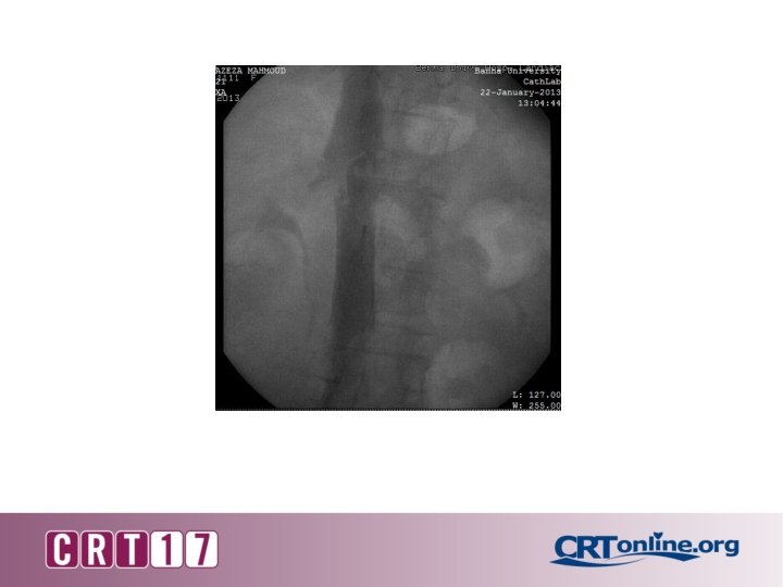
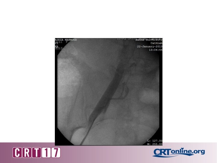
Procedure data • We decided to insert IVC filter for prevention of recurrent pulmonary embolism. • Through right femoral vein, 6 F sheath was inserted , Celect TM (COOk) was inserted. • The procedure went smoothly & patient transferred to intermediate care room.
• Two hours later, patient became hypotensive (70/40), tachypenic, tachycardic with abdominal pain & localized tenderness. • Fluid resuscitation was started, however the patient did not improved. • CBC shows decrease hemoglobin(7 g/dl) & haematocrit value (24). • 4 units of packed RBCs & 4 unit of plasma were given.
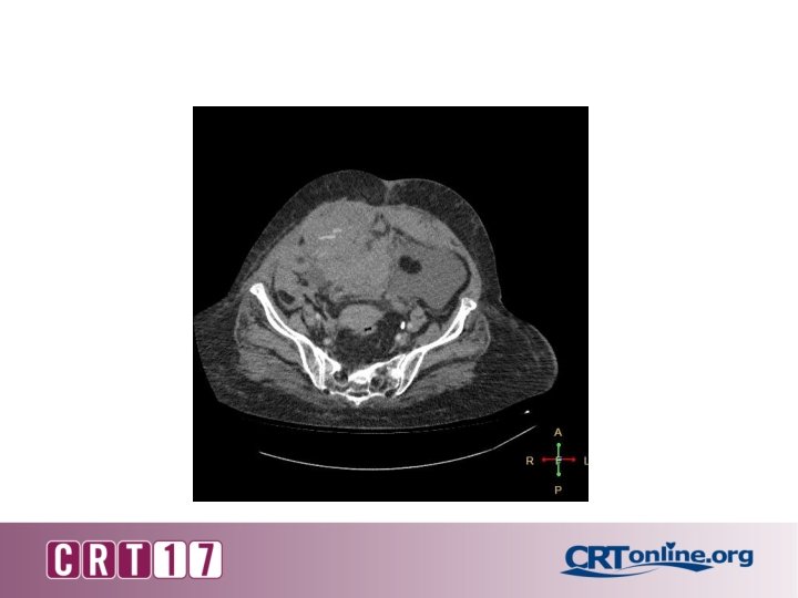
• Abdominal CT with contrast revealed type III rectus sheath haematoma extending into pelvis with active bleeding into haematoma.
Type III rectus sheath hematoma extending into pelvis with disruption of rectus sheath inferiorly.
Imaging study
• Discussion was made with vascular surgeons to decide whether to perform Gel Foam emblization or open surgical ligation of bleeder & evacuation of haemtoma. • Surgical ligation was done successfully with complete evacuation of haematoma.
• The patient was discharged to home after 7 days in stable condition.
Take Home Message • Rectus sheath hematoma is serious complication especially in anticoagulated patients. Laceration of inferior epigastric artery can occur during femoral puncture. Early diagnosis & management is of paramount importance as this condition can be fatal.