CLINICAL ANATOMY OF ANTERIOR ABDOMINAL WALL RECTUS SHEATH

CLINICAL ANATOMY OF ANTERIOR ABDOMINAL WALL & RECTUS SHEATH By: Dr. Mujahid Khan

Structure of Abdominal Cavity Ø Superiorly it is formed by diaphragm which separates the abdominal cavity from the thoracic cavity Ø Inferiorly the abdominal cavity is continuous with the pelvic cavity through the pelvic inlet

Structure of Abdominal Wall Anteriorly: Ø The abdominal wall is formed above by lower part of the thoracic cage Ø Below by the rectus abdominis, external oblique, internal oblique, and transversus abdominis muscles and fasciae

Structure of Ant. Abdominal Wall Ø It is made up of skin, superficial fascia, deep fascia, muscles, extraperitoneal fascia and parietal peritoneum Ø The abdominal walls are lined by a fascial envelope and the parietal peritoneum

Skin Ø Natural lines of cleavage in the skin are constant and run almost horizontally around the trunk Ø An incision along a cleavage line will heal as a narrow scar, while one that crosses the lines will heal as a wide scar

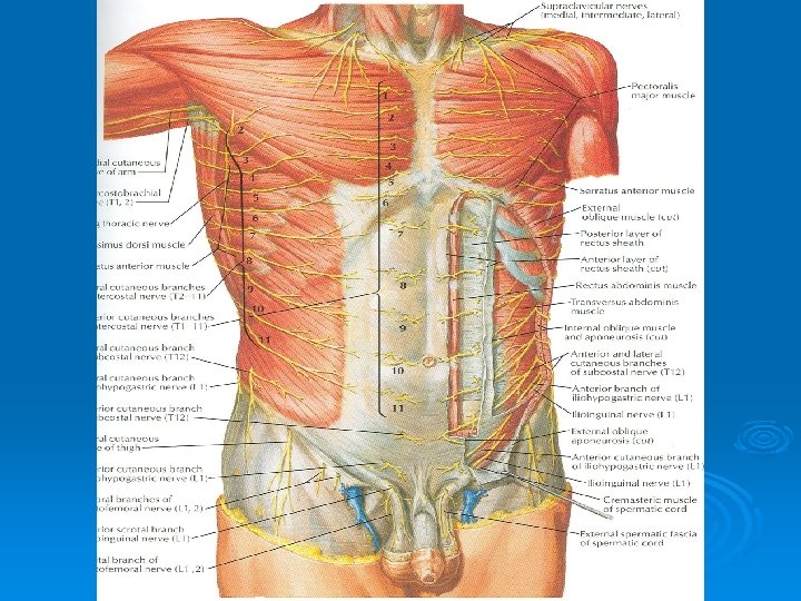
Cutaneous Nerve Supply Ø Is derived from the anterior rami of the lower six thoracic and first lumbar nerves Ø Thoracic nerves are the lower five intercostal and the subcostal nerves Ø First lumbar nerve is represented by the iliohypogastric and ilioinguinal nerves

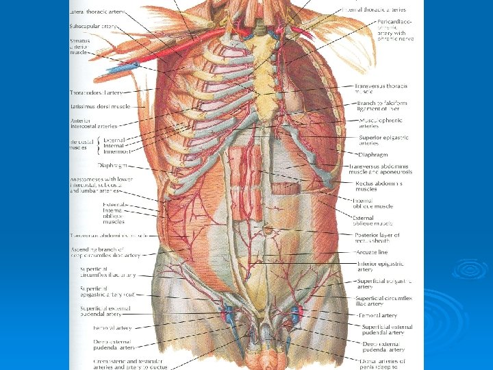
Blood Supply Ø Skin near the midline is supplied by branches of the superior epigastric artery (br. of int. thoracic artery) and the inferior epigastric artery ( br. of external iliac artery) Ø Skin of the flanks is supplied by branches from the intercostal, lumbar, and deep circumflex arteries

Superficial Fascia Ø Fatty layer or fascia of camper is continuous with the superficial fat over the rest of the body and may be extremely thick in obese patients Ø The membranous layer or scarpa’s fascia is thin and fades out laterally and above Ø Becomes continuous with the superficial fascia of the back and the thorax
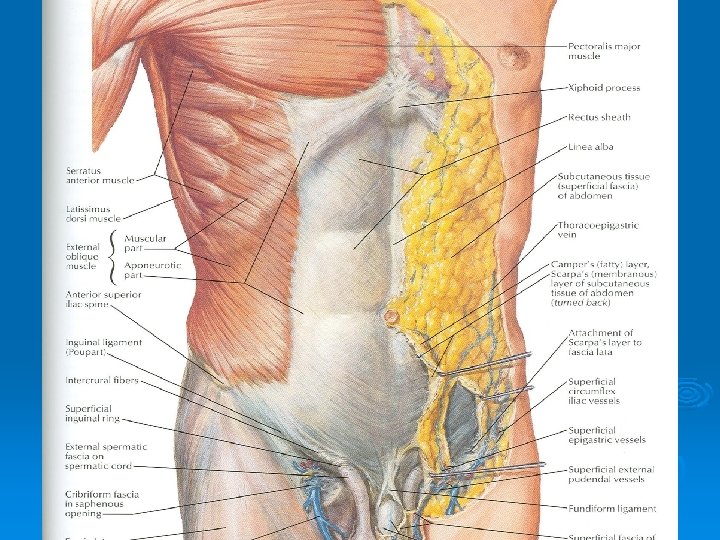

Superficial Fascia Ø Inferiorly the membranous layer passes onto the front of the thigh, where it fuses with the deep fascia Ø In the midline inferiorly forms a tubular sheath for the penis or clitoris Ø Below in the perineum, enters the wall of the scrotum or labia majora Ø From there it passes to be attached on each side to the margins of pubic arch, here it is called Colle’s fascia

Superficial Fascia Ø Posteriorly it fuses with the perineal body and the margin of the perineal membrane Ø The fatty layer is represented as a smooth muscle in the scrotum, the dartos muscle Ø The membranous layer persists as a separate layer

Deep Fascia Ø Deep fascia in the anterior abdominal wall is merely a thin layer of connective tissue covering the muscles Ø It lies immediately deep to the membranous layer of the superficial fascia

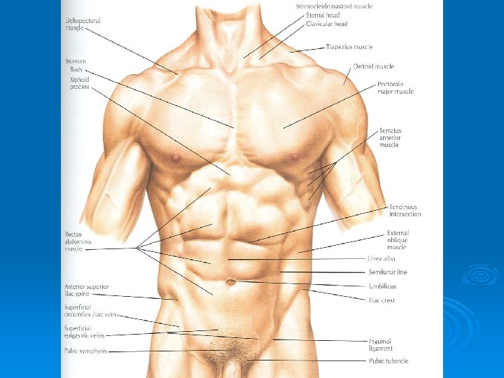
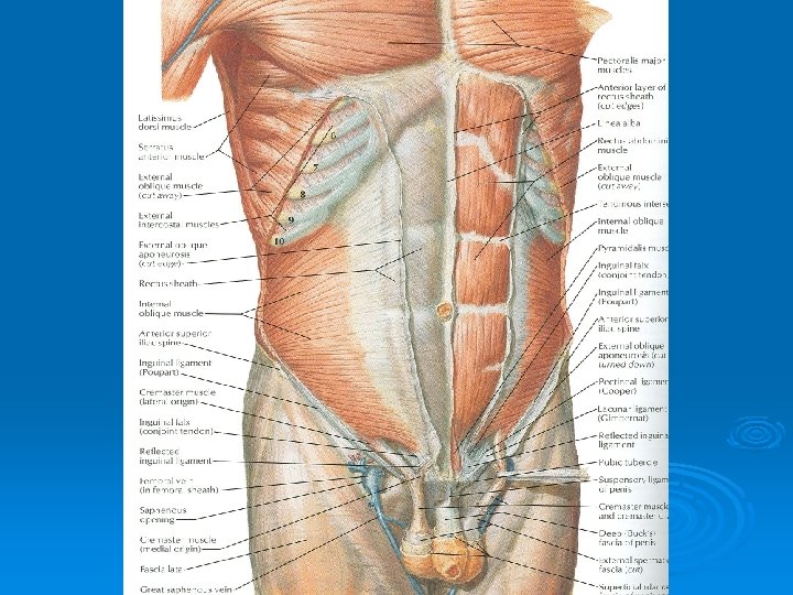
Muscles Ø Consists of Three broad thin sheets that are aponeurotic in front Ø From exterior to interior they are: Ø External oblique, internal oblique, and transverse Ø A wide vertical muscle, the rectus abdominis Ø They lie on either side of the midline anteriorly

Muscles Ø As the aponeurosis of three sheets pass forward, they enclose the rectus abdominis to form the rectus sheath Ø The cremaster muscle which is derived from the lower fibers of internal oblique, passes inferiorly as a covering of the spermatic cord and enters scrotum

External Oblique Muscle Ø Is a broad, thin, muscular sheet Ø Origin: Lower 8 ribs Ø Insertion: Xiphoid process, linea alba, pubic tubercle, iliac crest Ø Nerve Supply: Lower 6 thoracic nerves, iliohypogastric & ilioinguinal nerves Ø Action: Supports abdominal contents, assist in forced expiration, micturition, defecation, parturition, vomiting
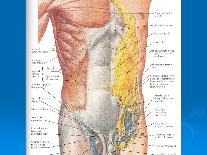

External Oblique Muscle Ø A triangular shaped defect in the external oblique aponeurosis lies immediately above and medial to the pubic tubercle, known as superficial inguinal ring Ø Between the anterosuperior iliac spine and the pubic tubercle, the lower border of the aponeurosis is folded backward on itself, forming the inguinal ligament

Internal Oblique Muscle Ø Origin: Lumbar fascia, iliac crest, lateral twothirds of inguinal ligament Ø Insertion: Lower three ribs and costal cartilages, xiphoid process, linea alba, symphysis pubis Ø Nerve Supply: Lower six thoracic nerves, iliohypogastric & ilioinguinal nerves Ø Action: Supports abdominal contents, assist in forced expiration, micturition, defecation, parturition, vomiting


Transversus Abdominis Ø Origin: Lower six costal cartilages, lumbar fascia, iliac crest, lateral third of inguinal ligament Ø Insertion: Xiphoid process, linea alba, symphysis pubis Ø Nerve Supply: Lower six thoracic nerves, iliohypogastric & ilioinguinal nerves Ø Action: Compresses abdominal contents
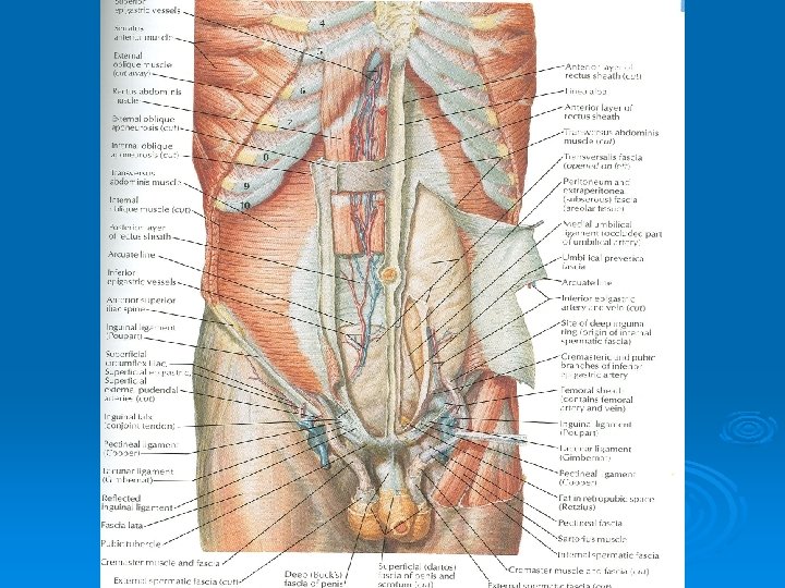

Rectus Abdominis Ø Origin: Symphysis pubis and pubic crest Ø Insertion: 5 th, 6 th and 7 th costal cartilages and xiphoid process Ø Nerve Supply: Lower six thoracic nerves Ø Action: Compresses abdominal contents, flexes vertebral column, accessory muscle of expiration

Lymph Drainage Ø Lymph drainage of the skin of the anterior abdominal wall above the umbilicus is upward to the anterior axillary (pectoral group of nodes) Ø Below the level of umbilicus drains downward and laterally to the superficial inguinal nodes Ø Swelling in the groin is may be due to enlarged superficial inguinal node

Venous Drainage Ø Venous blood is collected into a network of veins that radiate from the umbilicus Ø The network is drained above into the axillary vein via the lateral thoracic vein Ø Below into the femoral vein via the superficial epigastric and the great saphenous veins Ø Few small veins, the paraumbilical veins form a clinically important portal-system venous anastomosis
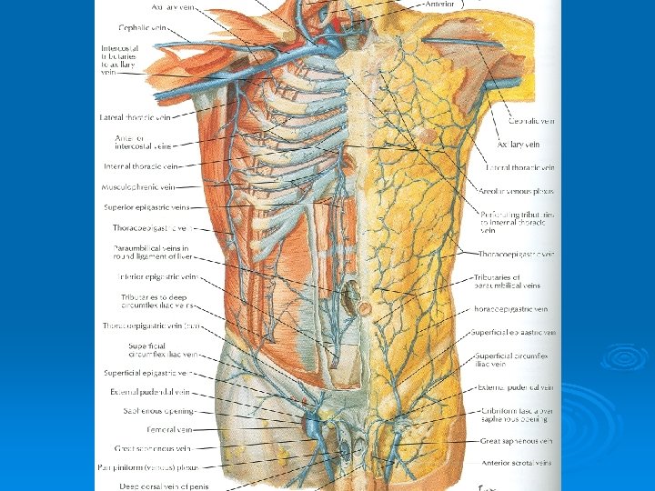

Caput Medusae Ø The superficial veins around the umbilicus and the paraumbilical veins connecting them to the portal vein may become grossly distended in case of portal vein obstruction Ø The distended subcutaneous veins radiate out from the umbilicus, producing in severe cases the clinical picture called Caput Medusae


Nerves Ø Nerves of the anterior abdominal wall supply the skin, muscles and the parietal peritoneum Ø They are derived from the anterior rami of lower six thoracic and the first lumbar nerves Ø Inflammation of parietal peritoneum causes pain in the overlying skin and also a reflex increase in tone of the abdominal musculature in the same area
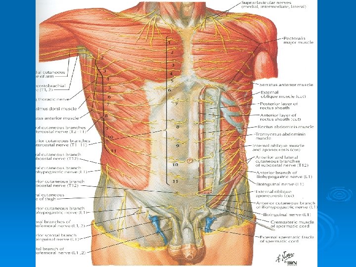

Rectus Sheath Ø Is a long fibrous sheath Ø Encloses the rectus abdominis and pyramidalis muscle (if present) Ø Contains the anterior rami of lower six thoracic nerves and the superior and inferior epigastric vessels and lymph vessels Ø Formed mainly by aponeurosis of three lateral abdominal muscles

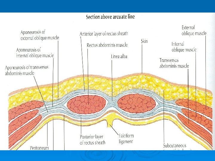
Rectus Sheath For description it is considered at three levels: Ø Above the costal margin the anterior wall is formed by the aponeurosis of the external oblique and posterior wall is formed by the thoracic wall Ø That is the 5 th , 6 th and 7 th costal cartilages and the intercostal spaces

Rectus Sheath Ø Between the costal margin and the level of the anterosuperior iliac spine, the aponeurosis of the internal oblique splits to enclose the rectus muscle Ø The external oblique aponeurosis is directed in front of the muscle Ø Transversus aponeurosis is directed behind the muscle

Rectus Sheath Ø Between the level of the anterosuperior iliac spine and the pubis, the aponeurosis of all three muscles form the anterior wall Ø The posterior wall is absent Ø The rectus muscle lies in contact with the fascia transversalis
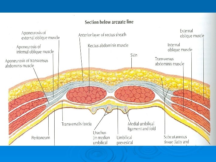

Rectus Sheath Ø The posterior wall of the rectus sheath is not attached to the rectus abdominis muscle Ø The anterior wall is firmly attached to it by the muscle’s tendinous intersections

Linea Alba Ø The rectus sheath is separated from its fellow on the opposite side by a fibrous band called the linea alba Ø Extends from the xiphoid process to the symphysis pubis
- Slides: 41