RECTUS SHEATH AND INGUINAL LIGAMENT Dr Tabrez Rectus

RECTUS SHEATH AND INGUINAL LIGAMENT Dr. Tabrez

Rectus Sheath n Is a long fibrous sheath n Formed by aponeurosis of three anterior abdominal muscles

Boundaries 2 walls Anterior wall n Posterior wall n 2 margins Medial margin n Lateral margin n

Formation of the Rectus Sheath For description it is considered at three levels 1. At the level of Xiphisternum/ Above the costal margin 2. From Xiphisternum to- midpoint of umbilicus and symphysis pubis (arcuate line) 3. From the above point to symphysis pubis n

At the level of Xiphisternum / Above the costal margin - anterior wall is formed by the aponeurosis of the external oblique and - posterior wall is formed by the thoracic wall, that is the 5 th , 6 th and 7 th costal cartilages and the intercostal spaces 1.

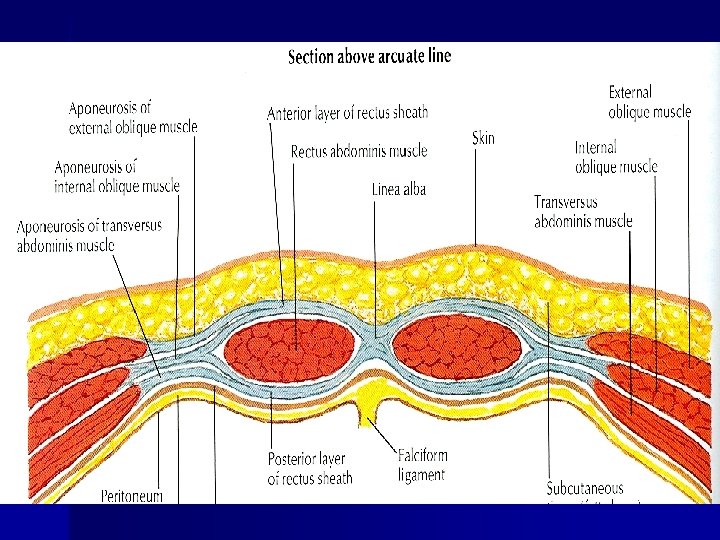
2. n From Xiphisternum to midpoint of umbilicus and symphysis pubis/ above arcuate line the aponeurosis of the internal oblique splits to enclose the rectus muscle n The external oblique aponeurosis is directed in front of the muscle n Transversus aponeurosis is directed behind the muscle

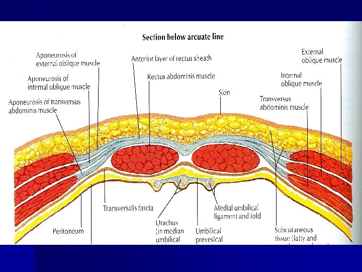
3. n From point 2 to symphysis pubis/below arcuate line the aponeurosis of all three muscles form the anterior wall n The posterior wall is absent n The rectus muscle lies in contact with the fascia transversalis

n The posterior wall of the rectus sheath is not attached to the rectus abdominis muscle n The anterior wall is firmly attached to it by the muscle’s tendinous intersections
 1. tip of the xiphoid process 2.")
Rectus abdominis muscle’s tendinous intersections (6 Abs) 1. tip of the xiphoid process 2. Midway between xiphoid process and umbilicus 3. Umbilicus Sometimes 4 th intersection below the umbilicus

Linea Alba/medial margin n The rectus sheath is separated from its fellow on the opposite side by a fibrous band called the linea alba n Extends from the xiphoid process to the symphysis pubis

Linea semilunaris/lateral margin n Curved fibrous line extending from the pubic tubercle to the tip of 9 th costal cartilage

Arcuate Line n Extent- from costal margin to midway between umbilicus and pubic symphysis n the line distal to which the posterior wall of rectus sheath is absent and presents as a concave free margin

Contents of Rectus Sheath n n n Muscles: Rectus Abdominis & Pyramidalis Vessels: Superior & Inferior Epigastric Vs. Nerves: 7 th to 11 th Intercostal nerves Subcostal Nerve

Applied Haematoma of rectus sheath n Epigastric hernia- weakness of linea alba n

INGUINAL LIGAMENT n n It is a thick, fibrous band, formed by the lower free border of the aponeurosis of the external oblique. It extends from ASIS to the pubic tubercle and its edge is curved back on itself to form a groove on its abdominal aspect

n n The strong deep fascia of the thigh, the fascia lata is attached to the rounded lower aspect of the entire length of the ligament, which makes it convex inferiorly by its pull due to tension. On the surface, inguinal ligament is marked by the inguinal fold which demarcates the abdomen from the lower limb.
 n n From the")
EXTENSIONS/EXPANSIONS OF INGUINAL LIGAMENT 1. LACUNAR LIGAMENT (OR GIMBERNAT’S LIGAMENT) n n From the medial end the deep fibers of the inguinal ligament curve horizontally backward to the medial part of the pecten pubis forming lacunar ligament. Its sharp lateral edge forms the medial boundary of the femoral canal, which is the site of production of a femoral hernia.

2. REFLECTED PART OF INGUINAL LIGAMENT n The superficial fibers from the medial end of the inguinal ligament expand upward and medially to form this ligament.

3. ILIOINGUINAL LIGAMENT n It is a fibrous band extending from the inferior aspect of the inguinal ligament to the iliopectineal eminence.
 n n The space between the inguinal ligament and the")
SUBINGUINAL SPACE (PELVIFEMORAL SPACE) n n The space between the inguinal ligament and the hip bone is called pelvifemoral/subinguinal space. This space is divided by ilioinguinal ligament/arch into two parts: (a) Large lateral part called lacuna musculorum. (b) Small medial part called lacuna vasculorum.

LOVE NATURE Thank You
- Slides: 23