https www yenepoya edu in 2272021 11 40



- Slides: 57
https: //www. yenepoya. edu. in 2/27/2021 11: 40 PM Yenepoya Nursing College (NAME) 1
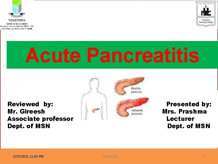
Acute Pancreatitis Reviewed by: Mr. Gireesh Associate professor Dept. of MSN 2/27/2021 11: 40 PM Presented by: Mrs. Prashma Lecturer Dept. of MSN (Prashma) 2
Objectives • • • Introduction Definition Epidemiology Aetiology & Pathogenesis Signs & Symptoms Investigations Management Complications Mortality
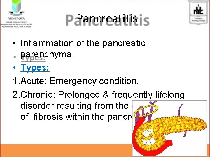
Pancreatitis • Inflammation of the pancreatic parenchyma. • Types: 1. Acute: Emergency condition. 2. Chronic: Prolonged & frequently lifelong disorder resulting from the development of fibrosis within the pancreas.
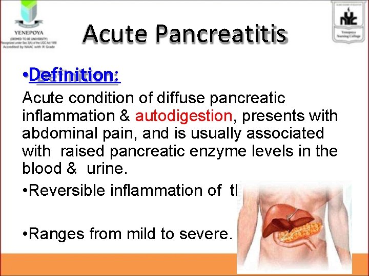
Acute Pancreatitis • Definition: Acute condition of diffuse pancreatic inflammation & autodigestion, presents with abdominal pain, and is usually associated with raised pancreatic enzyme levels in the blood & urine. • Reversible inflammation of the pancreas • Ranges from mild to severe.
Epidemiology • Acute pancreatitis accounts for 3% of all cases of abdominal pain among patients admitted to hospital in the UK. • Affect 2 – 28 per 100 000 of population. • It may occur at any age, peak incidence is between 50 and 60 years. • Women are affected more the men, but men are more likely to suffer recurrent attacks.
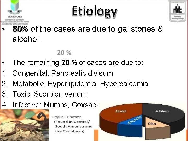
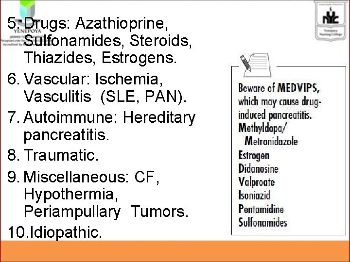
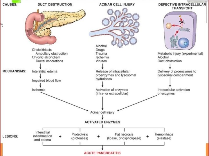
Etiology • 80% of the cases are due to gallstones & alcohol. • 1. 2. 3. 4. The remaining 20 % of cases are due to: Congenital: Pancreatic divisum Metabolic: Hyperlipidemia, Hypercalcemia. Toxic: Scorpion venom Infective: Mumps, Coxsackie B, EBV, CMV.
5. Drugs: Azathioprine, Sulfonamides, Steroids, Thiazides, Estrogens. 6. Vascular: Ischemia, Vasculitis (SLE, PAN). 7. Autoimmune: Hereditary pancreatitis. 8. Traumatic. 9. Miscellaneous: CF, Hypothermia, Periampullary Tumors. 10. Idiopathic.
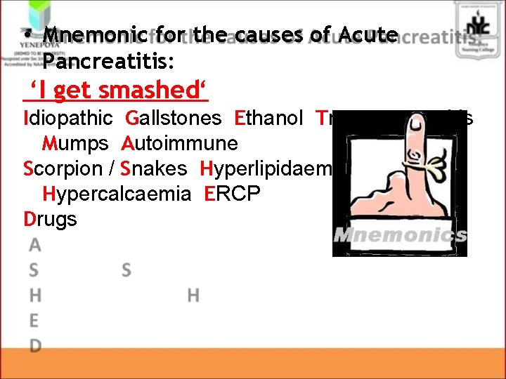
• Mnemonic for the causes of Acute Pancreatitis: ‘I get smashed‘ Idiopathic Gallstones Ethanol Trauma Steroids Mumps Autoimmune Scorpion / Snakes Hyperlipidaemia / Hypercalcaemia ERCP Drugs
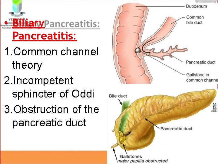
• Biliary Pancreatitis: 1. Common channel theory 2. Incompetent sphincter of Oddi 3. Obstruction of the pancreatic duct
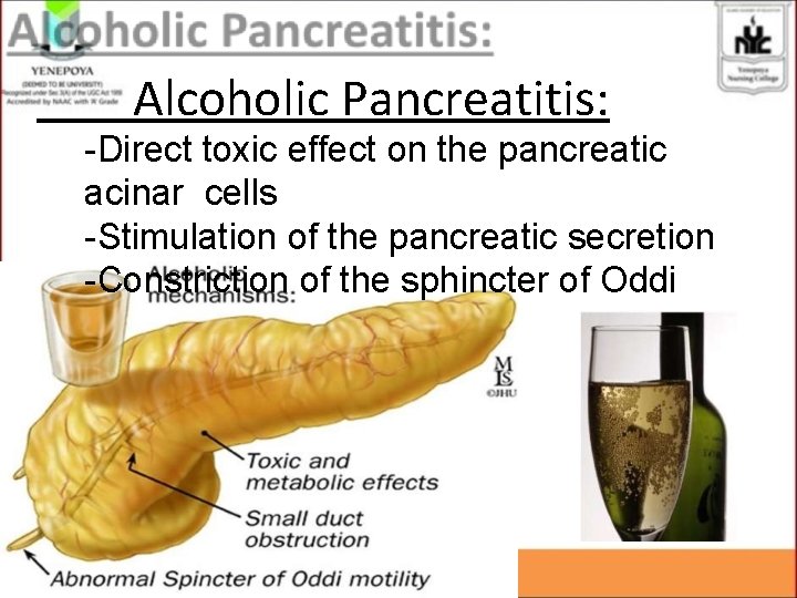
Alcoholic Pancreatitis: -Direct toxic effect on the pancreatic acinar cells -Stimulation of the pancreatic secretion -Constriction of the sphincter of Oddi
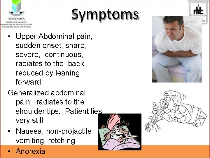
Symptoms • Upper Abdominal pain, sudden onset, sharp, severe, continuous, radiates to the back, reduced by leaning forward. Generalized abdominal pain, radiates to the shoulder tips. Patient lies very still. • Nausea, non-projactile vomiting, retching • Anorexia
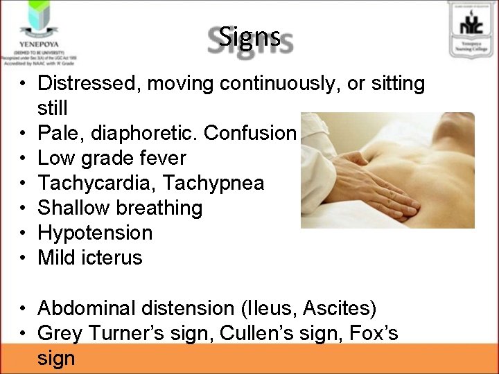
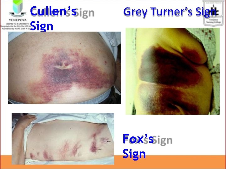
Signs • Distressed, moving continuously, or sitting still • Pale, diaphoretic. Confusion • Low grade fever • Tachycardia, Tachypnea • Shallow breathing • Hypotension • Mild icterus • Abdominal distension (Ileus, Ascites) • Grey Turner’s sign, Cullen’s sign, Fox’s sign
Cullen’s Sign Grey Turner’s Sign Fox’s Sign
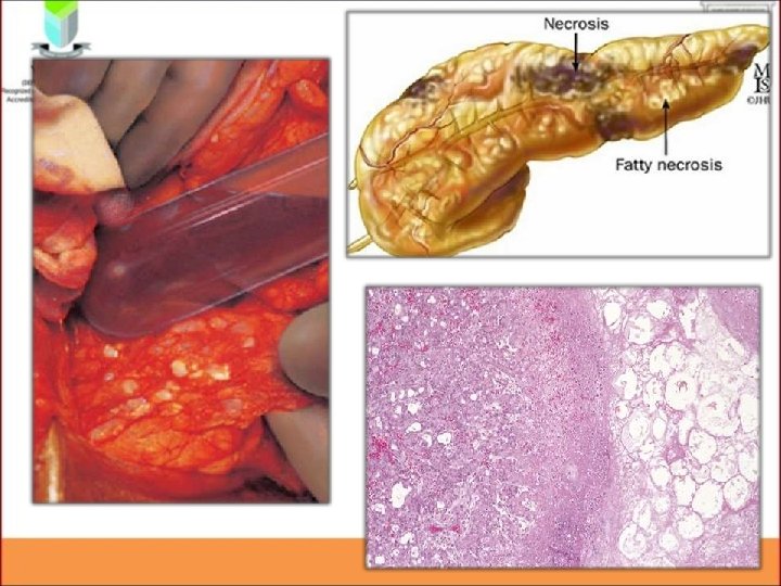
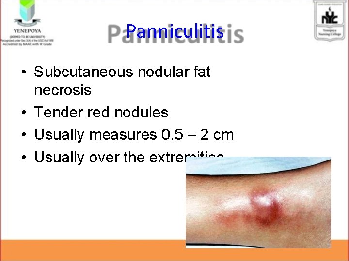
Panniculitis • Subcutaneous nodular fat necrosis • Tender red nodules • Usually measures 0. 5 – 2 cm • Usually over the extremities
Investigations Should be aimed at answering three questions: 1. Is a diagnosis of acute pancreatitis correct ? 2. How severe is the attack ? 3. What is the aetiology ?
Investigations Blood tests: • • • Complete Blood Count Serum amylase & lipase C-reactive Protein Serum electrolytes Blood glucose Renal Function Tests Liver Function Tests LDH Coagulation profile Arterial Blood Gas Analysis
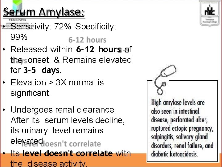
Serum Amylase: • Sensitivity: 72% Specificity: 99% • Released within 6 -12 hours of the onset, & Remains elevated for 3 -5 days. • Elevation ˃ 3 X normal is significant. • Undergoes renal clearance. After its serum levels decline, its urinary level remains elevated. • Its level doesn't correlate with the disease activity.
Serum Lipase: • More pancreatic-specific than s. Amylase. • Sensitivity: about 100% Specificity: 96% • Remains elevated longer than amylase (up to week). • Useful in patients presenting late to the physician. • S. Amylase tends to be higher in gallstone pancreatitis • S. Lipase tend to be higher in alcoholic pancreatitis
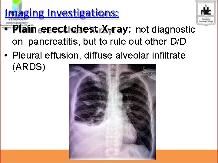
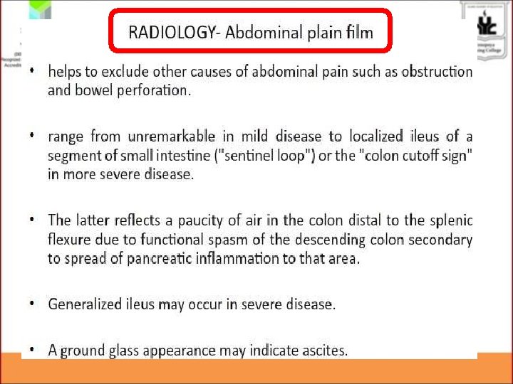
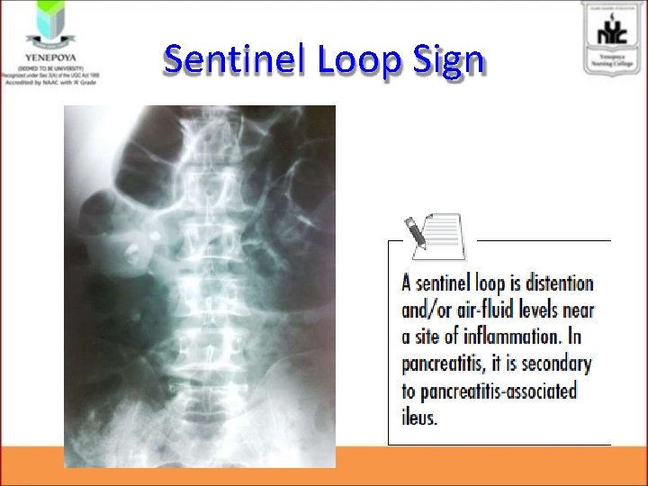
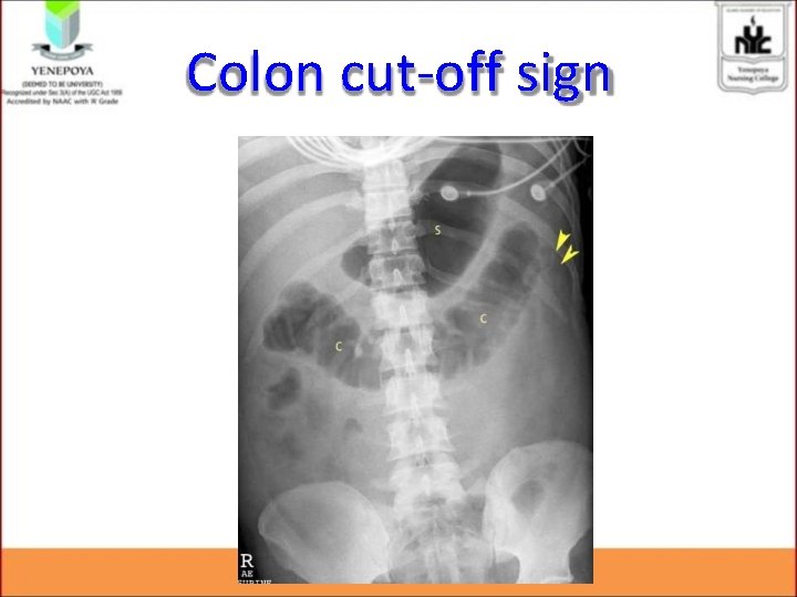
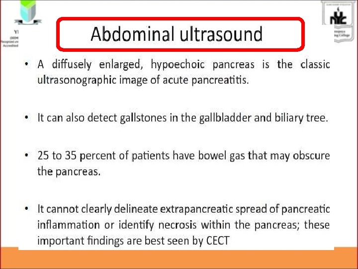
Imaging Investigations: • Plain erect chest X-ray: not diagnostic on pancreatitis, but to rule out other D/D • Pleural effusion, diffuse alveolar infiltrate (ARDS)
Sentinel Loop Sign
Colon cut-off sign
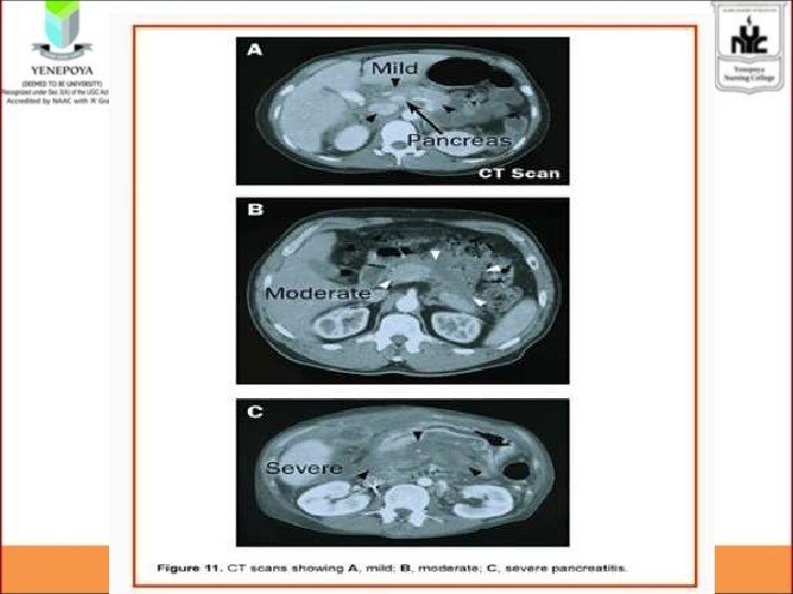
• CT Scan: not indicated in every patient, only in: 1. Diagnostic uncertainty. 2. Severe acute pancreatitis. 3. Clinical deterioration, with multi-organ failure, sepsis, progressive deterioration. 4. Local complications occurs (fluid collection, pseuodocyst, pseudo-aneurysm).
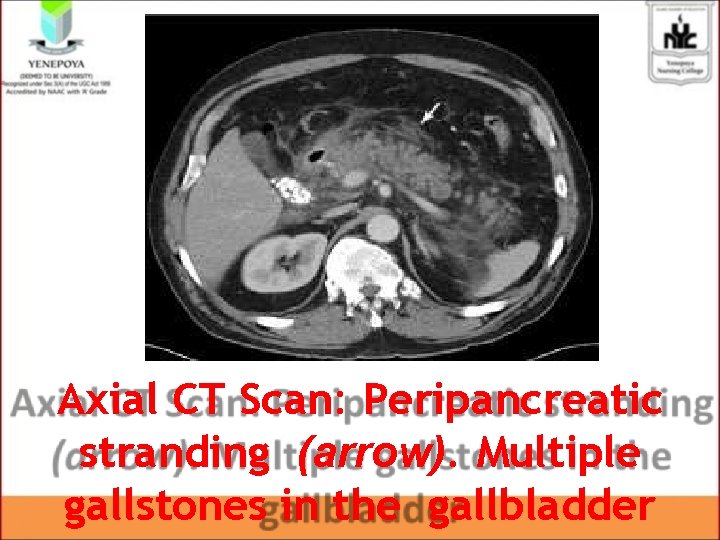
Axial CT Scan: Peripancreatic stranding (arrow). Multiple gallstones in the gallbladder
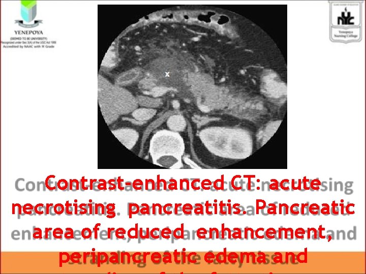
Contrast-enhanced CT: acute necrotising pancreatitis. Pancreatic area of reduced enhancement, peripancreatic edema and
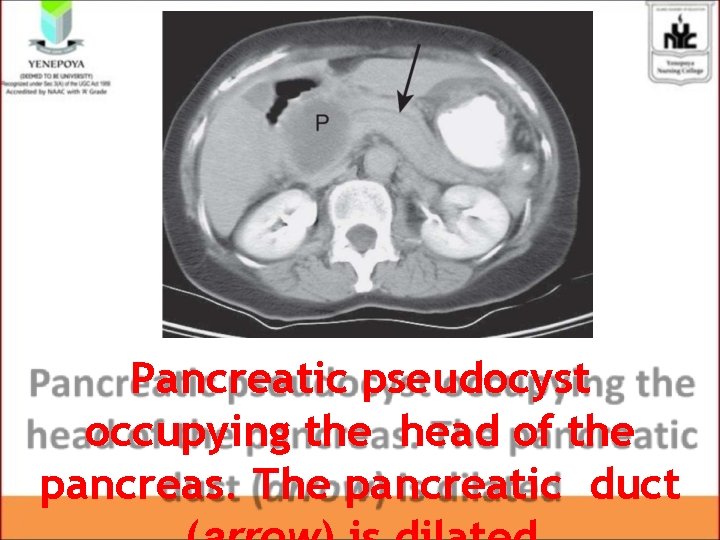
Pancreatic pseudocyst occupying the head of the pancreas. The pancreatic duct
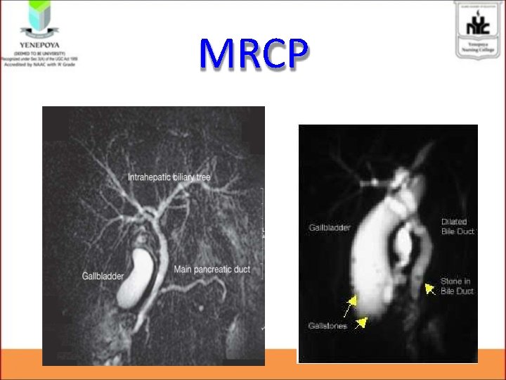
MRCP
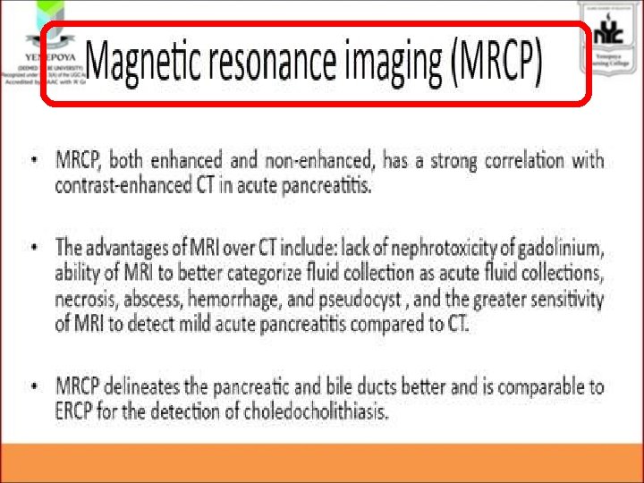
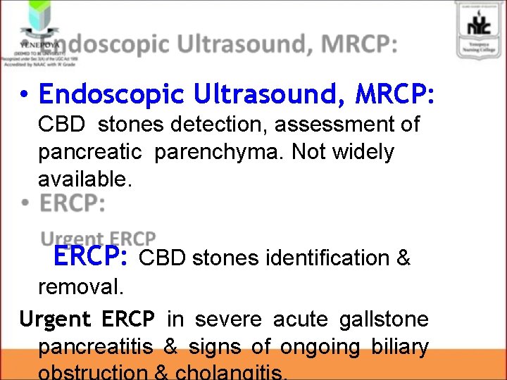
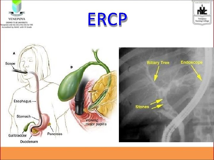
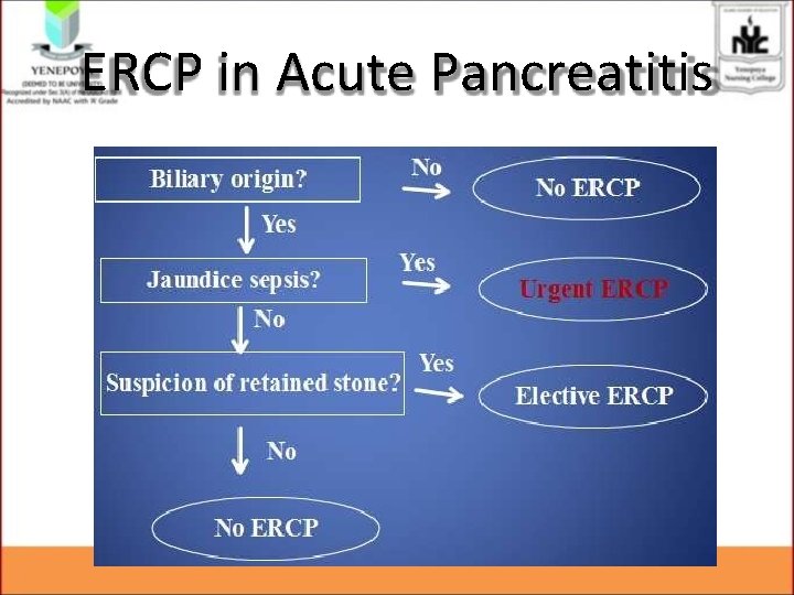
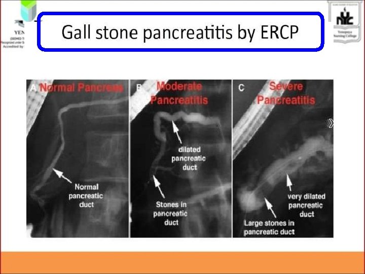
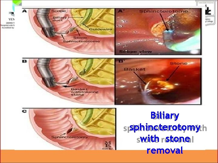
• Endoscopic Ultrasound, MRCP: CBD stones detection, assessment of pancreatic parenchyma. Not widely available. ERCP: CBD stones identification & removal. Urgent ERCP in severe acute gallstone pancreatitis & signs of ongoing biliary
ERCP
ERCP in Acute Pancreatitis
Biliary sphincterotomy with stone removal
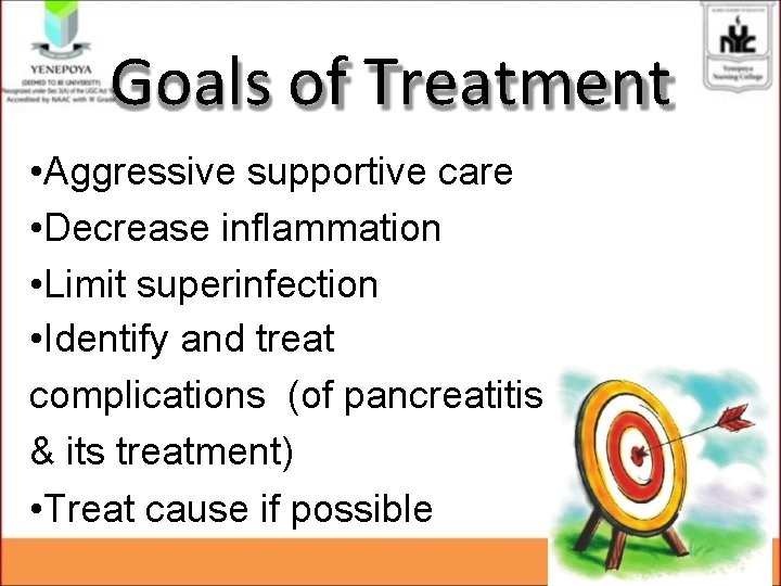
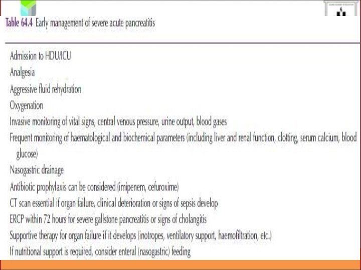
Goals of Treatment • Aggressive supportive care • Decrease inflammation • Limit superinfection • Identify and treat complications (of pancreatitis & its treatment) • Treat cause if possible
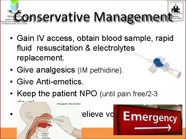
Conservative Management • Gain IV access, obtain blood sample, rapid fluid resuscitation & electrolytes replacement. • Give analgesics (IM pethidine). • Give Anti-emetics. • Keep the patient NPO (until pain free/2 -3 days). • NGT insertion to relieve vomiting.
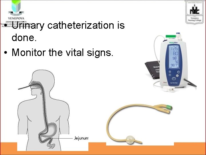
• Urinary catheterization is done. • Monitor the vital signs.
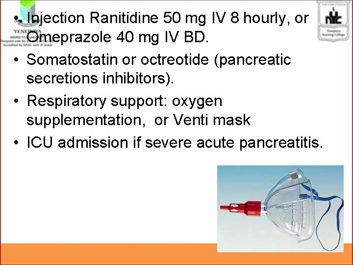
• Injection Ranitidine 50 mg IV 8 hourly, or Omeprazole 40 mg IV BD. • Somatostatin or octreotide (pancreatic secretions inhibitors). • Respiratory support: oxygen supplementation, or Venti mask • ICU admission if severe acute pancreatitis.
Role of Antibiotics • Prophylactic antibiotics have shown No decrease in mortality in severe acute pancreatitis. • Antibiotics are justified if: 1. Gas in retroperitoneal space 2. Needle aspiration of necrotic material confirms infection 3. Sepsis 4. CRP of ˃ 120 mg/L 5. Peri-pancreatic fluid collection 6. Organ dysfunction
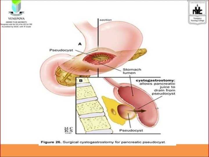
Operative Management • Surgery has no immediate role in acute pancreatitis. • Aggressive surgical pancreatic debridement (Necrosectomy) should be undertaken soon after confirmation of the presence of infected necrosis. • Pseudocyst: Cystogastrostomy, Cystodudenostomy, Roux-en-Y cystojejunostomy.
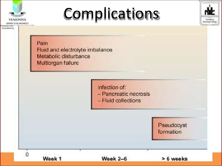
Complications
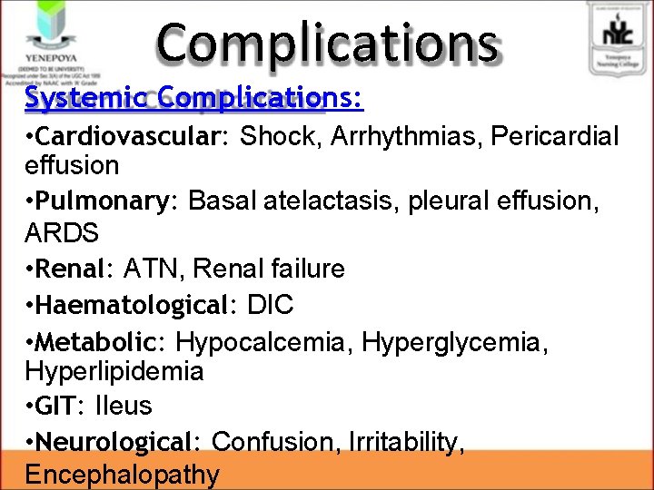
Complications Systemic Complications: • Cardiovascular: Shock, Arrhythmias, Pericardial effusion • Pulmonary: Basal atelactasis, pleural effusion, ARDS • Renal: ATN, Renal failure • Haematological: DIC • Metabolic: Hypocalcemia, Hyperglycemia, Hyperlipidemia • GIT: Ileus • Neurological: Confusion, Irritability, Encephalopathy
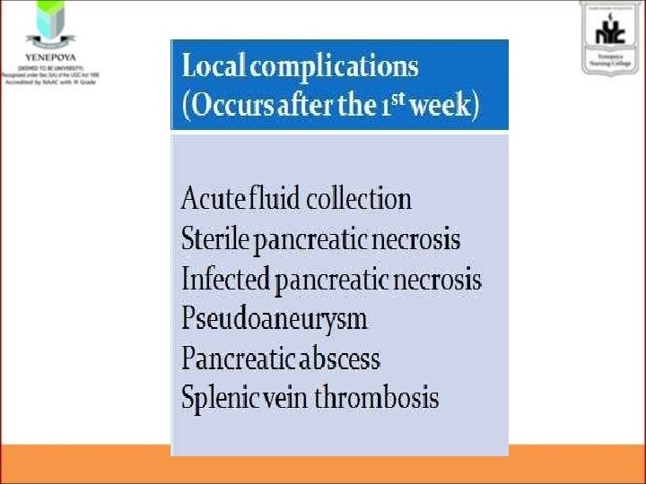
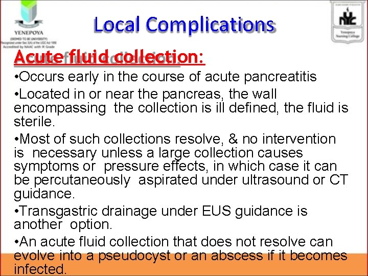
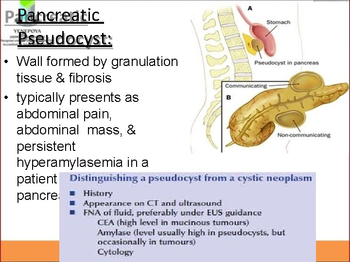
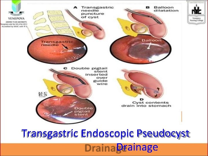
Local Complications Acute fluid collection: • Occurs early in the course of acute pancreatitis • Located in or near the pancreas, the wall encompassing the collection is ill defined, the fluid is sterile. • Most of such collections resolve, & no intervention is necessary unless a large collection causes symptoms or pressure effects, in which case it can be percutaneously aspirated under ultrasound or CT guidance. • Transgastric drainage under EUS guidance is another option. • An acute fluid collection that does not resolve can evolve into a pseudocyst or an abscess if it becomes infected.
Pancreatic Pseudocyst: • Wall formed by granulation tissue & fibrosis • typically presents as abdominal pain, abdominal mass, & persistent hyperamylasemia in a patient with prior pancreatitis.
Transgastric Endoscopic Pseudocyst Drainage
Mortality • Mild acute pancreatitis: Mortality rate of 1% • Severe pancreatitis: Mortality rate of 7590% • Overall mortality rate of 15 -20%
References • Bailey’s & Love’s Short Practice of Surgery, 25 th Edition, Page 1138 – 1146. • Essential Surgery, Burkitt, 4 th Edition, Page 380 – 388. • Robbins & Cotran Pathologic Basis of Disease, 8 th Edition. • http: //www. aafp. org/afp/2007/0515/p 1513. ht ml • http: //www. aafp. org/afp/2000/0701/p 164. html