Genetic Tests Oral Histology Kristine Krafts M D

- Slides: 22
Genetic Tests Oral Histology | Kristine Krafts, M. D.
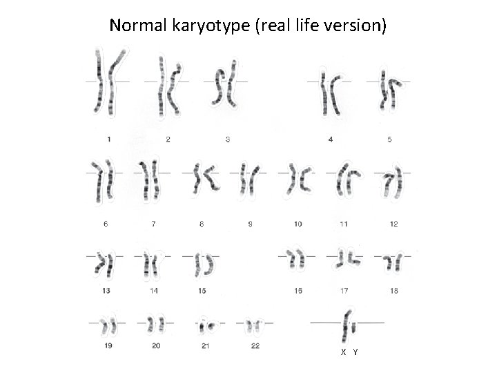
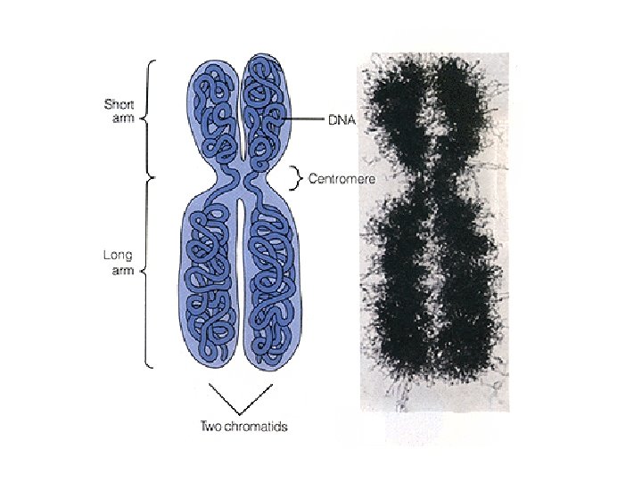
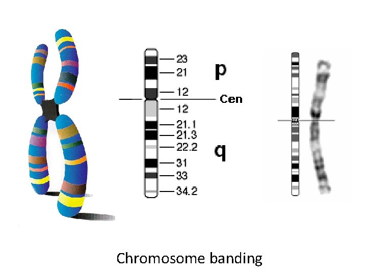
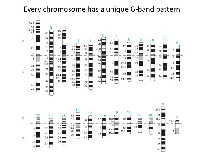
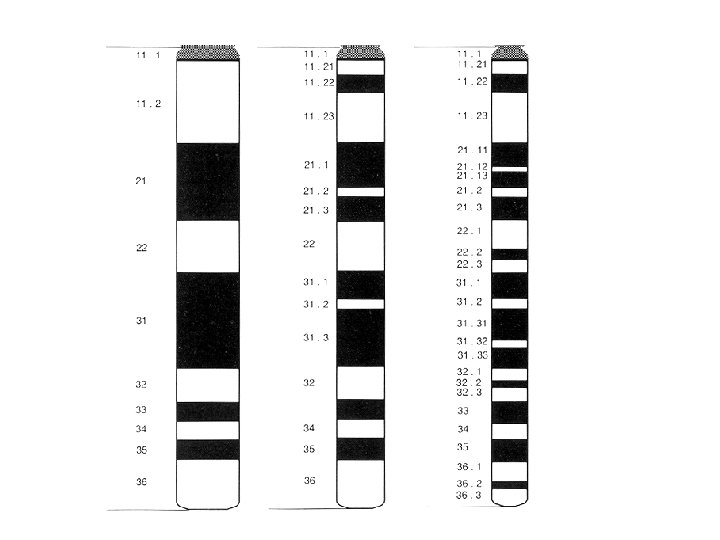
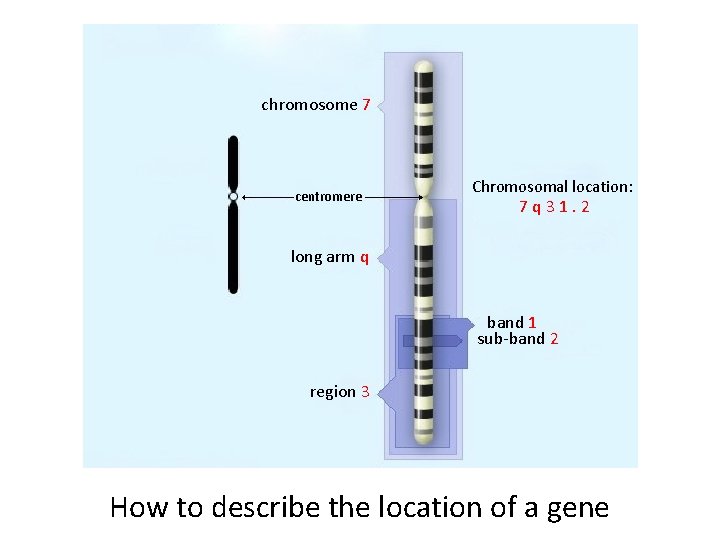
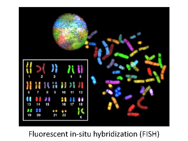
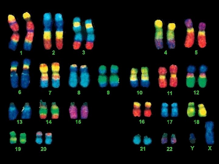
Objectives • Describe (in a general way) the basic cytogenetics procedure. • Be familiar with the layout and organization of a typical karyotype. • Understand the numbering system for G-banded chromosomes (region, band, sub-band, etc. ) • Describe what FISH is, and compare and contrast it with traditional cytogenetics (karyotyping).
Genetic tests are used in a wide variety of clinical settings. • • Diagnostic testing Carrier testing Predictive and presymptomatic testing Preimplantation testing Prenatal testing Newborn screening Pharmacogenomic testing Other stuff (forensics, paternity, geneology)
Genetic tests can look at: • • Chromosomes DNA RNA Proteins
Q. What is cytogenetics? A. The study of chromosomes (their structure and inheritance) and how they relate to diseases.
What tissues can you use for cytogenetics? Pretty much anything that can grow in culture. • • • Blood Skin Bone marrow Chorionic villi Amniotic fluid
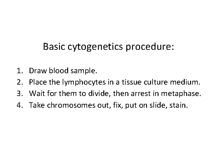
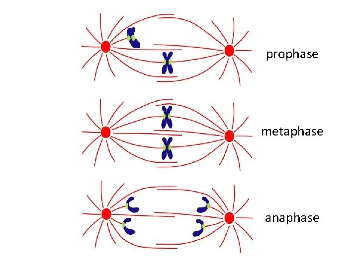
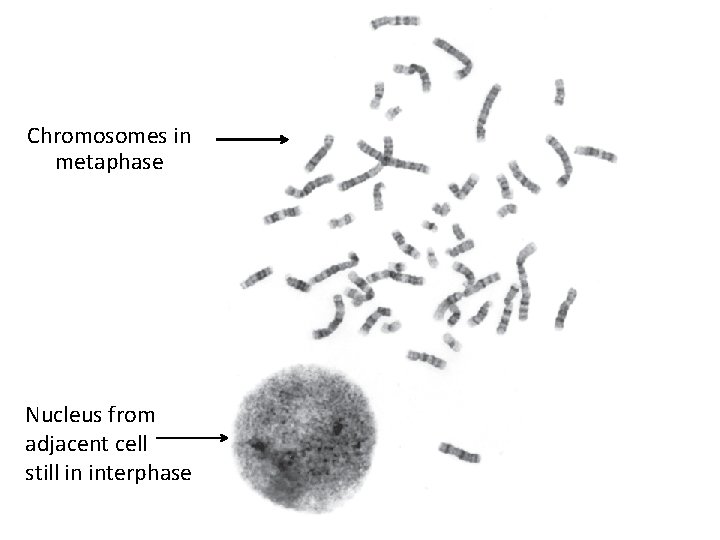
Basic cytogenetics procedure: 1. 2. 3. 4. Draw blood sample. Place the lymphocytes in a tissue culture medium. Wait for them to divide, then arrest in metaphase. Take chromosomes out, fix, put on slide, stain.
prophase metaphase anaphase
Chromosomes in metaphase Nucleus from adjacent cell still in interphase
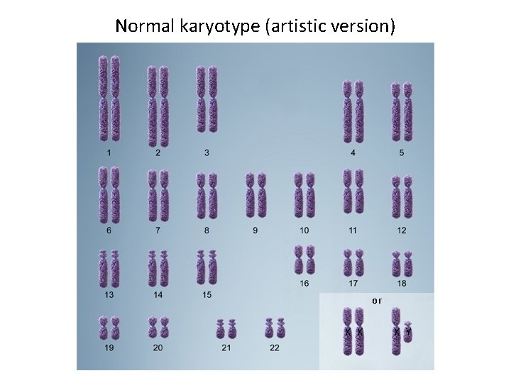
Normal karyotype (artistic version) or X X X Y
Normal karyotype (real life version) X Y
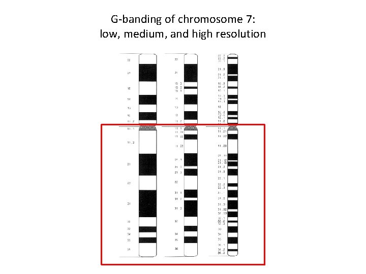
Chromosome banding
Every chromosome has a unique G-band pattern
G-banding of chromosome 7: low, medium, and high resolution
chromosome 7 centromere Chromosomal location: 7 q 31. 2 long arm q band 1 sub-band 2 region 3 How to describe the location of a gene
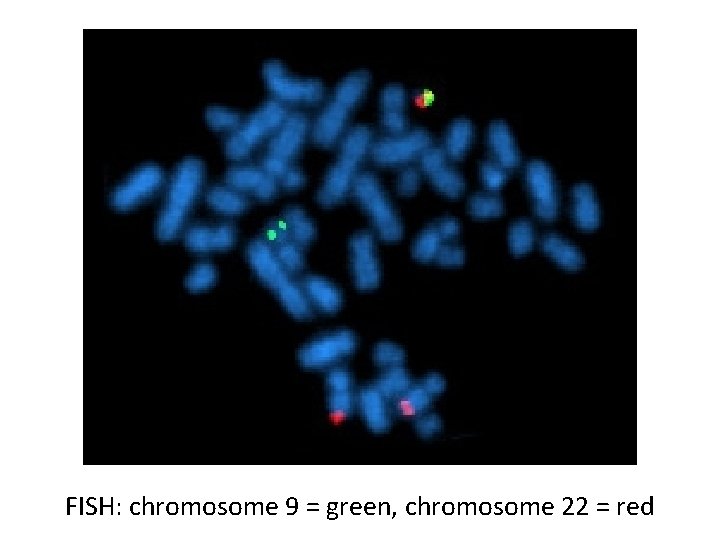
Fluorescent in-situ hybridization (FISH)
FISH: chromosome 9 = green, chromosome 22 = red
Normal Trisomy 18 FISH: chromosome 18 = blue, X = green, Y = red