Genesis and Regeneration of Skeletal Muscle Class V


- Slides: 21
Genesis and Regeneration of Skeletal Muscle Class V M. Sc. -Semester IV Dr. Hifzur R Siddique Section of Genetics Department of Zoology ALIGARH MUSLIM UNIVERSITY
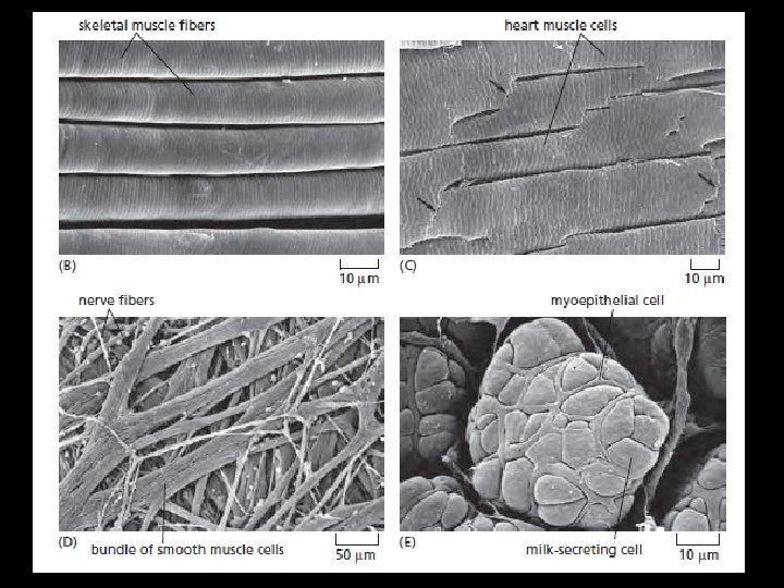
Muscle v “Muscle” includes many cell types, all specialized for contraction but in other respects dissimilar. Mammals possess FOUR MAIN categories of cells specialized for contraction: 1. Skeletal muscle cells, 2. Heart (cardiac) muscle cells, 3. Smooth muscle cells, 4. Myoepithelial cells. v These differ in function, structure, and development. v Although all of them generate contractile forces by using organized filament systems based on actin and myosin II. v The actin and myosin molecules employed have somewhat different amino acid sequences, are differently arranged in the cell, and are associated with different sets of proteins that control contraction.
v We focus in this section on skeletal muscle cells, which are responsible for practically all movements that are under voluntary control. v These cells can be very large (2 -3 cm long and 100 μm in diameter in an adult human) and are often called muscle fibers because of their highly elongated shape. v Each one is a syncytium, containing many nuclei within a common cytoplasm. v In an intact muscle, they are bundled tightly together, with fibroblasts (and some fat cells) in the interstices between them and blood vessels and nerve fibers running through the tissue. v Here, we consider the unusual strategy by which the multinucleate skeletal muscle cells are generated and maintained. v Giant cells with many nuclei and are formed BY CELL FUSION. The other types of muscle cells are more conventional, generally having only a single nucleus.
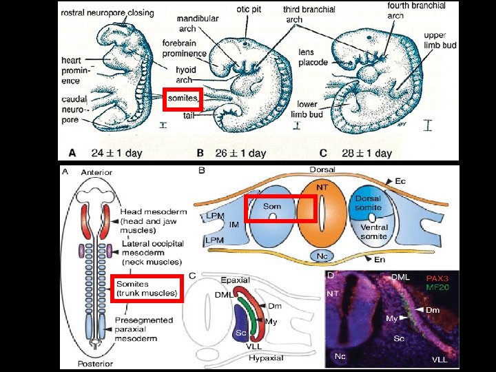
Myoblasts Fuse to Form New Skeletal Muscle Fibers v. During development, certain cells, originating from the SOMITES of a vertebrate embryo at a very early stage, become determined as MYOBLASTS, the precursors of skeletal muscle fibers. v. After a period of proliferation, the myoblasts undergo a dramatic change of state: Øthey stop dividing, switch on the expression of a whole battery of muscle -specific genes required for terminal differentiation. Øand fuse with one another to form multinucleate skeletal muscle fibers. v. Once differentiation and cell fusion have occurred, the cells do not divide and the nuclei never again replicate their DNA.
Myoblast fusion in culture.
Some Myoblasts Persist as Quiescent Stem Cells in the Adult • Humans do not normally generate new skeletal muscle fibers in adult life, they still have the capacity to do so, and existing muscle fibers can resume growth when the need arises. • Cells capable of serving as myoblasts are retained as small, flattened, and inactive cells lying in close contact with the mature muscle cell and contained within its sheath of basal lamina. Satellite cells repair skeletal muscle fibers.
• Satellite cells, or some subset of the satellite cells, are thus the stem cells of adult skeletal muscle, NORMALLY HELD IN RESERVE IN A QUIESCENT STATE but available when needed as a self-renewing source of terminally differentiated cells. • The process of muscle repair by means of satellite cells is, however, limited in what it can achieve. • In one form of MUSCULAR DYSTROPHY, for example, a genetic defect in the cytoskeletal protein dystrophin damages differentiated skeletal muscle cells. • As a result, satellite cells proliferate to repair the damaged muscle fibers. • This regenerative response is, however, unable to keep pace with the damage, and connective tissue eventually replaces the muscle cells, blocking any further possibility of regeneration. • A decline of capacity for repair likewise contributes to the weakening of muscle in the elderly.
Blood Vessels, Lymphatics, and Endothelial Cells v. Almost all tissues depend on a blood supply, and the blood supply depends on endothelial cells, which form the linings of the blood vessels. v. Endothelial cells have a remarkable capacity to adjust their number and arrangement to suit local requirements. v. They create an adaptable life-support system, extending by cell migration into almost every region of the body. v. If it were not for endothelial cells extending and remodeling the network of blood vessels, tissue growth and repair would be impossible. v. Cancerous tissue is as dependent on a blood supply as is normal tissue, and this has led to a surge of interest in endothelial cell biology, in the hope that it may be possible to block the growth of tumors by attacking the endothelial cells that bring them nourishment.
Endothelial Cells Line All Blood Vessels and Lymphatics v. The largest blood vessels are arteries and veins, which have a thick, tough wall of connective tissue and many layers of smooth muscle cells (Figure 22– 21). v. The inner wall is lined by an exceedingly thin single sheet of endothelial cells, the endothelium, separated from the surrounding outer layers by a basal lamina. v. The amounts of connective tissue and smooth muscle in the vessel wall vary according to the vessel’s diameter and function, but the endothelial lining is always present. Fig: Diagram of a small artery in cross section
In the finest branches of the vascular treethe CAPILLARIES and sinusoids-the walls consist of nothing but ENDOTHELIAL CELLS and a BASAL LAMINA, together with a few scattered PERICYTES. Pericytes (also k/a Rouget cells, or mural cells) are contractile cells THAT WRAP AROUND THE ENDOTHELIAL CELLS that line the capillaries and venules throughout the body & strengthen cells. v Less obvious than the blood vessels are the LYMPHATIC VESSELS. These carry no blood and have much thinner and more permeable walls than the blood vessels. v They provide a drainage system for the fluid (lymph) that SEEPS OUT OF THE BLOOD VESSELS, AS WELL AS AN EXIT ROUTE FOR WBCS that have migrated from blood vessels into the tissues. v Less happily, they can also provide the path by which CANCER CELLS escape from a primary tumor to invade other tissues.
v The lymphatics form a branching system of tributaries, all ultimately discharging into a single large lymphatic vessel- the thoracic duct, which opens into a large vein close to the heart. v Like blood vessels, LYMPHATICS ARE LINED WITH ENDOTHELIAL CELLS. v Thus, endothelial cells line the entire blood and lymphatic vascular system, and they control the passage of materials-and the transit of WBCs-into and out of the bloodstream. v Arteries, veins, capillaries, and lymphatics all develop from small vessels constructed primarily of (1) endothelial cells and (2) a basal lamina. v Connective tissue and smooth muscle are added later where required, under the influence of signals from the endothelial cells.
Endothelial Tip Cells Pioneer Angiogenesis v. To understand how the vascular system comes into being and how it adapts to the changing needs of tissues, we have to understand endothelial cells. v. How do they become so widely distributed, and how do they form channels that connect in just the right way for blood to circulate through the tissues and for lymph to drain back to the bloodstream? v. Endothelial cells originate at specific sites in the early embryo from precursors that also give rise to blood cells. v. From these sites, the early embryonic endothelial cells migrate, proliferate, and differentiate to form the first rudiments of blood vessels-a process called VASCULOGENESIS. v. Subsequent growth and branching of the vessels throughout the body occurs mainly by proliferation and movement of the endothelial cells of these first vessels, in a process called ANGIOGENESIS.
ANGIOGENESIS v Angiogenesis occurs in a broadly similar way in the young organism as it grows and in the adult during tissue repair and remodeling. v We can watch the behavior of the cells in naturally transparent structures, such as the cornea of the eye or the fin of a tadpole, or in tissue culture, or in the embryo. v The embryonic retina, which blood vessels invade according to a predictable timetable, provides a convenient example for experimental study. v Each new vessel originates as a capillary sprout from the side of an existing capillary or small venule (Figure 22– 24). v At the tip of the sprout, leading the way, is an endothelial cell with a distinctive character. v This tip cell has a pattern of gene expression somewhat different from that of the endothelial stalk cells following behind it, and while they divide, it does Thenot. tip cell’s most striking feature is that it puts out many long filopodia, resembling those of a neuronal growth cone.
v The endothelial tip cells that pioneer the growth of normal capillaries not only look like neuronal growth cones, but also respond similarly to signals in the environment. ØIn fact, many of the same guidance molecules are involved, including the NETRINS, SLITS, AND EPHRINS mentioned in our account of neural development ØThe corresponding receptors are expressed in the tip cells and guide the vascular sprouts along specific pathways in the embryo, often in parallel with nerves. v Perhaps the most important of the guidance molecules for endothelial cells, however, is one that is chiefly dedicated to the control of vascular development: vascular endothelial growth factor, or VEGF
Tissues Requiring a Blood Supply Release VEGF v. Almost every cell, in almost every tissue of a vertebrate, is located within 50 -100 μm of a blood capillary. 1. What mechanism ensures that the system of blood vessels branches into every nook and cranny? 2. How is it adjusted so perfectly to the local needs of the tissues, not only during normal development but also in pathological circumstances? v Wounding, for example, induces a burst of capillary growth in the neighborhood of the damage, to satisfy the high metabolic requirements of the repair process.
v. Local irritants and infections also cause a proliferation of new capillaries, most of which regress and disappear when the inflammation subsides. v. Less benignly, a small sample of tumor tissue implanted in the cornea, which normally lacks blood vessels, causes blood vessels to grow quickly toward the implant from the vascular margin of the cornea; the growth rate of the tumor increases abruptly as soon as the vessels reach it. v. In all these cases, the invading endothelial cells respond to signals produced by the tissue that they invade. The signals are complex, but a key part is played by VEGF. v The regulation of blood vessel growth to match the needs of the tissue depends on the control of VEGF production, through changes in the stability of its m. RNA and in its rate of transcription. v A shortage of oxygen, in practically any type of cell, causes an increase in the intracellular level of a transcription factor called hypoxia-inducible factor 1α (HIF 1α). v HIF 1α stimulates transcription of Vegf (and of other genes whose products are needed when oxygen is in short supply).
v. The VEGF protein is secreted, and acts on nearby endothelial cells. v. Stimulating them to proliferate, to produce proteases to help them digest their way through the basal lamina of the parent capillary or venule, and to form sprouts. v The tip cells of the sprouts detect the VEGF gradient and move toward its source. As the new vessels form, bringing blood to the tissue, the oxygen concentration rises. v The HIF 1α activity then declines, VEGF production is shut off, and angiogenesis comes to a halt
Signals from Endothelial Cells Control Recruitment of Pericytes and Smooth Muscle Cells to Form the Vessel Wall v. The vascular network is continually remodeled as it grows and adapts. A newly formed vessel may enlarge; or it may sprout side branches; or it may regress. v. Smooth muscle and other connective-tissue cells that pack themselves around the endothelium help to stabilize vessels as they enlarge. This process of vessel wall formation begins with recruitment of pericytes. v. The recruitment and proliferation of pericytes and smooth muscle cells to form a vessel wall depend on PLATELET-DERIVED GROWTH FACTOR-B (PDGF-B) secreted by the endothelial cells and on PDGF receptors in the pericytes and smooth muscle cells.
K N A H T U O Y