DNA Chromosomes and Replication DNA and Chromosomes In


- Slides: 19
DNA Chromosomes and Replication
DNA and Chromosomes In prokaryotic cells, DNA is located in the cytoplasm. Most prokaryotes have a single DNA molecule containing nearly all of the cell’s genetic information. Copyright Pearson Prentice Hall
DNA and Chromosomes Chromosome E. Coli Bacterium Bases on the Chromosomes Copyright Pearson Prentice Hall
DNA and Chromosomes Many eukaryotes have 1000 times the amount of DNA as prokaryotes. Eukaryotic DNA is located in the cell nucleus inside chromosomes. The number of chromosomes varies widely from one species to the next. *The # is not related to complexity. For instance humans have 46 chromosomes while a Chicken has 70. Copyright Pearson Prentice Hall
DNA and Chromosomes Chromosome Structure Eukaryotic chromosomes contain DNA and protein, tightly packed together to form chromatin. Chromatin consists of DNA tightly coiled around proteins called histones. DNA and histone molecules form nucleosomes. Nucleosomes pack together, forming a thick fiber. Copyright Pearson Prentice Hall
DNA and Chromosomes Eukaryotic Chromosome Structure Chromosome Nucleosome DNA double helix Coils Supercoils Histones Copyright Pearson Prentice Hall
DNA Replication • Origins of replication 1. Replication Forks: Forks hundreds of Y-shaped regions of replicating DNA molecules where new strands are growing. 5’ Parental DNA Molecule 3’ 3’ Replication Fork 5’
DNA Replication • Origins of replication 2. Replication Bubbles: Bubbles a. Hundreds of replicating bubbles (Eukaryotes) b. Single replication fork (bacteria). Bubbles
DNA Replication • Strand Separation: Separation 1. Helicase: Helicase enzyme which catalyze the unwinding and separation (breaking HBonds) of the parental double helix. 2. Single-Strand Binding Proteins: Proteins proteins which attach and help keep the separated strands apart.
DNA Replication • Strand Separation: Separation 3. Topoisomerase: Topoisomerase enzyme which relieves stress on the DNA molecule by allowing free rotation around a single strand. Enzyme DNA Enzyme
DNA Replication • Priming: 1. RNA primers: primers before new DNA strands can form, there must be small pre-existing primers (RNA) present to start the addition of new nucleotides (DNA Polymerase) 2. RNA Primase: Primase enzyme that polymerizes (synthesizes) the RNA Primer.
DNA Replication • Synthesis of the new DNA Strands: 1. DNA Polymerase: Polymerase with a RNA primer in place, DNA Polymerase (enzyme) catalyze the synthesis of a new DNA strand in the 5’ to 3’ direction 5’ 3’ Nucleotide DNA Polymerase RNA Primer 5’
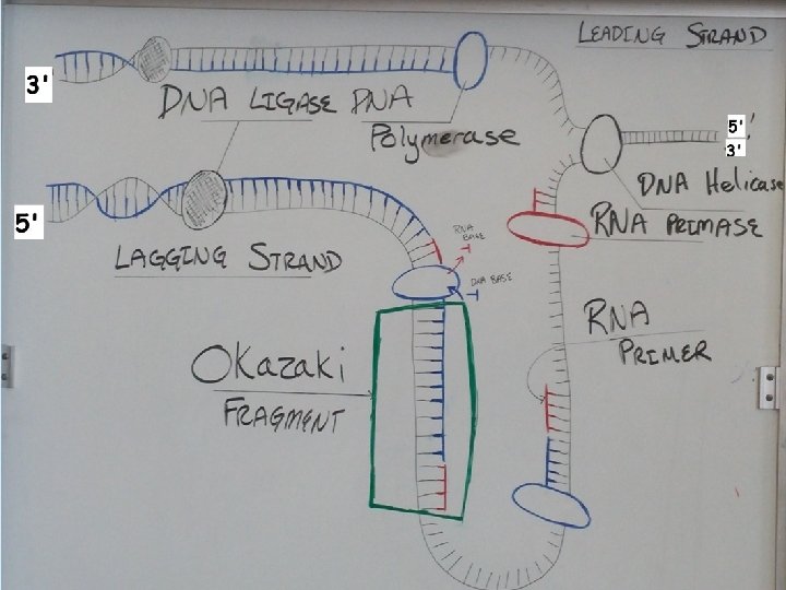
DNA Replication 2. Leading Strand: Strand synthesized as a single polymer in the 5’ to 3’ direction 5’ 3’ 5’ Nucleotides DNA Polymerase RNA Primer
DNA Replication 3. Lagging Strand: Strand also synthesized in the 5’ to 3’ direction, direction but discontinuously against overall direction of replication. Leading Strand 5’ 3’ DNA Polymerase RNA Primer 3’ 5’ 5’ 3’ 3’ 5’ Lagging Strand
DNA Replication 4. Okazaki Fragments: Fragments series of short segments on the lagging strand. DNA Polymerase Okazaki Fragment RNA Primer 5’ 3’ Lagging Strand 3’ 5’
DNA Replication 5. DNA ligase: ligase a linking enzyme that catalyzes the formation of a covalent bond from the 3’ to 5’ end of joining stands. Example: joining two Okazaki fragments together. DNA ligase 5’ 3’ Okazaki Fragment 1 Lagging Strand Okazaki Fragment 2 3’ 5’
Question: • What would be the complementary DNA strand for the following DNA sequence? DNA 5’-GCGTATG-3’
Answer: DNA 5’-GCGTATG-3’ DNA 3’-CGCATAC-5’