DISORDERS AND EXAMINATION OF PULMONARY VENTILATION Department of


- Slides: 14

DISORDERS AND EXAMINATION OF PULMONARY VENTILATION Department of Pathophysiology Faculty of Medicine in Pilsen Charles University

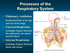
Pulmonary ventilation • provides change of air between atmosphere and lung alveoli • depends on airways patency, lung volume, lung and thoracic wall elasticity, respiratory centre activity and motor innervation of respiratory muscles

Static lung volumes • Tidal volume (VT) = 0, 5 l – amount of air inhaled or exhaled with each breath during quiet respiration • Inspiratory reserve volume (IRV) = 3 l – amount of air that can be forcefully inhaled after a normal tidal volume inhalation • expiratory reserve volume (ERV) = 1, 1 l – amount of air that can be forcefully exhaled after a normal tidal volume exhalation • residual volume (RV) = 1, 2 l – amount of air left in the lungs after a forced exhalation (it can not be expired) • dead space (VD)= the volume of air in the conducting airways (about 1 ml per 1 pound of body weight) that does not exchanges with pulmonary blood (it can not be measured directly with classical spirometry) – anatomical dead space (150 -200 ml) – physiological, airways to terminal bronchioli – total (functional) death space – anatomical death space + pathologically changed parts of lung, which are ventilated but in which change of respiratory gasses is restricted (decrease of perfusion of diffusion across the alveolocapillary membrane)

Static lung capacities • Vital capacity (VC): volume of air, which can be expired with maximal effort after a maximal inspiration VC=VT+IRV+ERV calculation of normal value of vital capacity in ml: man: VC = [27, 63 - (0, 112 x age years))] x height (cm) woman: VC = [21, 78 - (0, 101 x age years))] x height (cm) • total lung capacity (TLC): amount of air contained in the lungs after a maximal inspiration TLC=VT+IRV+ERV+RV • functional residual capacity (FRC): volume of air, remaining in the lungs after a normal tidal volume expiration FRC=ERV+RV

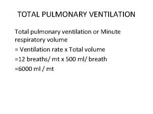
Dynamic lung volumes • forced expiratory volume (FEV 1): volume of air that can be exhaled during the first second of a forced expiration after a maximal inspiration • FEV 1% (Tieffeneau‘s index): FEV 1 expressed as the ratio in percentage terms FEV 1% = (FEV 1 / VC) x 100 % normal value: 80 % • minute lung ventilation = 8 l: volume of air expired during 1 minute of quiet breathing • minute alveolar ventilation: minute ventilation minus minute ventilation of the death space • maximal minute ventilation (MVV): maximal amount of air expired during 1 minute of forced breathing indirect calculation: MVV = VC x 30 breath/min calculation of normal values in l: man: MVV = [86, 5 – (0, 522 x age (years))] x body surface area(m 2) woman: MVV = [71, 3 – (0, 474 x age (years))] x body surface area(m 2)

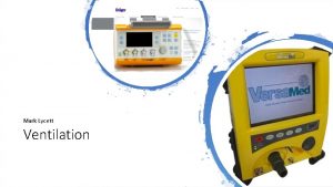
Examination of respiratory functions spirometers - devices measuring primarily volumes e. g. EUTEST - devices measuring primarily air flow e. g. Power. Lab

Examination of respiratory functions – physiological recording 7

Obstructive disorders of pulmonary ventilation • • reduction of patency of airways constriction of upper airways – inspiratory dyspnoea constriction of lower airways – expiratory dyspnoea diagnosis according to spirometry: normal VC , decreased FEV 1 → FEV 1% < 80 % • examples: asthma bronchial, bronchitis, corpus alien in the airways, partial obstruction or compression of bronchial tubes by tumors, struma etc.

Obstructive disorders of pulmonary ventilation 9

Restrictive disorders of pulmonary ventilation • restriction of lung capacity • diagnosis according to spirometry: decreased VC (pathological is the decrease under 80 % of normal value), FEV 1% often > 80 % examples: state after lung resection, lung atelectasis, lung edema, pneumonia, pneumothorax, hydrothorax, lung fibrosis, thoracic deformities, disorders of respiratory muscles (their innervation or neuromuscular junction)

Restrictive disorders of pulmonary ventilation 11

normal state obstructive disorder restrictive disorder 12

Assessment of the disorder severity -according to FEV 1 – in obstructive disorder decreased primarily - in restrictive disorder decreased secondarily (due to decrease of vital capacity) Decrease of FEV 1 under 80 % of the normal value is considered as pathological. FEV 1 60 -80 % of the normal value = moderate disorder FEV 1 40 -60 % of the normal value = middle disorder FEV 1 < 40 % of the normal value = severe disorder 13

THE END 14
 Difference between right and left bronchus
 Muscles of inspiration and expiration
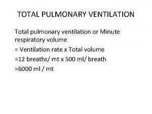
 Total pulmonary ventilation
 Pulmonary ventilation
 Types of respiration
 Pulmonary ventilation
 Mode of ventilation
 What is the physiology of respiration
 Pulmonary artery and aorta
 Bronchiole
 Aorta inferior vena cava
 Aorta and pulmonary artery
 Structure of the heart
 Circulatory system labeled
 Pulmonary volumes and capacities
