Content Defination n Discovery n Classification n Morphology

- Slides: 23
Content Defination n Discovery n Classification n Morphology n Structure n Cultural character n Compassion with L-formed Bacterium n
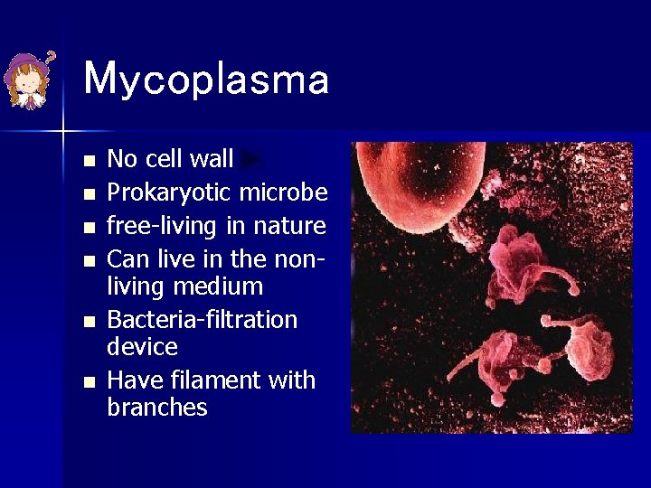
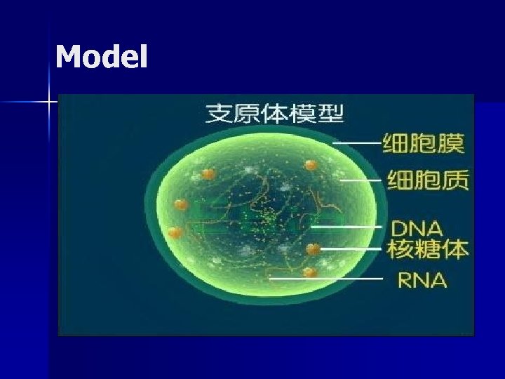
Mycoplasma n n n No cell wall Prokaryotic microbe free-living in nature Can live in the nonliving medium Bacteria-filtration device Have filament with branches
Model
Discovery n n In 1898, Nocard found Mycoplasma from the cattle that had pleuropneumonia. In 1937, Dienes isolated Mycoplasma from the pus of patients. Once they were named pleuropneumonialike organism (类胸膜肺炎微生物) In 1962, Chanock succeeded in cultivating Mycoplasma aitificially.
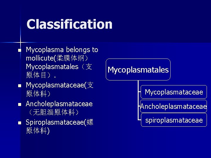
Classification n n Mycoplasma belongs to mollicute(柔膜体纲) Mycoplasmatales(支 原体目). Mycoplasmataceae(支 原体科) Ancholeplasmataceae (无胆淄原体科) Spiroplasmataceae(螺 原体科) Mycoplasmatales Mycoplasmataceae Ancholeplasmataceae spiroplasmataceae
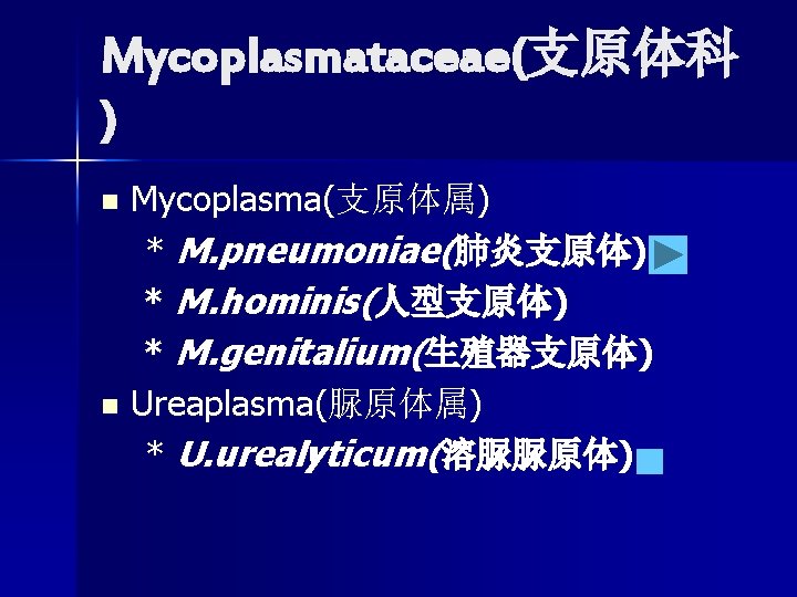
Mycoplasmataceae(支原体科 ) Mycoplasma(支原体属) * M. pneumoniae(肺炎支原体) * M. hominis(人型支原体) * M. genitalium(生殖器支原体) n Ureaplasma(脲原体属) * U. urealyticum(溶脲脲原体) n
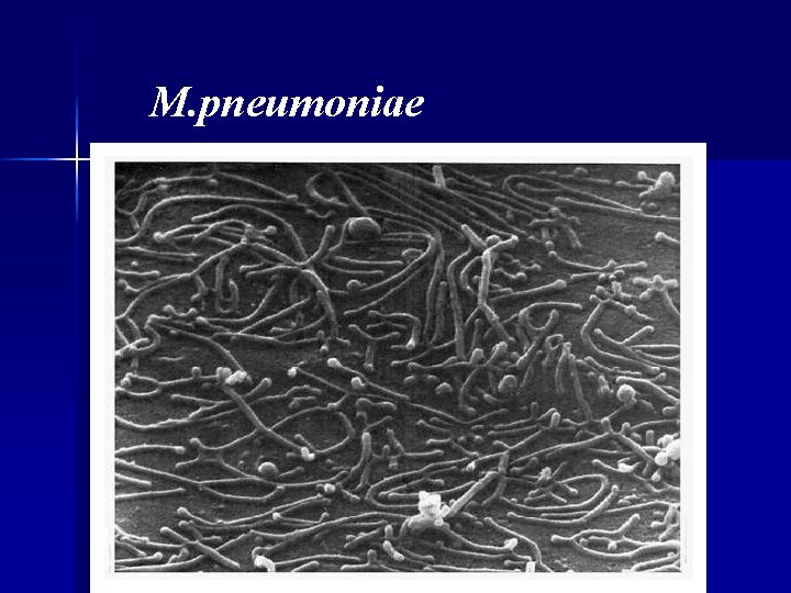
M. pneumoniae
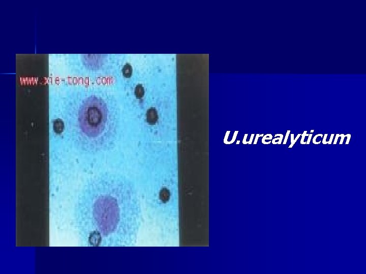
U. urealyticum
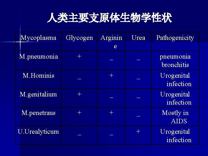
人类主要支原体生物学性状 Mycoplasma Glycogen Urea Pathogenicity + Arginin e _ M. pneumonia _ pneumonia bronchitis M. Hominis _ + _ Urogenital infection M. genitalium + _ _ M. penetraus + + _ Urogenital infection Mostly in AIDS U. Urealyticum _ _ + Urogenital infection
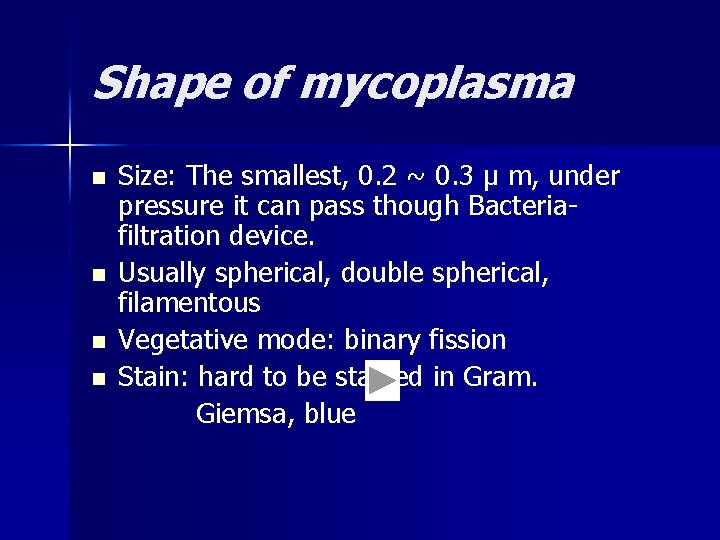
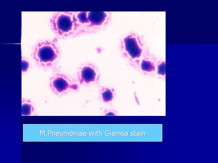
Shape of mycoplasma n n Size: The smallest, 0. 2 ~ 0. 3 μ m, under pressure it can pass though Bacteriafiltration device. Usually spherical, double spherical, filamentous Vegetative mode: binary fission Stain: hard to be stained in Gram. Giemsa, blue
M. Pneumoniae with Giemsa stain
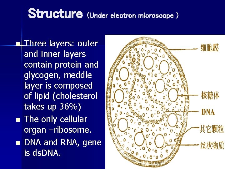
Structure (Under electron microscope ) n n n Three layers: outer and inner layers contain protein and glycogen, meddle layer is composed of lipid (cholesterol takes up 36%) The only cellular organ –ribosome. DNA and RNA, gene is ds. DNA.
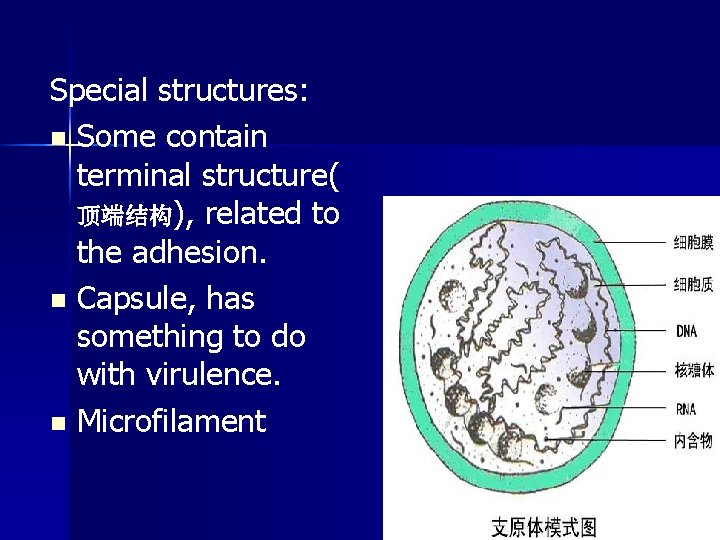
Special structures: n Some contain terminal structure( 顶端结构), related to the adhesion. n Capsule, has something to do with virulence. n Microfilament
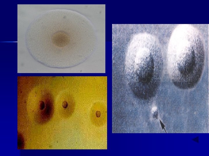
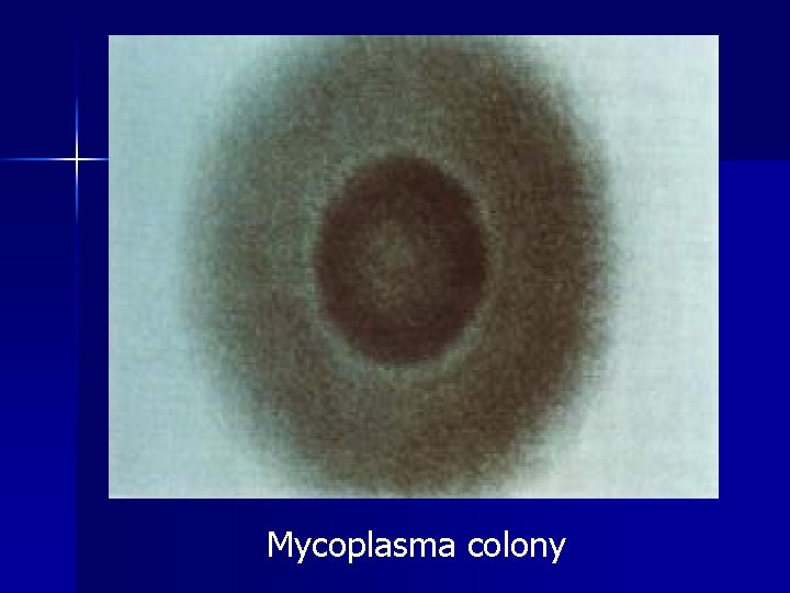
Cultivation n n n high Nutritional requirements : cardin (牛心浸液 ), serum, yeast steep(酵母浸液) In p. H 7. 8 ~ 8. 0 growth below 7. 0 deaths (Ureaplasma urealyticum p. H 6. 0 ~ 6. 5) Need 5%~10%CO 2 Slow Growth,life cycle: 1~3 h liquid medium: Difficult to see the turbid Solid medium: A typical colony was fried egg shape. U. urealyticum: 10~40 μ m, like "T" line
Antigen structure: membrane protein ( use ELISA to detect) and glycolipids. Glycolipids have strong antigens, but have poor specificity. n Part of M. pneumoniae has capsule, which is mainly polysaccharide. n Some have mitogen, cause autoimmune reaction. n
Resistance n n Death 50℃ 30 minutes or 55℃ 5 -15 minutes. Sensitive to UV, dry, heat, Cleanser and disinfectant. Strong resistance to: low temperature(-70 ℃), thallium acetate(铊酸), crystal violet(结 晶紫). Resistance to penicillin, etc. The antibiotic-sensitive protein synthesis interference.
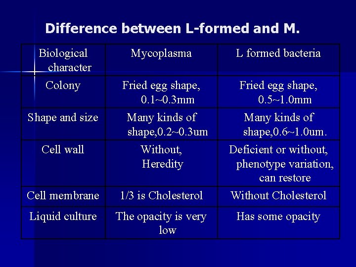
Both the mycoplasma and the L – formed bacterium don't have the cell wall, their biological characters are similar. But what’s the Differences between them?
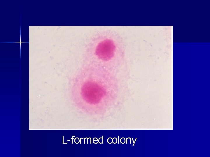
Difference between L-formed and M. Biological character Colony Shape and size Mycoplasma L formed bacteria Fried egg shape, 0. 1~0. 3 mm Fried egg shape, 0. 5~1. 0 mm Many kinds of shape, 0. 2~0. 3 um Cell wall Without, Heredity Cell membrane 1/3 is Cholesterol Liquid culture The opacity is very low Many kinds of shape, 0. 6~1. 0 um. Deficient or without, phenotype variation, can restore Without Cholesterol Has some opacity
Mycoplasma colony
L-formed colony
Thank you!!!~