Chapter 7 Lecture Chapter 1 Lecture HUMAN ANATOMY






- Slides: 30
Chapter 7 Lecture Chapter 1 Lecture HUMAN ANATOMY Chapter 7 The Skeletal System: Appendicular Division
Introduction • The appendicular skeleton is involved in changing your position in the external environment. – Standing – Walking – Sitting – Dressing – Driving a car
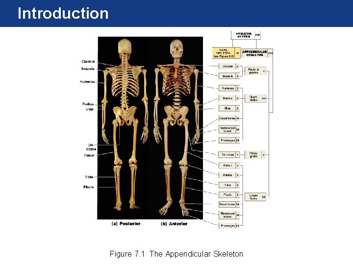
Introduction Figure 7. 1 The Appendicular Skeleton
The Pectoral Girdle and the Upper Limb • Includes the S-shaped clavicle (collarbone) and the flattened scapula (shoulder blade). • The clavicle articulates with the sternum’s manubrium; is the only direct connection between the axial skeleton and the pectoral girdle. • The scapula is attached to the clavicle anteriorly but has no connection to the actual axial skeleton; instead skeletal muscles and ligaments support it.
The Clavicle Figure 7. 3 The Clavicle
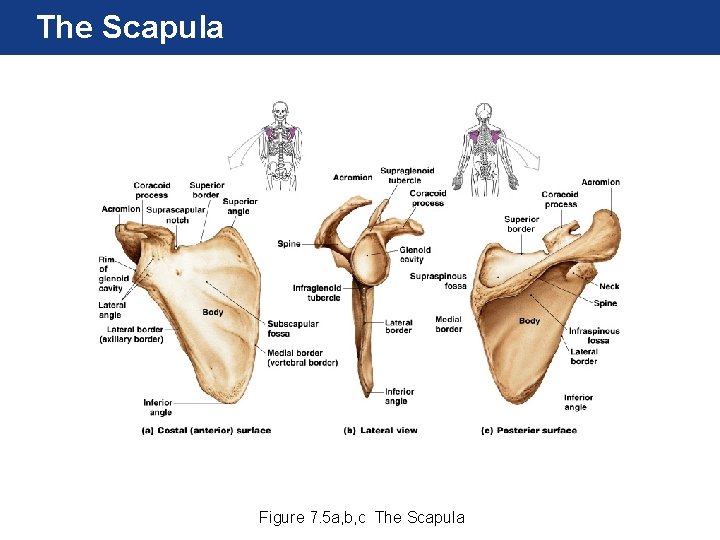
The Scapula Figure 7. 5 a, b, c The Scapula
Scapula
The Scapula supraspinous fossa superior border subscapular fossa acromion coracoid process glenoid fossa spine medial border infraspinous fossa lateral border inferior angle infraglenoid tubercle supraglenoid tubercle Figure 7. 5 d, e, f The Scapula
The Upper Limb • Consists of the: – Brachium (humerus) – Antebrachium (ulna and radius) – Wrist (carpals) – Hand (metacarpals and phalanges)
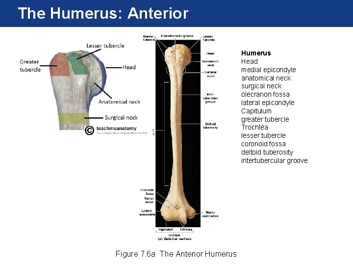
The Humerus: Anterior Humerus Head medial epicondyle anatomical neck surgical neck olecranon fossa lateral epicondyle Capitulum greater tubercle Trochlea lesser tubercle coronoid fossa deltoid tuberosity intertubercular groove Figure 7. 6 a The Anterior Humerus
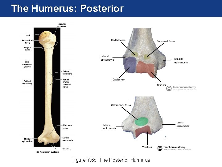
The Humerus: Posterior Figure 7. 6 d The Posterior Humerus
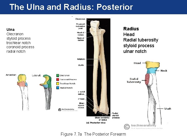
The Ulna and Radius: Posterior Ulna Olecranon styloid process trochlear notch coronoid process radial notch Radius Head Radial tuberosity styloid process ulnar notch Figure 7. 7 a The Posterior Forearm
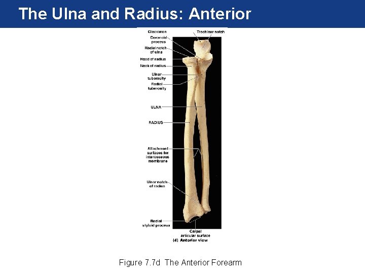
The Ulna and Radius: Anterior Figure 7. 7 d The Anterior Forearm
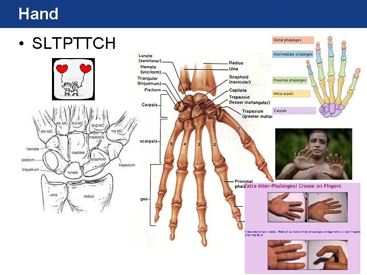
The Wrist and Hand • The carpal bones are the eight bones of the wrist. • The five metacarpal bones articulate with the distal carpal bones and make up the palm of the hand. • The fourteen phalanges of the hand make up the finger bones.
Hand • SLTPTTCH
The Pelvic Girdle and Lower Limb • The pelvic girdle supports and protects the lower viscera and developing fetus in females. • The bones of the pelvic girdle and lower limb are much more massive than their homologues of the upper limb. • Consists of two ossa coxae bones. • The lower limb includes the thigh (femur), kneecap (patella), leg, (tibia and fibula), ankle (tarsals), and foot (metatarsals and phalanges).
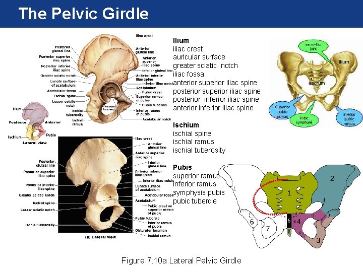
The Pelvic Girdle Ilium iliac crest auricular surface greater sciatic notch iliac fossa anterior superior iliac spine posterior inferior iliac spine anterior inferior iliac spine Ischium ischial spine ischial ramus ischial tuberosity Pubis superior ramus inferior ramus symphysis pubic tubercle Figure 7. 10 a Lateral Pelvic Girdle
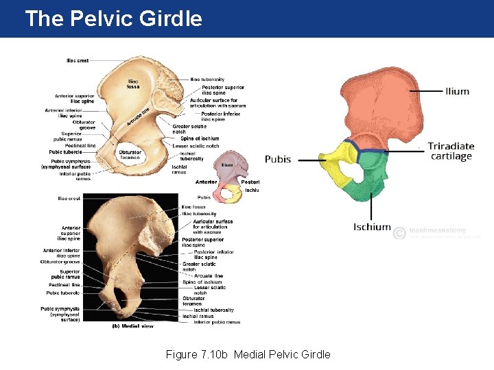
The Pelvic Girdle Figure 7. 10 b Medial Pelvic Girdle
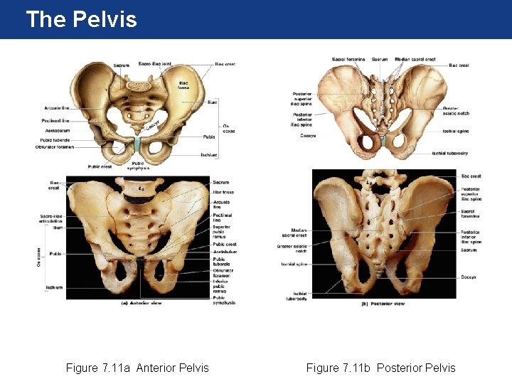
The Pelvis Figure 7. 11 a Anterior Pelvis Figure 7. 11 b Posterior Pelvis
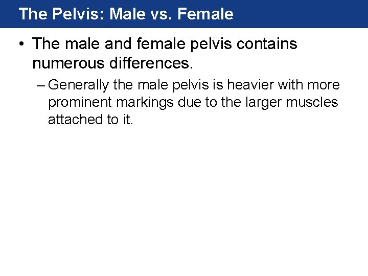
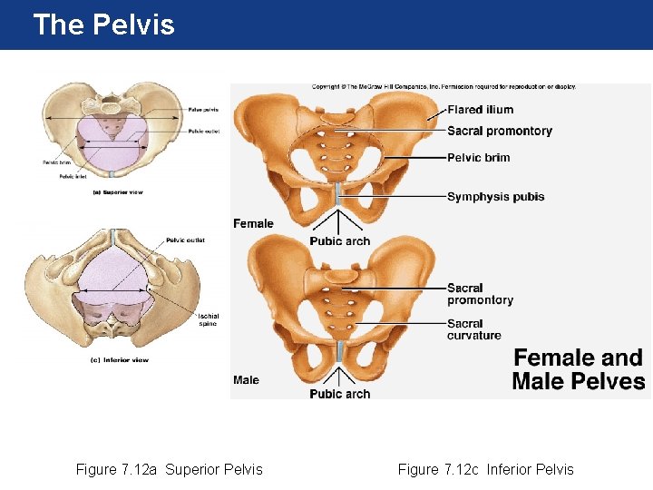
The Pelvis: Male vs. Female • The male and female pelvis contains numerous differences. – Generally the male pelvis is heavier with more prominent markings due to the larger muscles attached to it.
The Pelvis Figure 7. 12 a Superior Pelvis Figure 7. 12 c Inferior Pelvis
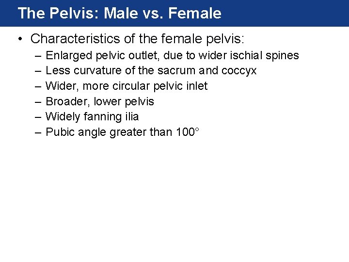
The Pelvis: Male vs. Female • Characteristics of the female pelvis: – – – Enlarged pelvic outlet, due to wider ischial spines Less curvature of the sacrum and coccyx Wider, more circular pelvic inlet Broader, lower pelvis Widely fanning ilia Pubic angle greater than 100°
The Femur: Anterior Femur Head Neck greater trochanter lesser trochanter intercondylar fossa patellar surface linea aspera fovea capitis medial condyle lateral condyle intertrochanteric line intertrochanteric crest Figure 7. 14 a The Anterior Femur
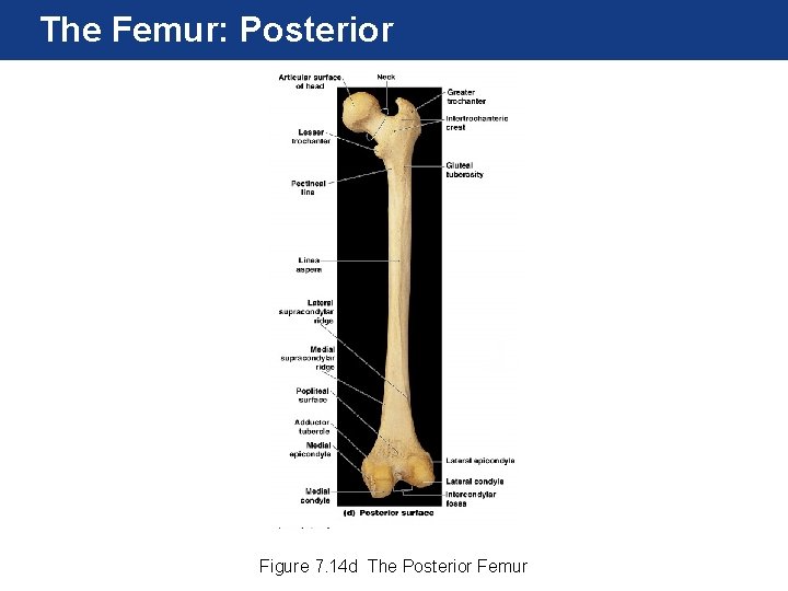
The Femur: Posterior Figure 7. 14 d The Posterior Femur
The Patella Figure 7. 15 The Patella
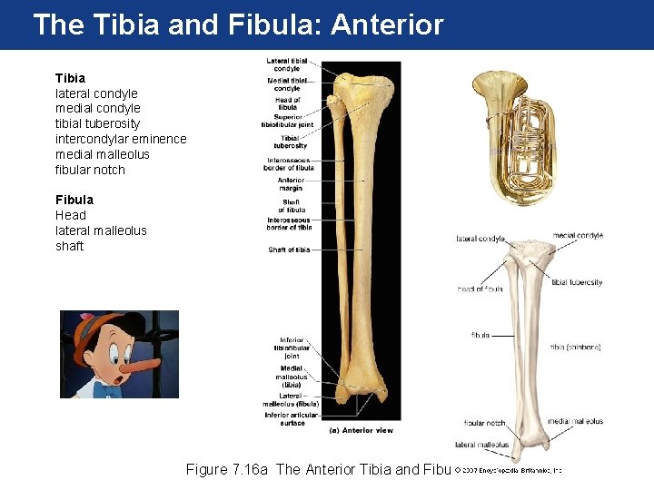
The Tibia and Fibula: Anterior Tibia lateral condyle medial condyle tibial tuberosity intercondylar eminence medial malleolus fibular notch Fibula Head lateral malleolus shaft Figure 7. 16 a The Anterior Tibia and Fibula
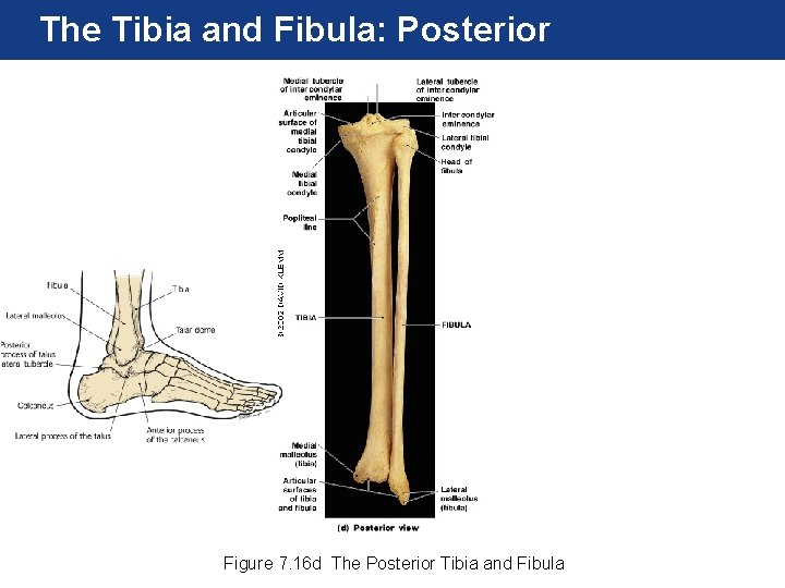
The Tibia and Fibula: Posterior Figure 7. 16 d The Posterior Tibia and Fibula
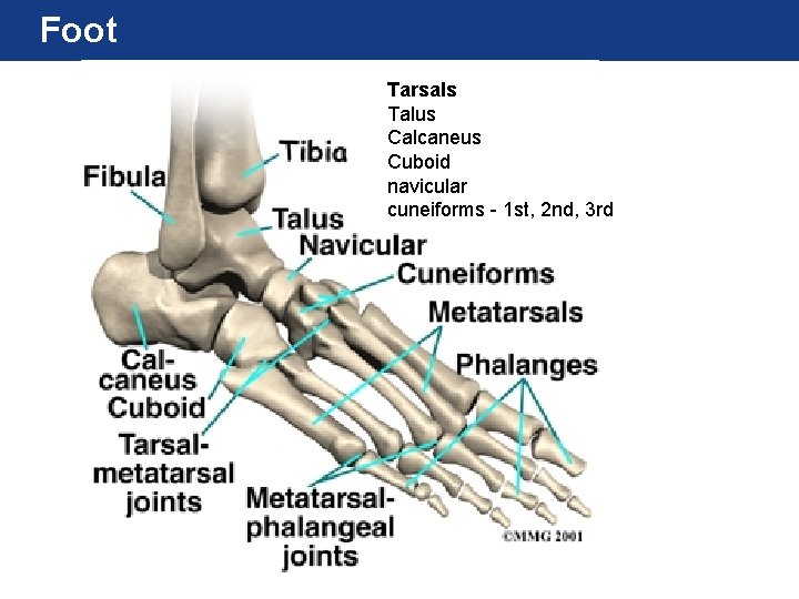
The Ankle and Foot • Seven tarsal bones make up the ankle. • The five metatarsal bones articulate with the distal tarsal bones and make up the arches of the foot. • The fourteen phalanges of the foot make up the toe bones.
Foot Tarsals Talus Calcaneus Cuboid navicular cuneiforms - 1 st, 2 nd, 3 rd
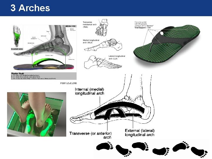
3 Arches