Ch 3 Cell Cycle 1 Fig 3 21

- Slides: 24
Ch 3 Cell Cycle 1
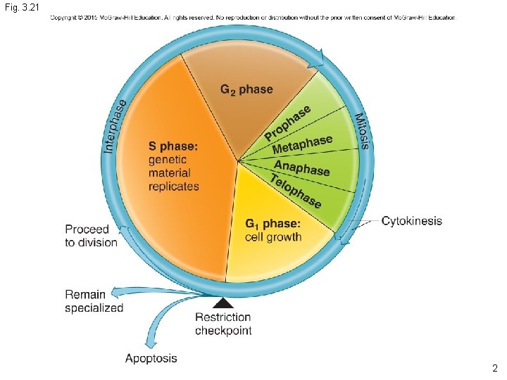
Fig. 3. 21 2
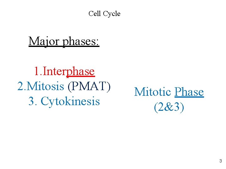
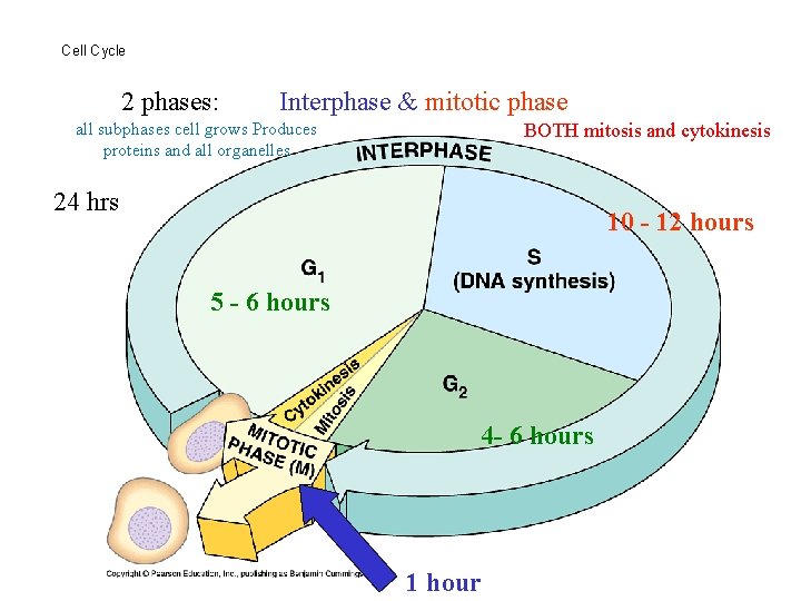
Cell Cycle Major phases: 1. Interphase 2. Mitosis (PMAT) 3. Cytokinesis Mitotic Phase (2&3) 3
Please note that due to differing operating systems, some animations will not appear until the presentation is viewed in presentation mode (Slide Show view). You may see blank slides in the “Normal” or “Slide Sorter” views. All animations will appear after viewing in Slide Show mode and playing each animation. Most animations will require the latest version of the Flash Player, which is available at http: //get. adobe. com/flashplayer. 4
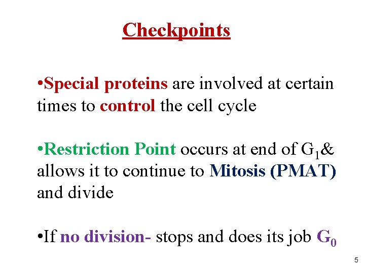
Checkpoints • Special proteins are involved at certain times to control the cell cycle • Restriction Point occurs at end of G 1& allows it to continue to Mitosis (PMAT) and divide • If no division- stops and does its job G 0 5
Cell Cycle 2 phases: Interphase & mitotic phase all subphases cell grows Produces proteins and all organelles BOTH mitosis and cytokinesis 24 hrs 10 - 12 hours 5 - 6 hours 4 - 6 hours 1 hour
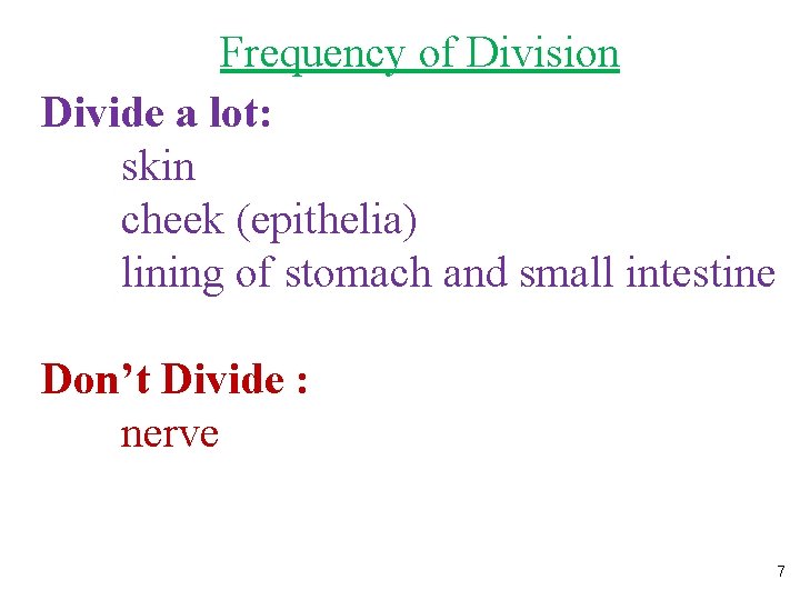
Frequency of Division Divide a lot: skin cheek (epithelia) lining of stomach and small intestine Don’t Divide : nerve 7
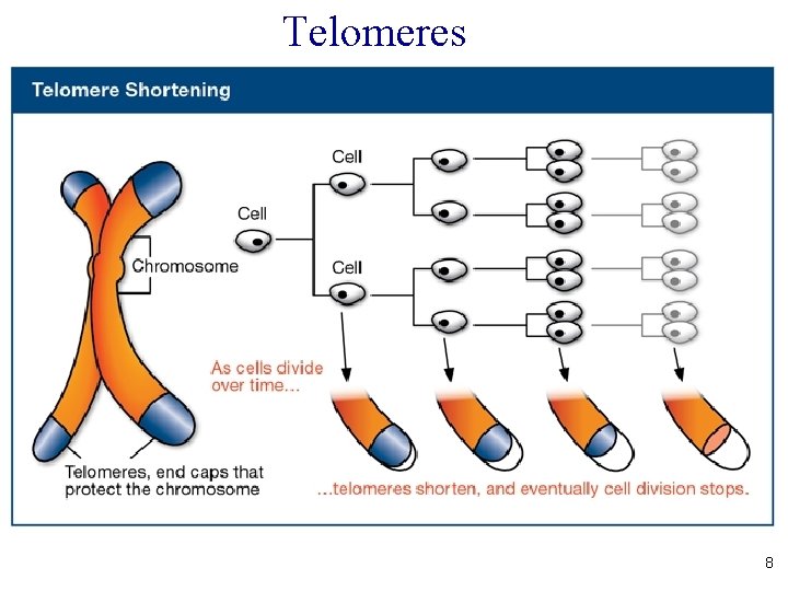
Telomeres 8
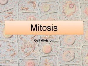
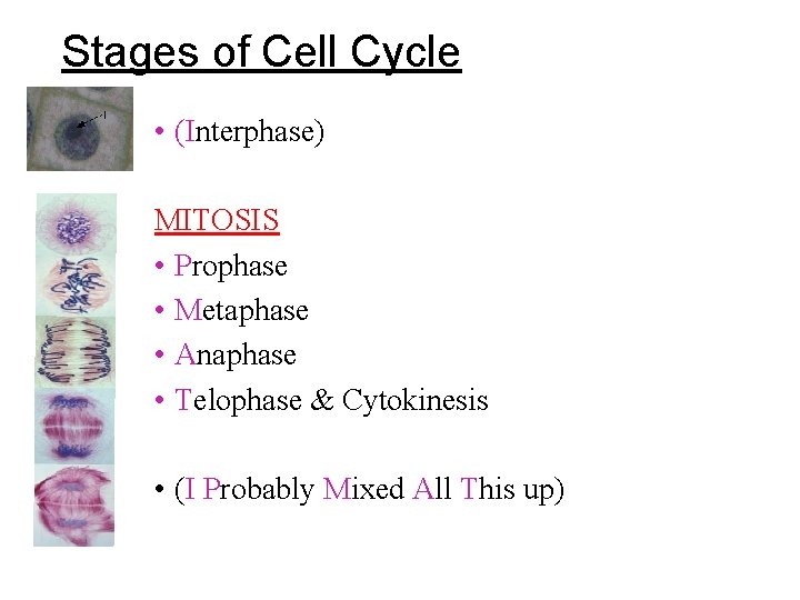
Stages of Cell Cycle • (Interphase) MITOSIS • Prophase • Metaphase • Anaphase • Telophase & Cytokinesis • (I Probably Mixed All This up)
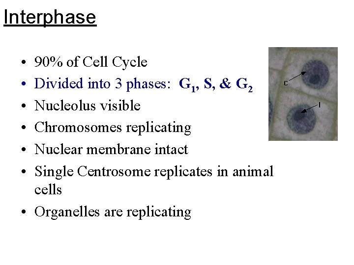
Interphase • • • 90% of Cell Cycle Divided into 3 phases: G 1, S, & G 2 Nucleolus visible Chromosomes replicating Nuclear membrane intact Single Centrosome replicates in animal cells • Organelles are replicating
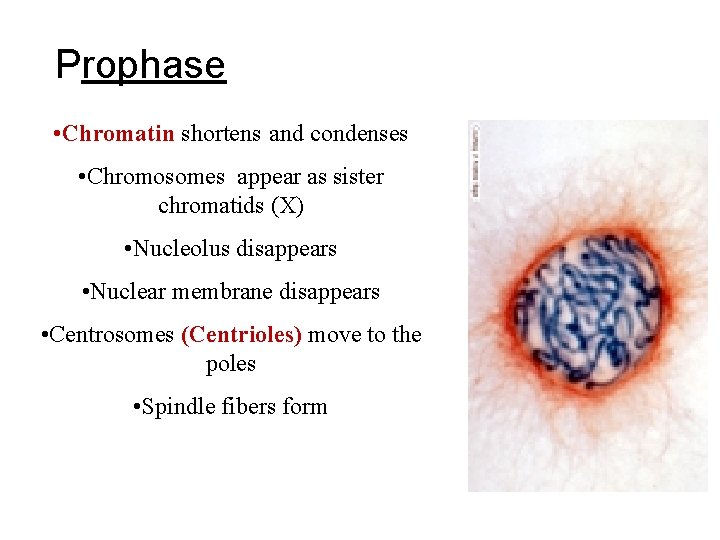
Prophase • Chromatin shortens and condenses • Chromosomes appear as sister chromatids (X) • Nucleolus disappears • Nuclear membrane disappears • Centrosomes (Centrioles) move to the poles • Spindle fibers form
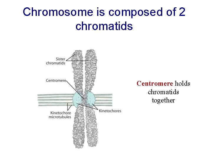
Chromosome is composed of 2 chromatids Centromere holds chromatids together
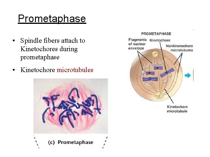
Prometaphase • Spindle fibers attach to Kinetochores during prometaphase • Kinetochore microtubules
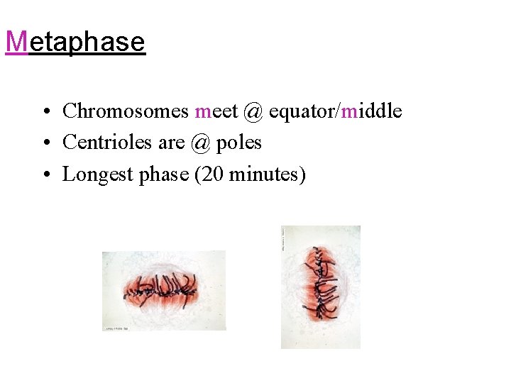
Metaphase • Chromosomes meet @ equator/middle • Centrioles are @ poles • Longest phase (20 minutes)
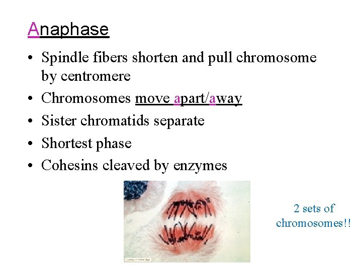
Anaphase • Spindle fibers shorten and pull chromosome by centromere • Chromosomes move apart/away • Sister chromatids separate • Shortest phase • Cohesins cleaved by enzymes 2 sets of chromosomes!!
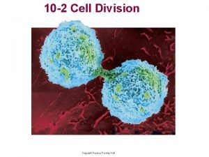
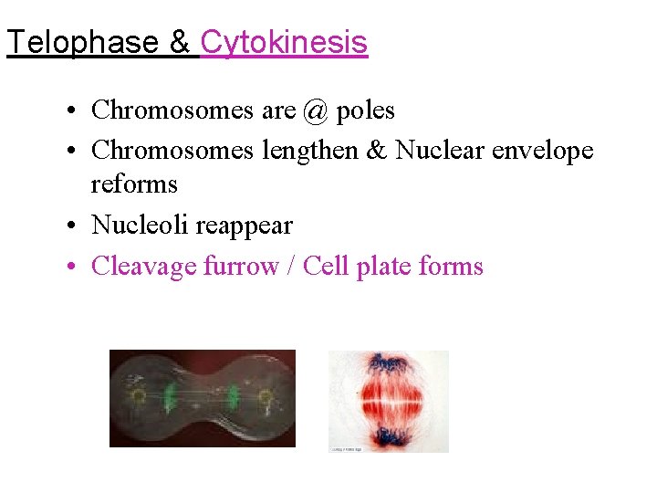
Telophase & Cytokinesis • Chromosomes are @ poles • Chromosomes lengthen & Nuclear envelope reforms • Nucleoli reappear • Cleavage furrow / Cell plate forms
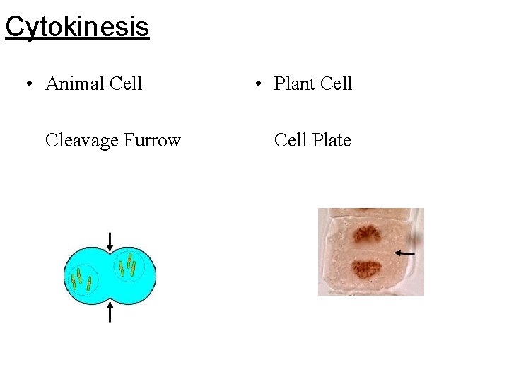
Cytokinesis • Animal Cell Cleavage Furrow • Plant Cell Plate
Please note that due to differing operating systems, some animations will not appear until the presentation is viewed in presentation mode (Slide Show view). You may see blank slides in the “Normal” or “Slide Sorter” views. All animations will appear after viewing in Slide Show mode and playing each animation. Most animations will require the latest version of the Flash Player, which is available at http: //get. adobe. com/flashplayer. 18
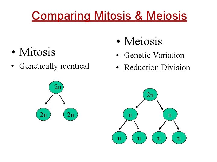
Comparing Mitosis & Meiosis • Mitosis • Genetically identical • Genetic Variation • Reduction Division 2 n 2 n n n n
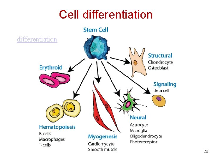
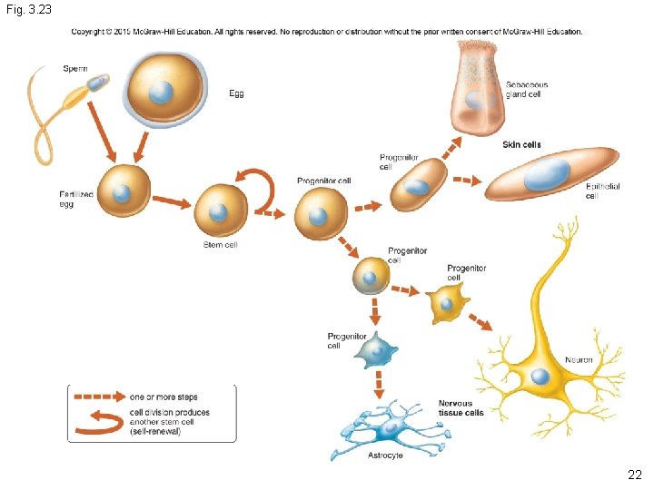
Cell differentiation 20
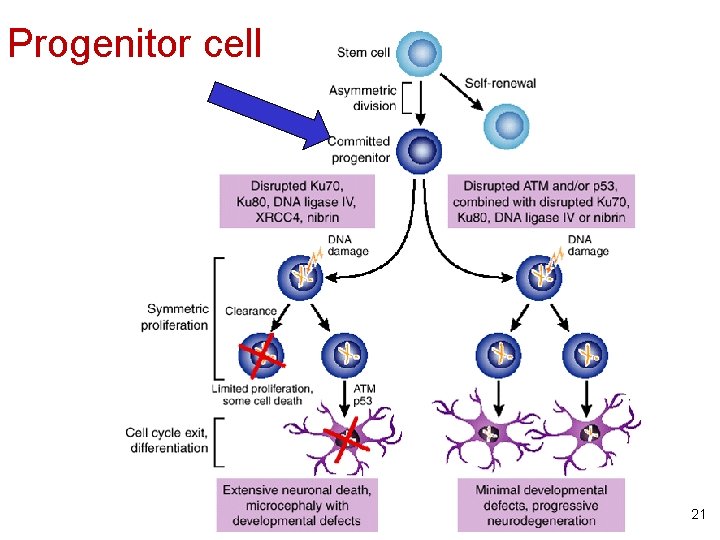
Progenitor cell 21
Fig. 3. 23 22
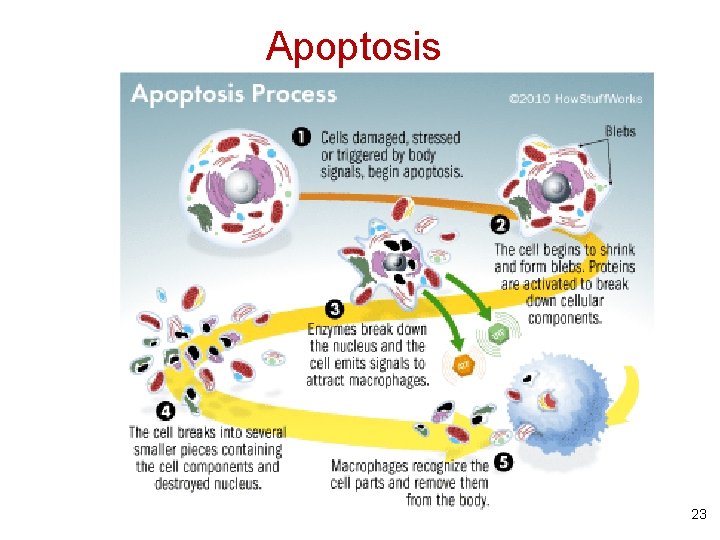
Apoptosis 23
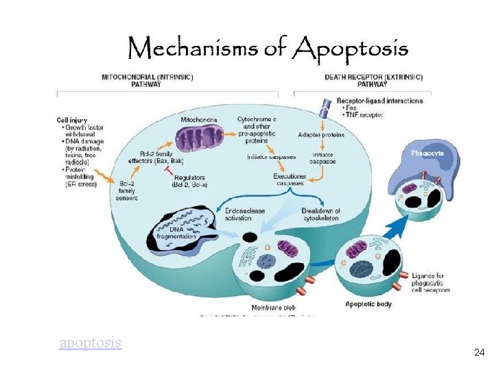
apoptosis 24