Cell Architecture CELL THEORY Mathias Schleiden and Theodore


- Slides: 36
Cell Architecture
CELL THEORY • Mathias Schleiden and Theodore Schwann • Prokaryotic cell • Eukaryotic cell – plant cell and animal cell • Figure 9 -1
Animal cell substructures
Single and Double Membrane Substructures • Single Membrane Structures – Plasma Membrane – Endoplasmic Reticulum – Golgi Apparatus – Lysosome – Peroxisome – Endosome • Double membrane structures – Nucleus and Mitochondria
Plant Cell Substructures
Single and Double Membrane Substructures • Single Membrane Structures – Plasma Membrane – Endoplasmic Reticulum – Golgi Apparatus – Lysosome – Peroxisome – Endosome • Double membrane structures – Nucleus and Chloroplast
Plasma Membrane Chapter 10 Fig. 10 -1
EM OF A THIN ERYTHROCYTE MEMBRANE
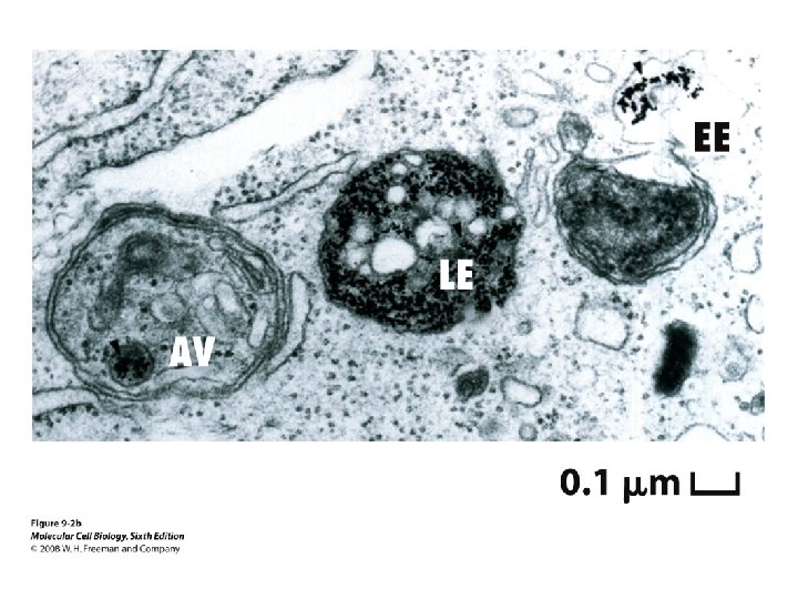
How do endosomes work?
Lysosomes • Internal compartment is very different form the cytosol –more acidic p. H • Degrades substances that are obsolete • Phagocytosis and Endocytosis • All lysosmal enzymes are acid hydrolases • Tays Sach’s disease – defect in enzyme catalyzing a step in the lysosomal breakdown of gangliosides.
Autophagy
Plant Vacuoles • Similar to lysosomes – degradative enzymes • Concentration of solutes is larger inside the vacuole than outside • Stores small molecules and is permeable to water • Elongation of the plant is related to water uptake by the vacuole
EM OF A PLANT CELL
Peroxisomes • 0. 2 -1. 0 micrometer in diameter • Oxidases • Catalase 2 H₂O₂ -----> 2 H₂O + O₂ • Main site of fatty acid peroxidation • Various toxic molecules that enter the body are degraded here
Mitochondrial vs. Peroxisomal Oxidation Fig. 12 -12
Endoplasmic Reticulum • Smooth ER: – Synthesis of Fatty Acids and Lipids – Metabolism of Carbohydrates – Detoxify Drugs and Poisons • Rough ER: – Synthesis of Secretory Proteins, Membrane and Organelle Proteins
ROUGH ER IS MADE OF RIBOSOMES
RIBOSOMES • Composed of r. RNA and protein • Site of protein synthesis • Proteins are marked for different organelles or for secretion • Ribosomes are assembled in the nucleolus • Free Ribosomes
PROTEIN MODIFICATION IN THE ER SECRETED PROTIENS – GLYCOSYLATION – DISULFIDE BOND FORMATION
HORMONE SECRETING CELL FROM RAT PITUITARY
Golgi Apparatus
Golgi - Function • Flattened membrane vesicles or sacs • Cis, medial and trans -Golgi • Proteins targeted for different points in the cell are modified differently • Secretory proteins • Plasma membrane proteins • Membrane or soluble proteins to other organelles
HOW SECRETORY PROTEINS ARE TRANSPORTED?
ORGANELLES WITH DOUBLE MEMBRANES • MITOCHONDRIA – Power house of the cell – Site of cellular respiration – Organic molecules are used fro ATP synthesis NUCLEUS contains chromosomes, DNA and RNA CHLOROPLAST Site of Photosynthesis
EM OF A MITOCHONDRION
STRUCTURE OF A MITOCHONDRION
OUTER MEMBRANE AND INNER MEMBRANE • OUTER MEMBRANE – 50% lipid and 50% protein – Porin proteins (MW of 10, 000) INNER MEMBRANE - CRISTAE
POWER HOUSE OF THE CELL
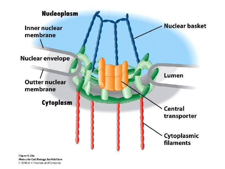
Nucleus • Inner membrane and outer membrane • Outer membrane is continuous with rough ER • The intermembrane space is continuous with the lumen of the ER • Nuclear pores and nucleoporins • Heterochromatin • Ribosomal RNA assembly
HETEROCHROMATIN
CHLOROPLAST • • • Double membraned organelle Length is 10µm and thickness of 0. 5 -2 µm Sacs - Thylakoids Stacks are grana Matrix space called Stroma Photosynthesis
MITOCHONDRIA AND CHLOROPLAST • • ATP production Move around in the cell Contain their own DNA Some of their proteins are encoded in the nucleus
EM OF A PLANT CHLOROPLAST