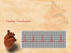
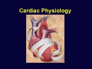
Cardiac Physiology Relation to Cardiac Anatomy Conduction System


- Slides: 12

Cardiac Physiology Relation to Cardiac Anatomy & Conduction System of the Heart

The heart Definition : • It is a hollow muscular organ located in the center of the chest cavity (mediastinum) and rests on the diaphragm

The heart Size of the heart : • Heart weights approximately 300 gm • It’s size are influenced by q Age q Gender q. Body weight q Also influenced by the extent of exercises and heart diseases

Coverings and Wall Consist of three layers: Pericardium: Tightly hugs the external surface of the heart and It’s part of the heart wall Myocardium: consist of thick bundles of cardiac muscles, It is the layer that actually contracts Endocardium: consist of endothelial tissue and lines the inside of the heart & valves

Chambers of the Heart The heart consist of four hollow chambers: • The two atria ( singular atrium ) • The two ventricles

-Right atrium : Received non Oxygenated blood by the vena cava from all body parts - Left atrium : Received Oxygenated blood from the lungs by four pulmonary veins Right ventricle : Received non Oxygenated blood from the right atrium and pumped it through of the two pulmonary arteries to the lungs - - Left ventricle : Has wall that are three times thickness of the right ventricle. Received Oxygenated blood from the left atrium has to be pumped through out the body

• Great vessels • Superior and inferior vena cava : Hold relatively oxygen poor blood from all body parts to the right atrium • Pulmonary arteries: Carry blood from right ventricle to the lungs where oxygen is picked up and carbon dioxide is unloaded • Four pulmonary veins : Carry the Oxygen rich blood from the lungs to the left atrium • Aorta ( Main Great Vessel ): Oxygenated blood pumped through it to the all body parts

Coronary Arteries • The left and right coronary arteries and their branches supply arterial blood to the heart • These arteries originate from the aorta just above the aortic valve leaflets • The heart has large metabolic requirement, extracting approximately 70 - 80% of the oxygen delivered

Coronary Veins • The veins that drains blood from the muscular tissue of the heart and empties into the coronary sinus

The Coronary Sinus • Located posteriorly in the coronary sulcus, enters the right atrium near the tricuspid valve, the entry point in guarded by an incomplete valvethe besius valve

The Heart Valves The tricuspid valve regulates blood flow between the right atrium and right ventricle The pulmonary valve controls blood flow from the right ventricle into the pulmonary arteries, which carry blood to your lungs to pick up oxygen The mitral valve (bicuspid) lets oxygen rich blood from your lungs pass from the left atrium into the left ventricle The aortic valve opens the way for oxygen rich blood to pass from the left ventricle into the aorta, your body's largest artery, where it is delivered to the rest of your body

By : Wissam Aqil Imad Abu Khamis
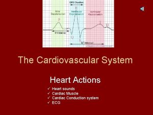
 Intrinsic cardiac conduction system
 Anatomy and physiology unit 7 cardiovascular system
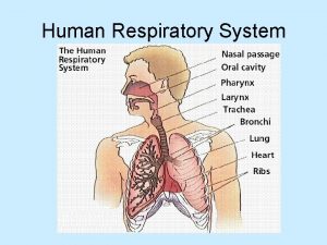
 Types of respiration in human
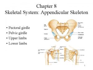
 Anatomy and physiology chapter 8 skeletal system
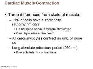
 Cardiac conduction
 Respiratory
 Tattoo anatomy and physiology
 Anatomy science olympiad
 Structure anatomy and physiology in agriculture
 Anatomy and physiology bone
 Anatomy and physiology of peptic ulcer
 Liver physiology and anatomy
 Epigastric region
