Approach to a child with weakness Dr Pushpa

- Slides: 26
Approach to a child with weakness Dr. Pushpa Raj Sharma Professor of Child Health Institute of Medicine
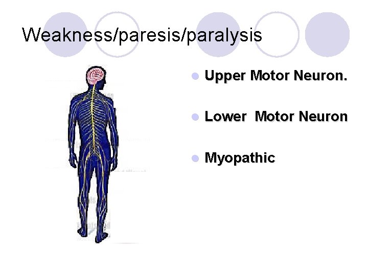
Weakness/paresis/paralysis l Upper Motor Neuron. l Lower Motor Neuron l Myopathic
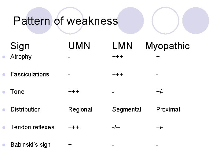
Pattern of weakness Sign UMN LMN Myopathic l Atrophy - +++ + l Fasciculations - +++ - l Tone +++ - +/- l Distribution Regional Segmental Proximal l Tendon reflexes +++ -/-- +/- l Babinski’s sign + - -
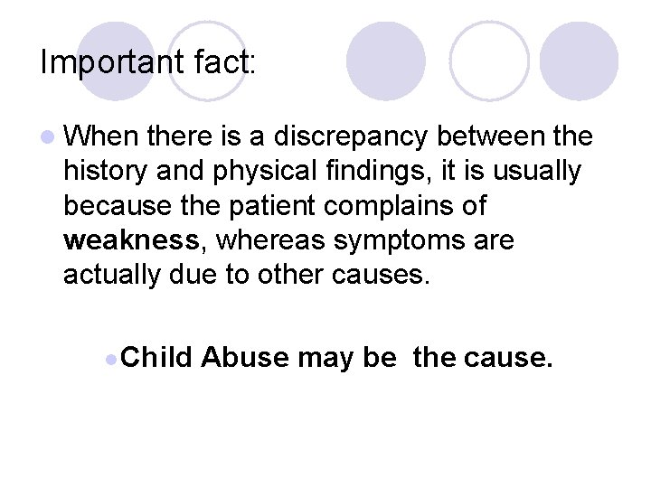
Important fact: l When there is a discrepancy between the history and physical findings, it is usually because the patient complains of weakness, whereas symptoms are actually due to other causes. l Child Abuse may be the cause.
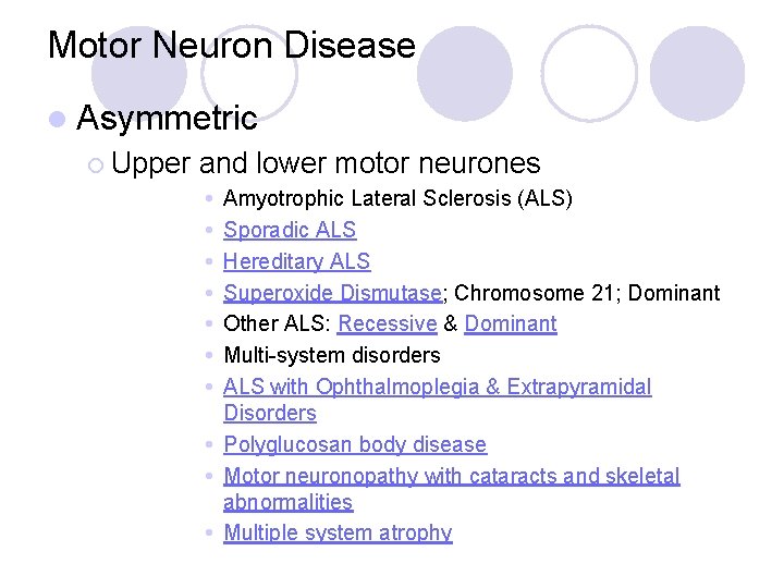
Motor Neuron Disease l Asymmetric ¡ Upper and lower motor neurones Amyotrophic Lateral Sclerosis (ALS) Sporadic ALS Hereditary ALS Superoxide Dismutase; Chromosome 21; Dominant Other ALS: Recessive & Dominant Multi-system disorders ALS with Ophthalmoplegia & Extrapyramidal Disorders Polyglucosan body disease Motor neuronopathy with cataracts and skeletal abnormalities Multiple system atrophy
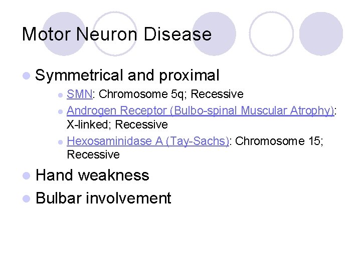
Motor Neuron Disease l Symmetrical and proximal SMN: Chromosome 5 q; Recessive l Androgen Receptor (Bulbo-spinal Muscular Atrophy): X-linked; Recessive l Hexosaminidase A (Tay-Sachs): Chromosome 15; Recessive l l Hand weakness l Bulbar involvement
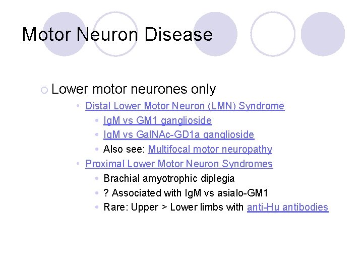
Motor Neuron Disease ¡ Lower motor neurones only • Distal Lower Motor Neuron (LMN) Syndrome Ig. M vs GM 1 ganglioside Ig. M vs Gal. NAc-GD 1 a ganglioside Also see: Multifocal motor neuropathy • Proximal Lower Motor Neuron Syndromes Brachial amyotrophic diplegia ? Associated with Ig. M vs asialo-GM 1 Rare: Upper > Lower limbs with anti-Hu antibodies
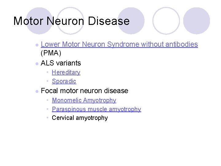
Motor Neuron Disease Lower Motor Neuron Syndrome without antibodies (PMA) l ALS variants l • Hereditary • Sporadic l Focal motor neuron disease • Monomelic Amyotrophy • Paraspinous muscle amyotrophy • Cervical amyotrophy
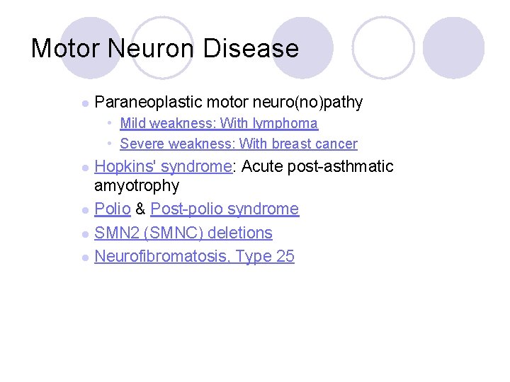
Motor Neuron Disease l Paraneoplastic motor neuro(no)pathy • Mild weakness: With lymphoma • Severe weakness: With breast cancer Hopkins' syndrome: Acute post-asthmatic amyotrophy l Polio & Post-polio syndrome l SMN 2 (SMNC) deletions l Neurofibromatosis, Type 25 l
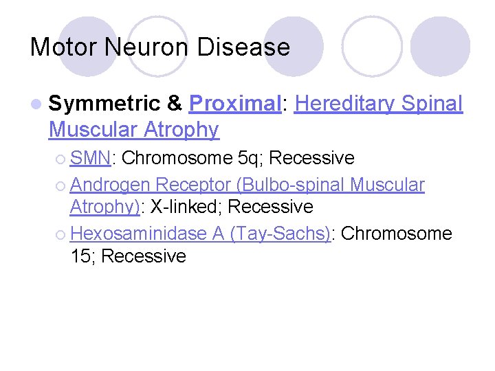
Motor Neuron Disease l Symmetric & Proximal: Hereditary Spinal Muscular Atrophy ¡ SMN: Chromosome 5 q; Recessive ¡ Androgen Receptor (Bulbo-spinal Muscular Atrophy): X-linked; Recessive ¡ Hexosaminidase A (Tay-Sachs): Chromosome 15; Recessive
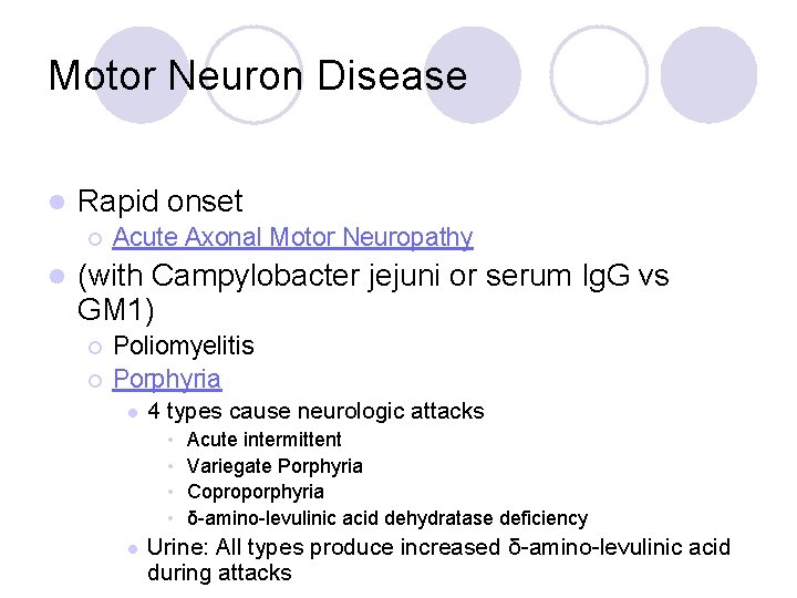
Motor Neuron Disease l Rapid onset ¡ l Acute Axonal Motor Neuropathy (with Campylobacter jejuni or serum Ig. G vs GM 1) ¡ ¡ Poliomyelitis Porphyria l 4 types cause neurologic attacks • • l Acute intermittent Variegate Porphyria Coproporphyria δ-amino-levulinic acid dehydratase deficiency Urine: All types produce increased δ-amino-levulinic acid during attacks
Motor Neuron Disease l Painful l Acquired l Others
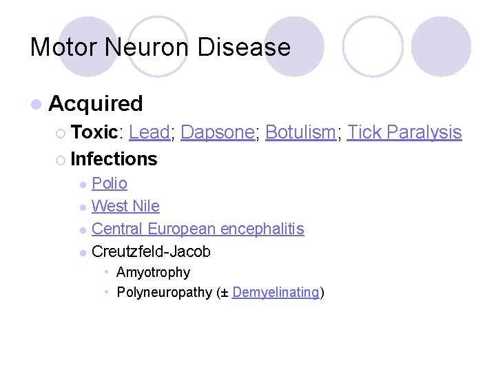
Motor Neuron Disease l Acquired ¡ Toxic: Lead; Dapsone; Botulism; Tick Paralysis ¡ Infections Polio l West Nile l Central European encephalitis l Creutzfeld-Jacob l • Amyotrophy • Polyneuropathy (± Demyelinating)
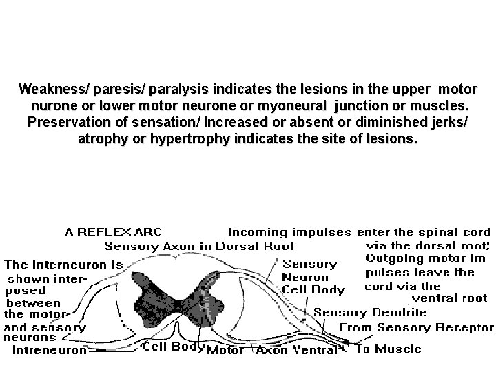
Weakness/ paresis/ paralysis indicates the lesions in the upper motor nurone or lower motor neurone or myoneural junction or muscles. Preservation of sensation/ Increased or absent or diminished jerks/ atrophy or hypertrophy indicates the site of lesions.
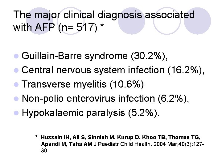
The major clinical diagnosis associated with AFP (n= 517) * l Guillain-Barre syndrome (30. 2%), l Central nervous system infection (16. 2%), l Transverse myelitis (10. 6%) l Non-polio enterovirus infection (6. 2%), l Hypokalaemic paralysis (5. 2%). * Hussain IH, Ali S, Sinniah M, Kurup D, Khoo TB, Thomas TG, Apandi M, Taha AM J Paediatr Child Health. 2004 Mar; 40(3): 12730
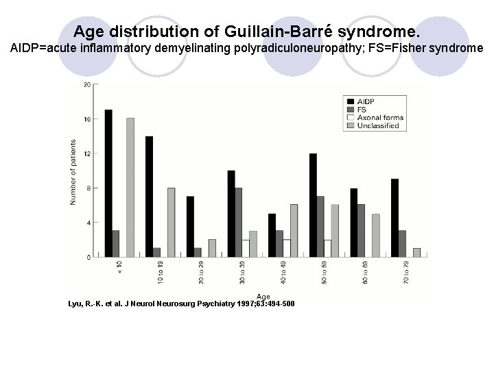
Age distribution of Guillain-Barré syndrome. AIDP=acute inflammatory demyelinating polyradiculoneuropathy; FS=Fisher syndrome Lyu, R. -K. et al. J Neurol Neurosurg Psychiatry 1997; 63: 494 -500
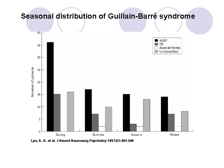
Seasonal distribution of Guillain-Barré syndrome Lyu, R. -K. et al. J Neurol Neurosurg Psychiatry 1997; 63: 494 -500
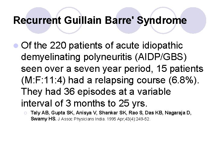
Recurrent Guillain Barre' Syndrome l Of the 220 patients of acute idiopathic demyelinating polyneuritis (AIDP/GBS) seen over a seven year period, 15 patients (M: F: 11: 4) had a relapsing course (6. 8%). They had 36 episodes at a variable interval of 3 months to 25 yrs. ¡ Taly AB, Gupta SK, Anisya V, Shankar SK, Rao S, Das KB, Nagaraja D, Swamy HS. J Assoc Physicians India. 1995 Apr; 43(4): 249 -52.
Follow-up of AGBS l At a follow-up of 1 year or more, 20 patients recovered and 3 had residua. ¡ Hung PL, Chang WN, Huang LT, Huang SC, Chang YC, Chang CJ, Chang CS, Wang KW, Cheng BC, Chang HW, Lu CH. Pediatr Neurol. 2004 Feb; 30(2): 86 -91. l permanent neurological defects in children under 15 years of age was 1. 4/10 million annually ¡ Rantala H, Uhari M, Niemela M. Arch Dis Child. 1991 Jun; 66(6): 706 -8; discussion 708 -9.
Subacute Inflammatory Demyelinating Polyneuropathy. l progressive motor and/or sensory dysfunction consistent with neuropathy in more than one limb with time to nadir between 4 and 8 weeks, l electrophysiologic evidence of demyelination in at least two nerves, l no other etiology of neuropathy, and l no relapse on adequate follow-up. Complete recovery was achieved in 69% of cases and partial recovery in others. ¡ Oh SJ, Kurokawa K, de Almeida DF, Ryan HF Jr, Claussen GC. Neurology. 2003 Dec 9; 61(11): 1507 -12.
Chronic Inflammatory Demyelinating Polyradiculoneuropathy l A low frequency of antecedent events. l Weakness accompanied by sensory loss and diminished tendon reflexes l Pain and cranial neuropathies are uncommon l Progressive weakness for two months. ¡ Sladky JT; Ashwal S: Peripheral neuropathies in childre. Pediatric Neurology Principles and Practice. Ed. Swaiman KS; Ashwal S. 3 rd ed. 1999. Publisher: Mosby
Causes Inflammatory Polyradiculoneuropathy l GB Syndrome 58% l Chronic inflammatory demyelinating polyneuropathy 31% l Associated with collagen vascular disease 7% l Other immune/infectous disorders 4%
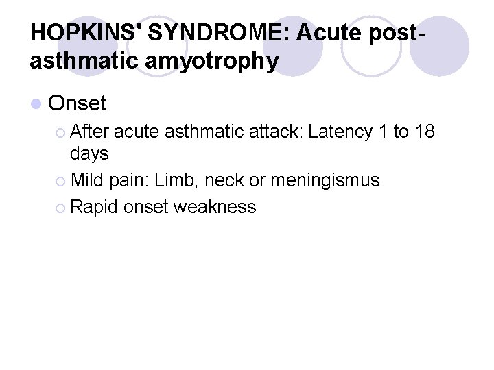
HOPKINS' SYNDROME: Acute postasthmatic amyotrophy l Onset ¡ After acute asthmatic attack: Latency 1 to 18 days ¡ Mild pain: Limb, neck or meningismus ¡ Rapid onset weakness
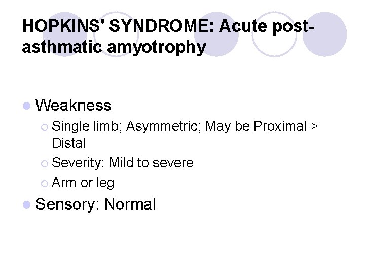
HOPKINS' SYNDROME: Acute postasthmatic amyotrophy l Weakness ¡ Single limb; Asymmetric; May be Proximal > Distal ¡ Severity: Mild to severe ¡ Arm or leg l Sensory: Normal
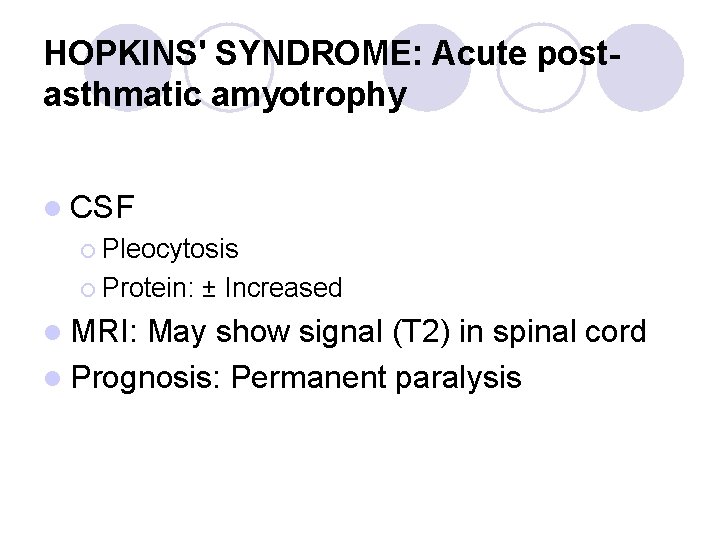
HOPKINS' SYNDROME: Acute postasthmatic amyotrophy l CSF ¡ Pleocytosis ¡ Protein: ± Increased l MRI: May show signal (T 2) in spinal cord l Prognosis: Permanent paralysis
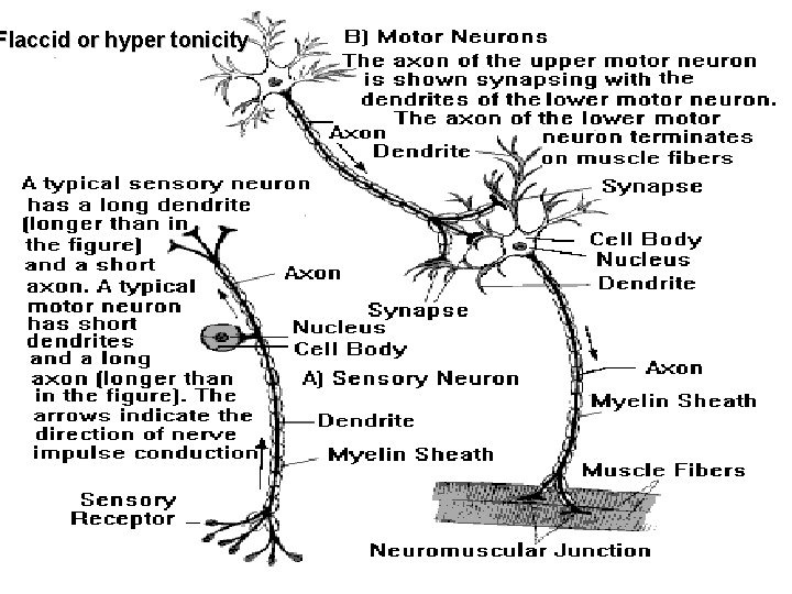
Flaccid or hyper tonicity