ANATOMY OF THE EXCRETORY SYSTEMS Kaan Ycel M


, each of which divides")


 with narrow lumina that carry urine from")


- Slides: 19
ANATOMY OF THE EXCRETORY SYSTEMS Kaan Yücel M. D. , Ph. D. 5. December. 2014 Friday
KIDNEYS Remove excess water, salts, and wastes of protein metabolism from the blood Return nutrients and chemicals to the blood. Lie retroperitoneally on the posterior abdominal wall, one on each side of the vertebral column.
Right kidney T 12 -L 3 Left kidney T 11 -L 2
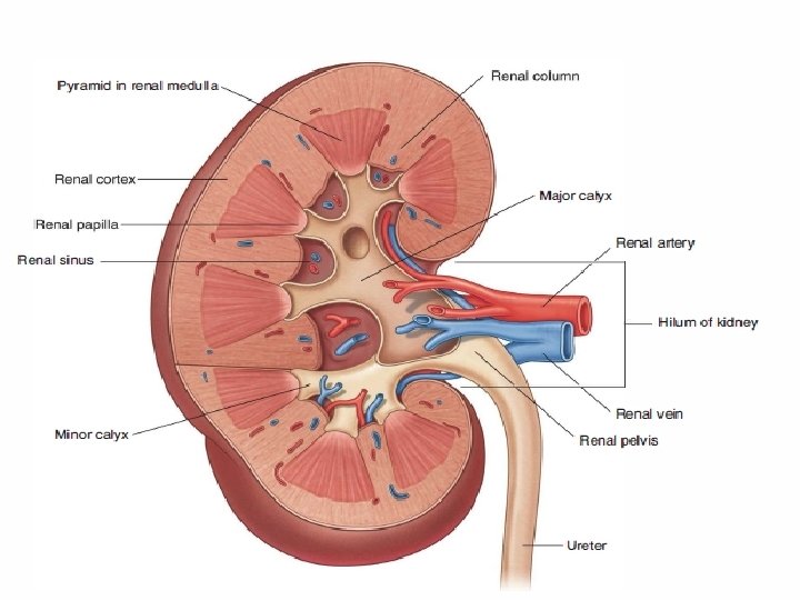
At the concave medial margin of each kidney is a vertical cleft renal hilum The renal hilum is the entrance to a space within the kidney renal sinus Structures that serve the kidneys (vessels, nerves, and structures that drain urine from the kidney) enter and exit the renal sinus through the renal hilum.
Each kidney has anterior and posterior surfaces, medial and lateral margins, and superior and inferior poles. The renal pelvis is the flattened, funnel-shaped expansion of the superior end of the ureter.
The renal pelvis receives two or three major calices (calyces), each of which divides into two or three minor calices Each minor calyx is indented by a renal papilla, papilla the apex of the renal pyramid, pyramid from which the urine is excreted. The pyramids and their associated cortex form the lobes of the kidney.
SUPRARENAL GLANDS Located between the superomedial aspects of the kidneys & diaphragm, where they are surrounded by connective tissue containing considerable perinephric fat.
Suprarenal cortex secretes corticosteroids androgens. These hormones cause the kidneys to retain sodium and water in response to stress, increasing the blood volume and blood pressure. They also affect muscles and organs such as the heart and lungs.
Suprarenal medulla is a mass of nervous tissue associated with the sympathetic nervous system. The chromaffin cells of the medulla secrete catecholamines (mostly epinephrine) into the bloodstream in response to signals from presynaptic neurons. The powerful medullary hormones epinephrine (adrenaline) and norepinephrine (noradrenaline) activate the body to a flight-or-fight status in response to traumatic stress. They also increase heart rate and blood pressure, dilate the bronchioles, and change blood flow patterns, preparing for physical exertion.
PELVIC VISCERA Pelvic viscera include the distal parts of the urinary system and digestive tract, and the reproductive system. The bladder and rectum—true pelvic viscera—are inferior continuations of systems encountered in the abdomen.
Except for features related to sharing of the male urethra by the urinary and reproductive tracts, and physical relationships to the respective reproductive organs, there are relatively few distinctions between the male and female pelvic urinary and digestive organs.
URINARY ORGANS Pelvic urinary organs: Pelvic portions of the ureters, which carry urine from the kidneys. Urinary bladder, which temporarily stores urine. Urethra, which conducts urine from the bladder to the exterior.
URETERS Muscular ducts (25 -30 cm long) with narrow lumina that carry urine from the kidneys to the urinary bladder. Run inferiorly from apices of the renal pelves at the hila of the kidneys. Along the lateral wall of the pelvis and enter the urinary bladder.
A hollow viscus with strong muscular walls Characterized by its distensibility Temporary reservoir for urine Varies in size, shape, position, and relationships according to its content and the state of neighboring viscera When empty, the adult urinary bladder is lies on the pubic bones and pubic symphysis anteriorly and the prostate (males) or anterior wall of the vagina posteriorly.
Toward the neck of the male bladder, the muscle fibers form the involuntary internal urethral sphincter. This sphincter contracts during ejaculation to prevent retrograde ejaculation (ejaculatory reflux) of semen into the bladder.
MALE URETHRA Muscular tube that conveys urine from internal urethral orifice of the urinary bladder to external urethral orifice, orifice located at the tip of the glans penis in males. Also provides an exit for semen (sperms and glandular secretions).
FEMALE URETHRA Passes anteroinferiorly from the internal urethral orifice of the urinary bladder, posterior and then inferior to the pubic symphysis, to the external urethral orifice.
The musculature surrounding the internal urethral orifice of the female bladder is not organized into an internal sphincter. In females, the external urethral orifice is located in the vestibule, vestibule the cleft between the labia minora of the external genitalia, genitalia directly anterior to the vaginal orifice. The urethra lies anterior to the vagina (forming an elevation in the anterior vaginal wall).