Adrenergic transmissionPartI Dr Rashmi Rekha Kumari Asstt Prof
 Dr. Rashmi Rekha Kumari Asstt. Prof, Deptt. Of Pharmacology & Toxicology, BVC,")


 • It is carried out by extraneuronal amine transporter")

- Slides: 14
Adrenergic transmission(Part-I) Dr. Rashmi Rekha Kumari Asstt. Prof, Deptt. Of Pharmacology & Toxicology, BVC, Patna-14
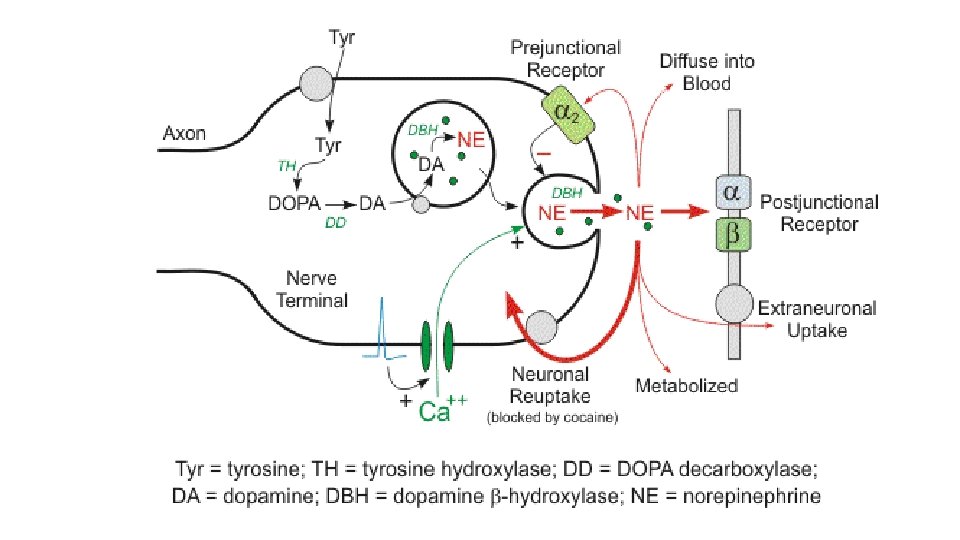
Adrenergic Transmission Adrenergic transmission include transmission at synapse or neuroeffector junction mediated by norepinephrine (post-ganglionic sympathetic nerve terminals and CNS), dopamine (CNS) and epinephrine (adrenal medulla) is in general called as adrenergic transmission. All these transmitters are also called as catecholamines. CATECHOLAMINES: Norepinephrine: It acts as transmitter at most peripheral sympathetic neuroeffector junctions and in the CNS. Epinephrine : It is the major hormone released from adrenal medulla. Dopamine : It is believed to transmit impulse information in specific areas within the CNS (basal ganglia, limbic system, CTZ, anterior pituitary etc. ).
Synthesis of catecholamines Ø L tyrosine, aromatic amine is taken up by adrenergic neurones. Ø. L-tyrosin is converted to dopa by tyrosine hydroxylase(rate limiting step). ØTyrosin hydroxylase occur only in catecholaminergic neurones ØA tyrosine analogue αmethyl tyrosine strongly inhibit tyrosin hydroxylase, used clinically in rare inoperable cases of pheochromocytoma. ØThe next step is convertion of DOPA to Dopamine which is catalysed by DOPA decarboxylase. ØDOPA decarboxylase is relatively non specific enzyme which is not confined to catecholaminergic neurones only and also catalyses covertion of Lhistidine and L-tryptophan to histamine and 5 HT respectively.
• DOPA decarboxylase is present in cytosol • Dopamine is converted into noradrenaline by dopamine β hydroxylase located in synaptic vesicle • DBH is also a relatively nonspecific enzyme but restricted to catecholamine synthesing cells. It is located in synaptic vesicle mainly in membrane bound form. • Phenylethanolamine N methyltransferase(PNMT) catalyses N-methylation of noradrenaline to adrenaline. • The main location of this enzyme is adrenal medulla but also found in brain at the site where adrenaline act as neurotransmitter • In adrenal medulla the norepinephrine thus formed within chromaffine granules diffuses out in the cytoplasm, is methylated and adrenaline is formed • This adrenaline is again taken up by separate set of granules and stored in separate vesicles.
Steps in synthesis of catecholamines
Noradrenaline storage ØNoradrenaline is stored at high concentration in synaptic vesicle, together with ATP, chromogranin and DBH all of which is released by exocytosis ØStorage within the granular vesicles is accomplished by complexation of the noradrenaline with ATP (in molecular ratio of 4: 1) which is adsorbed on a protein, chromogranin. This complexation renders the amine inactive until their release ØThe intra-granular pool of NE is the principal source of neurotransmitter released upon nerve stimulation. ØThe cytoplasmic pool of catecholamines is kept low by the enzyme monoamine oxidase (MAO) present in the outer surface of neuronal mitochondria
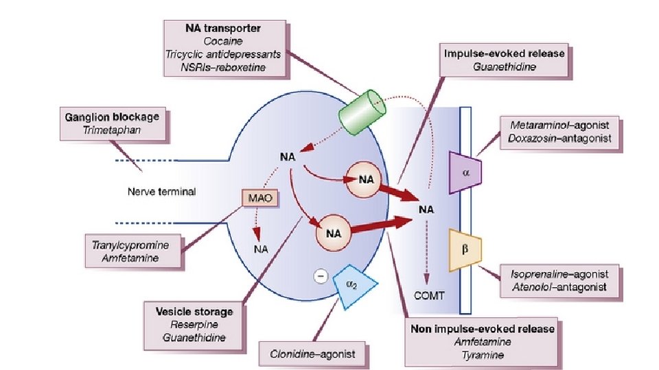
ØHigh concentration of noradrenaline is maintained by a transport mechanism similar to amine transporter responsible for noradrenaline uptake into the nerve terminal but using the transvascular proton gradient as its driving force. ØDrug such as reserpine blocks this transport and cause nerve terminals to be depleted of their noradrenaline store ØNoradrenaline content of cytosol is low owing to monoamineoxidase in nerve terminal.
Noradrenaline Release ØDepolarisation of the nerve terminal membrane opens calcium channel in nerve terminal membrane, and the resulting entry of ca 2+ promotes fusion and discharge of synaptic vesicle. ØNonexocytotic release occur in response to indirectly acting sympathomimeic amines(amphetamines) which displace noradrenaline from vesicle. Noradrenaline escape via uptake-1. ØNoradrenaline release is controlled by autoinhibitory feedback, mediated by α 2 adrenoreceptors. ØCotransmission occurs at many noradrenergic nerve terminals, ATP and neuropeptide Y being frequently coreleased with noradrenaline. ATP mediate the early phase of smooth muscle contraction in response to sympathetic nerve activity.
Uptake and degradation of catecholamines ØThe action of released noradrenaline is terminated mainly by reuptake of the noradrenaline into noradrenergic nerve terminal. ØThis is an active process and responsible for termination of action of nerve impulse in most of the tissue ØThis occurs in two steps: Axonal uptake and Vesicular uptake ØAxonal uptake : An active amine pump(NET) is present at neuronal membrane which is Na dependent and transport NA by a Na coupled mechanism. It takes up NA at a higher rate than adrenaline. This is called Uptake-1. Ø This uptake is the most important mechanism for terminating the postjunctional action of NA. ØThis pump is inhibited by cocaine, desipramine and few other drugs.
Vesicular uptake ØThe membrane of intracellular vesicles has another amine pump the ‘vesicular monoamine transporter’ (VMAT 2), which transports CA from the cytoplasm to within the storage vesicle. Ø The VMAT 2 transports monoamines by exchanging with H+ ions. Ø The vesicular NA is constantly leaking out into the axoplasm and is recaptured by this mechanism. ØThis carrier also takes up DA formed in the axoplasm for further synthesis to NA. ØThus, it is very important in maintaining the NA content of the neurone. Ø This uptake is inhibited by reserpine, resulting in depletion of CAs.
Extraneuronal uptake of CAs (uptake-2) • It is carried out by extraneuronal amine transporter (ENT or OCT 3) and other organic cation transporters OCT 1 and OCT 2 into cells of other tissues. • This uptake process is ubiquitous and is present in glial, hepatic, myocardial and other tissue. • In contrast to NET this uptake transports Adr at a higher rate than NA, is not Na+ dependent and is not inhibited by cocaine, but inhibited by corticosterone. • It is not of physiological or pharmacological importance unless the neuronal uptake mechanism is blocked. • It may be greater importance in disposition of circulating catecholamine than in removal of amines that has been released fromadrenergic nerve terminal