11 18 16 Respiration Mechanisms of Gas Exchange


- Slides: 15
11. 18. 16 Respiration
Mechanisms of Gas Exchange Gas exchange has three phases: - breathing - transport of gases - exchange of gases with tissue cells It provides O 2 for cellular respiration and removes its waste product, CO 2 Respiratory & Circulatory systems
1. Breathing • O 2 is taken into lungs and blood vessels The 3 Phases of Gas Exchange • CO 2 diffuses out and leaves body 2. Transport of gases by the circulatory system • O 2 attaches to hemoglobin and is carried in blood to body tissues • CO 2 is transported from tissues back to lungs 3. Exchange of gases with tissue cells • Body cells take up O 2 from blood and release CO 2 back to the blood • This is diffusion through the plasma membrane (mitochondria internally)
LE 22 -1 O 2 Breathing CO 2 Lung Circulatory system Transport of gases by the circulatory system Mitochondria Exchange of gases with body cells O 2 Capillary Cell
Human Air Pathway: Inhaled through the nostrils Passes through the pharynx and larynx (including vocal cords) Into the trachea, bronchi, and bronchioles* * Bronchioles end in clusters of tiny alveoli, where gas exchange occurs Cilia and mucus are the respiratory system's cleaning elements
Partner 1 - Describe how oxygen gets into your body. Partner 2 – Describe how carbon dioxide leaves.
LE 22 -5 b Oxygen-rich blood Oxygen-poor blood Bronchiole Alveoli Blood capillaries
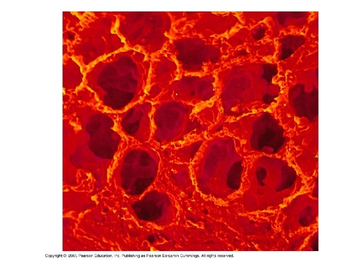
6. 4. U 2 Type I pneumocytes are extremely thin alveolar cells that are adapted to carry out gas exchange. AND 6. 4. U 3 Type II pneumocytes secrete a solution containing surfactant that creates a moist surface inside the alveoli to prevent the sides of the alveolus adhering to each other by reducing surface tension. Alveoli walls and gas exchange Connective tissue Type I pneumocytes Type II pneumocytes • A Single layer of cells form the • Secrete fluid to moisten the inner walls of an alveolus surface of the alveolus • Extremely thin – short diffusion • Fluid aids diffusion of gases distance • Fluid contains surfactant to prevent the • Permeable – aids diffusion walls sticking together – maintains the lumen • Can divide to form Type I pneumocytes – repair damage
Smoking is a deadly assault on our respiratory system – Tobacco smoke irritates cells lining the bronchi • Inhibits or destroys cilia • Kills defensive macrophages – Causes: lung cancer, emphysema, cardiovascular disease – Choosing not to smoke is the most important lifestyle choice for health
LE 22 -6 Lung Heart
Breathing* ventilates the lungs *alternation of inhalation and exhalation Maintains high O 2 and low CO 2 at the respiratory surface
Negative pressure breathing 1. During inhalation, changes in lungs, diaphragm, and rib cage reduce air pressure in alveoli 2. Air rushes in due to higher pressure outside 3. During exhalation, decreased volume of chest cavity forces air out A residual volume of air ("dead air") remains in lungs even after exhalation Vital capacity is the maximum volume of air we can inhale and exhale during forced breathing
Rib cage expands as rib muscles contract Air inhaled Rib cage gets smaller as rib muscles relax Air exhaled Lung Diaphragm contracts (moves down) Diaphragm relaxes (moves up) Inhalation Exhalation
6. 4. A 3 External and internal intercostal muscles, and diaphragm and abdominal muscles as examples of antagonistic muscle action. AND 6. 4. U 5 Muscle contractions cause the pressure changes inside thorax that force air in and out of the lungs to ventilate them. Expiration us pressure change decrease in pressure (draws air inwards) increase in pressure (pushes air outwards) volume change increase decrease ribcage movement up and outward down and inward external intercostal muscles contract relax internal intercostal muscles relax contract diaphragm Contract (flattens, moves downwards) relax abdominal muscles relax Contract (pushes diaphragm up) ca us ca es Inspiration es Summary of the mechanics of ventilation diagrams http: //media 1. shmoop. com/images/biology/biobook_animalmovement_graphik_36. png