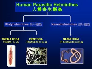
1 Class Trematoda flukes are classified into 4


- Slides: 30

1

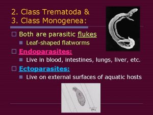
Class Trematoda (flukes) are classified into 4 - Lung fluke 1 - Liver fluke Paragonimus westermani Fasciola 2 - Intestinal flukes 1 - Heterophyes heterophyes 2 - Fasciolopsis busci 3 - Blood flukes 1 - Schistosoma mansoni 2 - Schistosoma haematobium 3 - Schistosoma japonicum 2

Paragonimus westermani (Oriental lung fluke) v. Geographical distribution : Endemic in Far East of Asia (Japan, Korea, China, Philippine and Central & South America. v. Habitat : Lung in cyst like pockets. v. D. H : Man, fish eating animals & carnivorous. v. I. H: 1 st: Fresh water snail (Semisulcospira). 2 nd: Fresh water crayfish or crabs. Diseases : Paragonimiasis 3

Egg (D. S) Size : 90 x 50 µm. Snail 1 st(I. H) Crabs (2 nd I. H) Semisulcospira Shape : Oval. Shell : Thick shell with operculum. Color : Golden brown. Content : Immature ovum. 5

6

Cercaria Microcercous cercaria with small tail Encysted metacercaria ( I. S) Mode of infection: Infection occurs by eating raw or undercooked crabs or crayfish containing encysted metacercaria (I. S) 7

Pathogenesis & Symptomatology Adult worms live Migration stage and granulomatous symptoms: reaction, diarrhea, fibrous capsule is formed abdominal allergic lung stimulate nonspecific chest in & pain, rxn, fever & chills. surrounding worms and eggs to form cyst containing blood tinged fluid. Small Blood vesseles in the capsule provide leakage of metabolites from the cystic cavity into the bronchioles, so the patient paroxysmal shows coughing Cont… due to the discharge of eggs and metabolites. worm Blood is also leacked out from the cyst mixed with ova to give blood tiged sputum. 8

Pathogenesis & Symptomatology Rupture of the cyst Complications into : causes bronchioles pulmonary pneumonia, Chronic cases bronchitis, lung resemble fever, chest pain and abscess pulmonary cough &pneumo-thorax tuberculosis. symptoms such as with rusty sputum (blood tinged and with eggs) effusion. pleural 9

Chronic Paragonimiasis 10 -20% of radiograph findings will be normal. Abnormal Findings including: Lobar infiltration. Cavities. Calcified nodules. Hilar enlargement. P. westermani is diagnosed after TB treatment has failed, therefore a careful differential diagnosis is key in determining Paragonimiasis infections 10

Extrapulmonaty Paragonimiasis Fluks that miss the lungs produce extrapulmonaty symptomes due to cysts, granulomas, and abcesses including: CNS : seizures, coma , paralysis. CNS Paragonimiasis GIT: abdominal pain & diarrhea. Skin: migratory allergic skin lesions. 11

Laboratory Diagnosis Direct Indirect §Detection of eggs & sometimes §Serological tests: adult in rusty sputum. CFT and ELISA §Detection of eggs in stool. § Eosinophilia. §Chest X-ray & CT. Treatment 1 - Praziquentel is the drug of choice 2 - Surgical excision of extrapulmonary lesions 12

Echinococcus granulosus (Hydatid worm) 13

Cestodes are classified according to habitat into Intestinal cestodes (Adult in the small intestine of man) (Man is the D. H) Tissue cestodes (Larvae in the tissues of man) (Man is the I. H) 1 - Diphyllobothrium latum 1 - Cysticercus cellulosa (larva of T. 2 - Taenia saginata solium) Cysticercosis 3 - Taenia solium 2 - Hydatid cyst (larva of Echinococcus 4 - Hymenolepis nana granulosus) Hydatidosis 3 - Cysticercoid nana (larva of H. nana) Cysticercoid nana Note: H. nana & T. solium are considered as intestinal and tissue cestodes 14

Echinococcus granulosus Ø Geographical distribution : Cosmopolitan. Ø Habitat: Small intestine of the D. H. Ø D. H: Dogs, foxes and other canines. Ø I. H: Sheep, cattle, pigs and occasionally man. 15

Morphological Characters 1 - Adult worm 16

2 -Egg of E. granulosus (I. S to man & herbivorous). Size: 30 -40 um. Shape: Spherical. Shell: Thick, radially striated emberyophore. Color: brownish. Content: Mature hexacanth embryo (onchosphere) 17

Lif cycle of the E. granulosus 18

3 -Hydatid cyst Simple unilocular hydatid cyst: Ø The most common type. Ø Size : Variable from pin's head to head of the foetus (1 mm - 20 cm). Ø Shape : More or less spherical. 19

Structure of Hydatid cyst Protoscolex 20

Structure of Hydatid cyst 21

22

Definition Ø It is a parasitic infection of both humans and other mammals such as sheep, cattle and pigs with hydatid cyst, the larval stage of different Echinococcus species. Mode of infection ØIngestion of eggs with food or drinks contaminated with dogs faeces or by handling dogs whose hair are usually contaminated with eggs. 23

Pathogenesis 1) Local inflammatory reaction around the hydatid cyst, ending in formation of a fibrous capsule which may become calcified or even ossified. 2) The symptoms depend on the size & site of the cyst. 3) Large sized cysts pressure atrophy of affected organs. 4) Liver is the commonest organ affected (70%) then lung (20%) & other organs (10%) as brain, bones, kidney, heart , muscles & eyes. 24

Pathogenesis 5) Spontaneous rupture of the cyst into peritoneal cavity or pleura may lead to severe allergic reaction (anaphylactic shock) or secondary cysts. 6) Bacterial infection may occur abscess formation. 25

Pulmonary Cystic Echinococcosis Ø Common in children than adult. Clinical pictures: q Mainly asymptomatic until the cyst enlarges to cause symptoms. q Complication occurs as a result of cyst enlargement & its rupture. It presented by: 1. Cough. 2. Chest pain. 3. Dyspnea. 4. Haemoptysis. 5. Pneumothorax, plural effusion & pulmonary abscess. 26

Diagnosis Clinical: A. Ø History of contact with dogs. Ø Slowly growing cystic tumour. Ø Hydatid thrill. Laboratory: B. Direct: 1) Ø X-ray for calcified cyst. Ø Ultrasonography, CT scan and MRI. Ø Scolices in sputum due to rupture of the cyst in bronchus. Ø Puncture & aspiration of hydatid fluid may lead to anaphylactic shock due to leakage of the fluid. 27

Diagnosis 2) Indirect: A. Intradermal test (Casoni test). B. Serological tests: Indirect hemagglutination test, CFT, immunofluorescence antibody test, ELISA. C. PCR: Nucleic acid detection. D. Eosinophilia. 28

Treatment Surgical removal of the cyst: The most efficient treatment but it may 1) cause mortality (2%) and recurrence of the disease (2 - 25%). Percutaneous treatment (PAIR): In three steps: 2) Puncture (P) and needle aspiration (A) of the cyst. Injection (I) of a scolicidal solution usually hypertonic sodium chloride solution or ethanol and left for 5 - 30 minutes. Cyst-re-aspiration (R) and final washing. It aimed to achieve safe aspiration of large symptomatic cysts and cysts with a danger of impending rupture. 29

Treatment 3) Medical treatment: Indications: In inoperable cases and before and after surgery. Ø Albendazole (ABZ) for 1 - 5 months. Ø Recently, the combination of ABZ and Praziquantil (PZQ) provides synergistic effect and better efficacy. ü Disadvantages: § It may lead to drug resistance. § It is used for long time in high dose. 30

Case Discussion 25 -year-old male presented with abdominal pain. US abdomen was performed which demonstrates a large cyst involving the right hepatic lobe with irrigular wall. CT abdomen shows a well defined cystic lesion involving the right hepatic lobe with multiple curvilinear hyperattenuating structures, this pathologicaly proven hydatid cyst. 31
 Mikael ferm
 Platyhelminthes and nemathelminthes
 Tapeworm class
 Where are liver flukes found
 Greg leos
 Proserkoid
 Filum platyhelminthes
 Classification of manufacturing process
 Carpentry tools and their uses with pictures
 Broadly classified
 Two categories of multimedia
 Slip gauges
 How are organisms classified into domains and kingdoms
 Lesson 2 prokaryotes 1
 Wolffia
 Classification of dienes
 Simetri tubuh platyhelminthes nemathelminthes annelida
 Usus trematoda
 Siklus hidup fasciola hepatica
 Morfologi paragonimus westermani
 Tapeworm pseudocoelomate
 Trematoda
 Telur fasciolopsis buski
 Klonorkiasis
 Contoh trematoda
 Taxonomic
 Trematoda
 Daur hidup platyhelminthes
 Filo platelmintos
 Ploštěnci
 How are volcanoes classified?

