What are we measuring with EEG and MEG















- Slides: 33
What are we measuring with EEG and MEG ? Isabel Zlobinski & Xavier De Tiège
Introduction Neurophysiological background EEG MEG
Introduction EEG and MEG are 2 functional cerebral imaging techniques that are closely related In both methods, the measured signals are generated by the same synchronized neuronal activity in the brain The main interest of M-EEG compared to other techniques TEMPORAL RESOLUTION
The temporal resolution of M-EEG Follow the rapid changes in cortical activity Reflect ongoing signal processing in the brain
Neurophysiological background Glial cells Structural support Metabolism Ions & NTT transport Myelin Neurons Information-processing units
Grey matter cell bodies & dendrites Cortex & basal ganglia White matter axones (myeline)
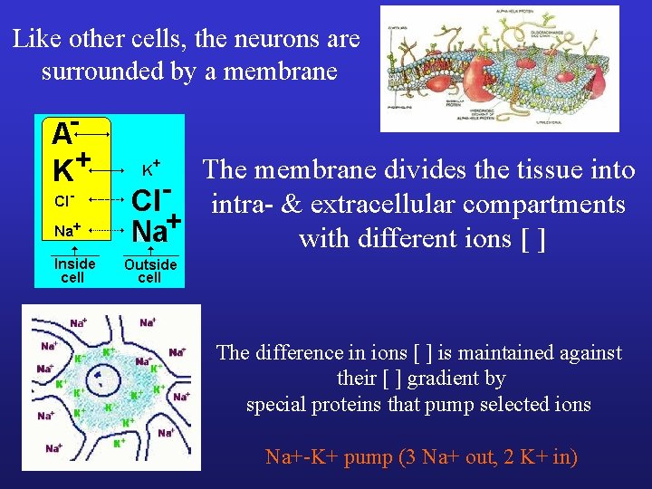
Like other cells, the neurons are surrounded by a membrane The membrane divides the tissue into intra- & extracellular compartments with different ions [ ] The difference in ions [ ] is maintained against their [ ] gradient by special proteins that pump selected ions Na+-K+ pump (3 Na+ out, 2 K+ in)
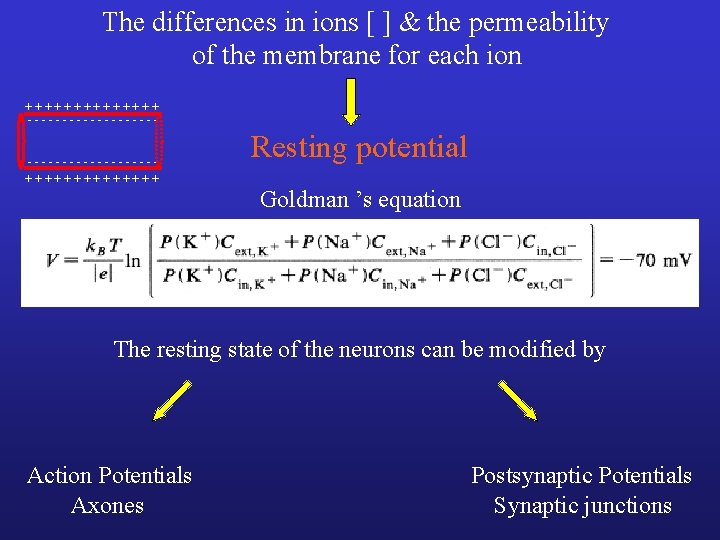
The differences in ions [ ] & the permeability of the membrane for each ion + + + + - - - - - - - - - - + + + + Resting potential Goldman ’s equation The resting state of the neurons can be modified by Action Potentials Axones Postsynaptic Potentials Synaptic junctions
Action Potentials Depolarization Repolarization Generated at the cell body/axone junction Hyperpolarization
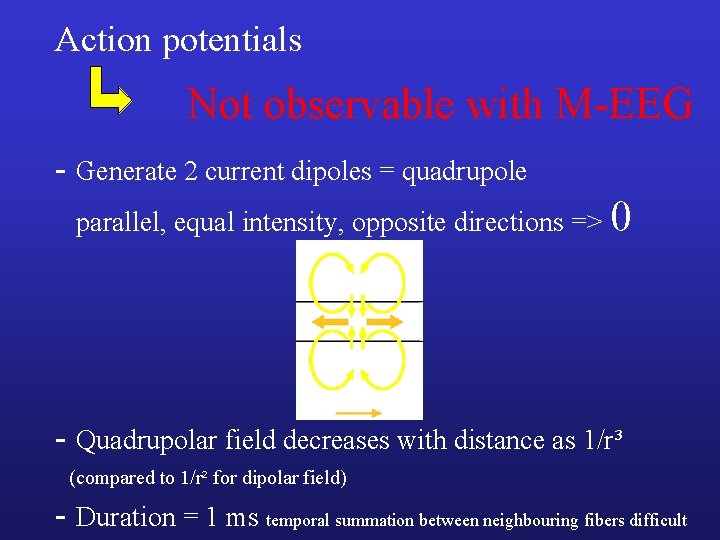
Action potentials Not observable with M-EEG - Generate 2 current dipoles = quadrupole parallel, equal intensity, opposite directions => 0 - Quadrupolar field decreases with distance as 1/r³ (compared to 1/r² for dipolar field) - Duration = 1 ms temporal summation between neighbouring fibers difficult
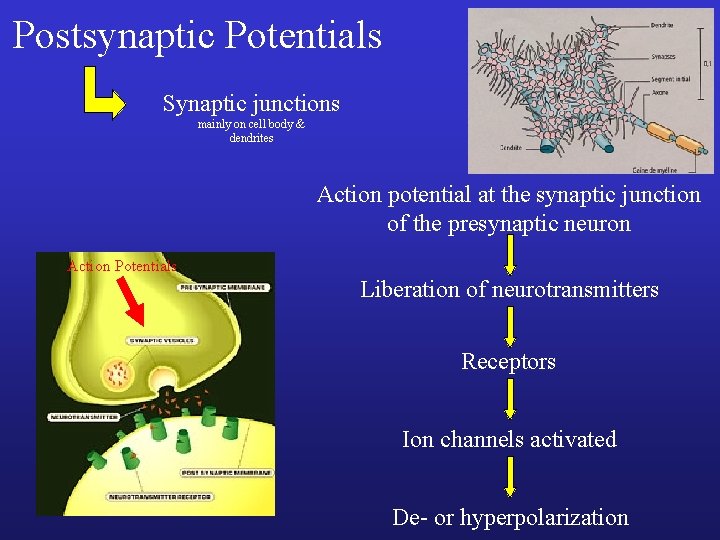
Postsynaptic Potentials Synaptic junctions mainly on cell body & dendrites Action potential at the synaptic junction of the presynaptic neuron Action Potentials Liberation of neurotransmitters Receptors Ion channels activated De- or hyperpolarization
Acetylcholine or glutamate Activate Na+ and Ca++ channels Depolarization Excitatory PSP Summation of EPSP Action potential at the cell body/axon junction GABA Activate Cl- channels Hyperpolarization Prevents action potential generation Inhibitory PSP
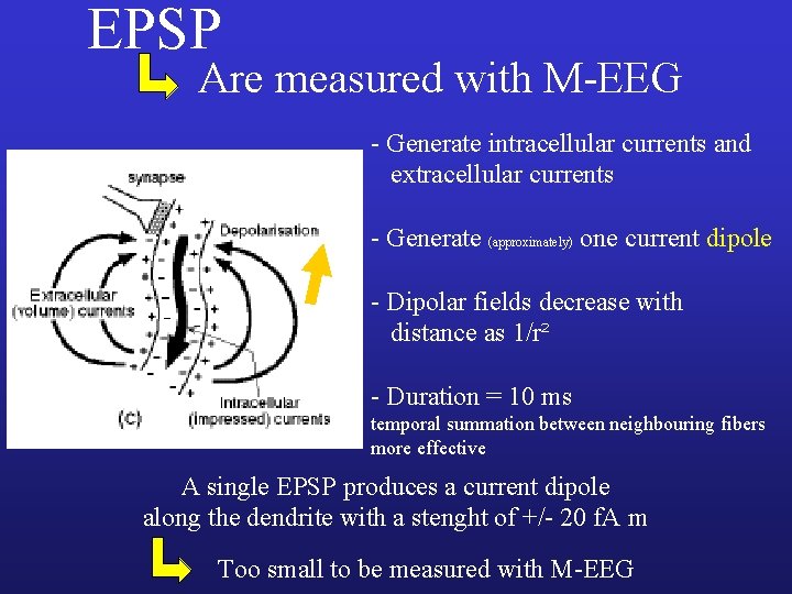
EPSP Are measured with M-EEG - Generate intracellular currents and extracellular currents - Generate (approximately) one current dipole - Dipolar fields decrease with distance as 1/r² - Duration = 10 ms temporal summation between neighbouring fibers more effective A single EPSP produces a current dipole along the dendrite with a stenght of +/- 20 f. A m Too small to be measured with M-EEG
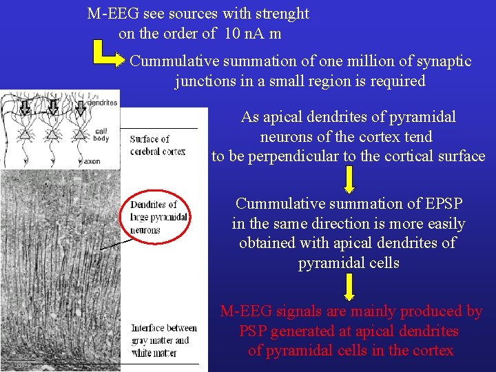
M-EEG see sources with strenght on the order of 10 n. A m Cummulative summation of one million of synaptic junctions in a small region is required As apical dendrites of pyramidal neurons of the cortex tend to be perpendicular to the cortical surface Cummulative summation of EPSP in the same direction is more easily obtained with apical dendrites of pyramidal cells M-EEG signals are mainly produced by PSP generated at apical dendrites of pyramidal cells in the cortex
What are we measuring with MEG ?
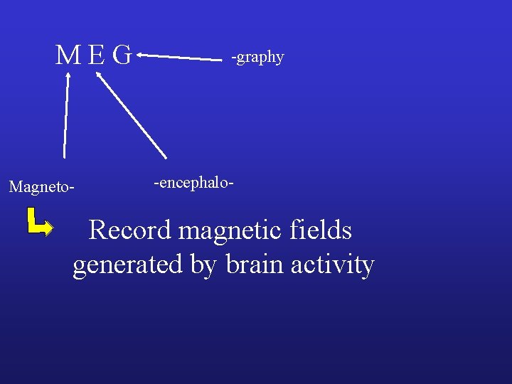
M E G Magneto- -graphy -encephalo- Record magnetic fields generated by brain activity
PSP induced intracellular currents (primary currents) and extracellular currents (secondary currents) Secondary currents yield potential differences on the scalp of the head that can be measured by EEG MEG measures magnetic fields induced mainly by primary currents Cummulative summation of PS primary currents of millions apical dendrites of pyramidal cells in one cortical area Generates a magnetic field measurable by MEG
Primary currents "Right Hand Law" Induced magnetic field Volume currents
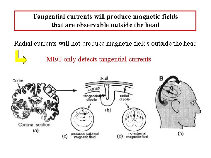
Tangential currents will produce magnetic fields that are observable outside the head Radial currents will not produce magnetic fields outside the head MEG only detects tangential currents
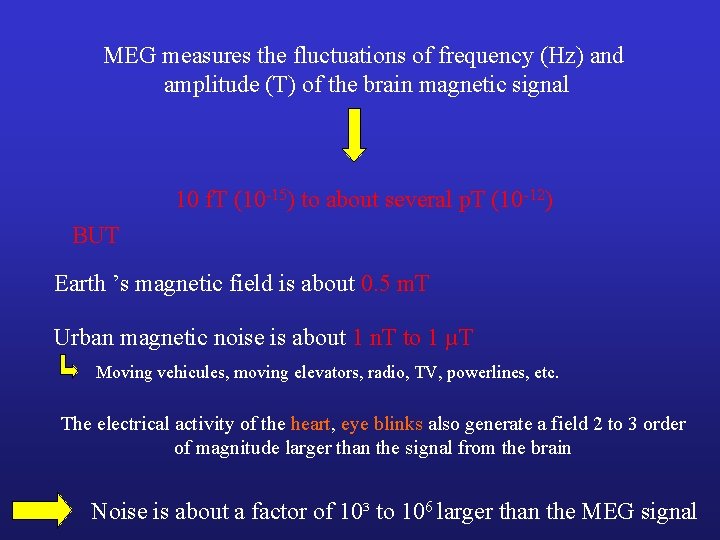
MEG measures the fluctuations of frequency (Hz) and amplitude (T) of the brain magnetic signal 10 f. T (10 -15) to about several p. T (10 -12) BUT Earth ’s magnetic field is about 0. 5 m. T Urban magnetic noise is about 1 n. T to 1 µT Moving vehicules, moving elevators, radio, TV, powerlines, etc. The electrical activity of the heart, eye blinks also generate a field 2 to 3 order of magnitude larger than the signal from the brain Noise is about a factor of 10³ to 106 larger than the MEG signal
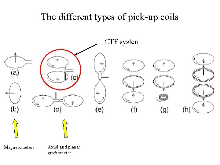
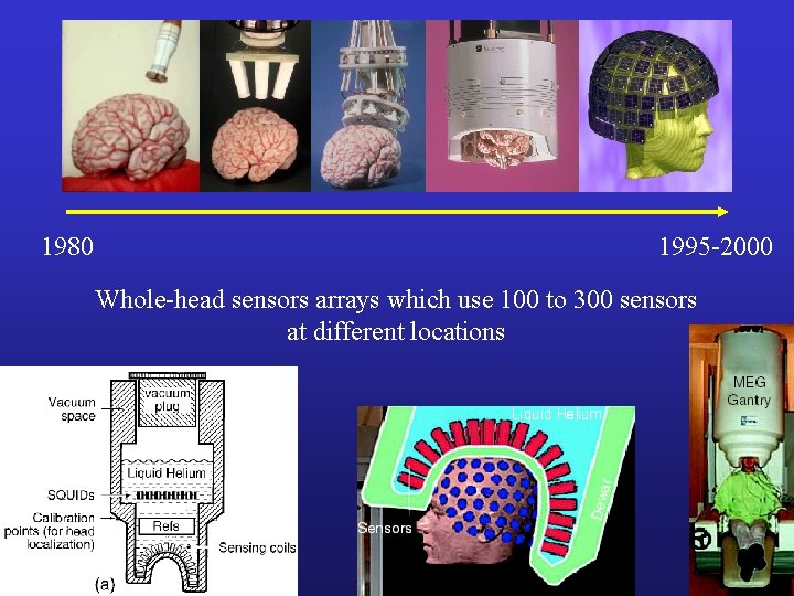
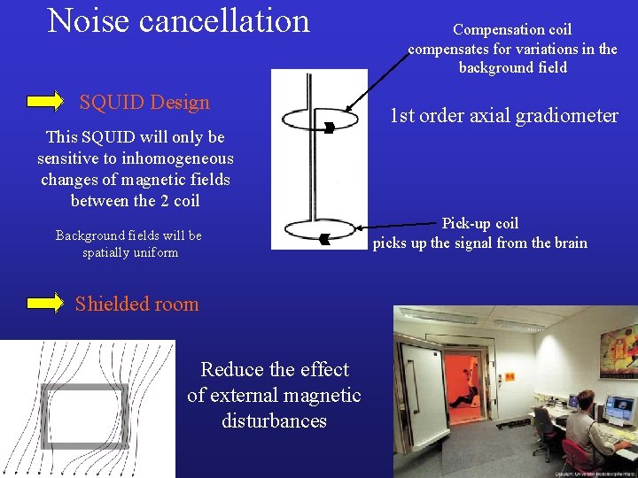
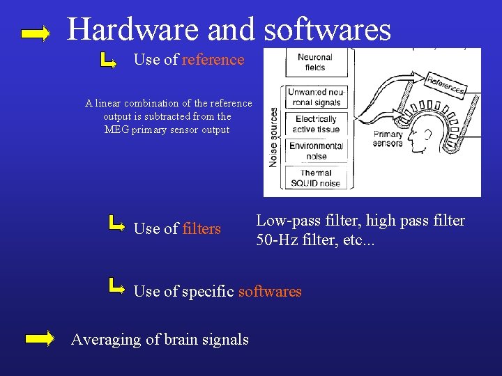
We need very sensitive MEG sensors to pick up the brain magnetic fields SQUIDs MEG measurements need noise cancellation with extraordinary accuracy Design of the SQUID Magnetic shielded room Hardware and software Averaging
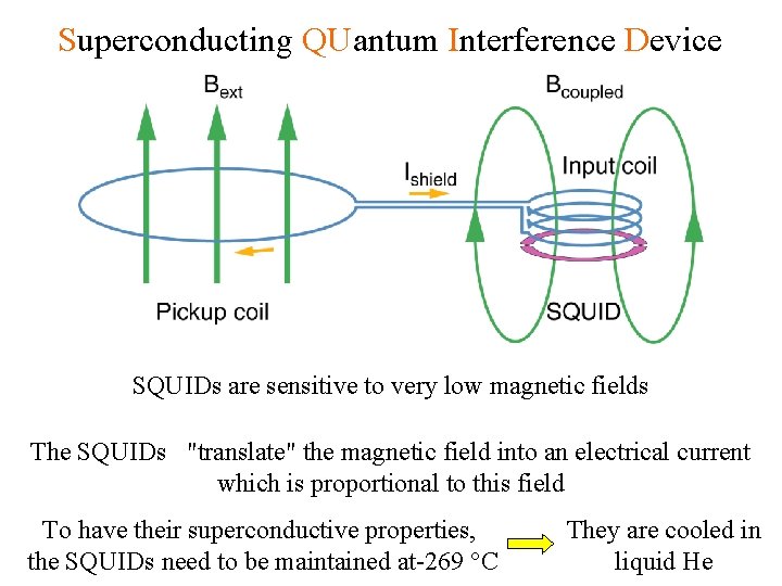
Superconducting QUantum Interference Device SQUIDs are sensitive to very low magnetic fields The SQUIDs "translate" the magnetic field into an electrical current which is proportional to this field To have their superconductive properties, the SQUIDs need to be maintained at-269 °C They are cooled in liquid He
The different types of pick-up coils CTF system Magnetometers Axial and planar gradiometer
1980 1995 -2000 Whole-head sensors arrays which use 100 to 300 sensors at different locations
Noise cancellation SQUID Design This SQUID will only be sensitive to inhomogeneous changes of magnetic fields between the 2 coil Background fields will be spatially uniform Shielded room Reduce the effect of external magnetic disturbances Compensation coil compensates for variations in the background field 1 st order axial gradiometer Pick-up coil picks up the signal from the brain
Hardware and softwares Use of reference A linear combination of the reference output is subtracted from the MEG primary sensor output Use of filters Low-pass filter, high pass filter 50 -Hz filter, etc. . . Use of specific softwares Averaging of brain signals
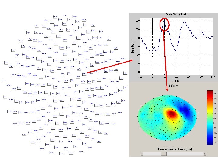
With MEG, you can make (as in EEG) : - Continuous acquisition of brain signals and study some events that appear « randomly » (Epileptic abnormalities, etc. ) - Evoked response: averaged MEG signals that are synchronous with an external stimulus or voluntary motor event
References : - Hämäläinen et al. , Reviews of Modern Physics, 1993 - Baillet et al. , IEEE Signal Processing Magazine, 2001 - Jeremie Mattout Ph. D thesis - Murakami & Okada, J Physiol, in press