
Basis of the MEEG Signal Methods for Dummies

Basis of the M/EEG Signal Methods for Dummies Jan 28 th 2015 Clare Palmer & Brianna Beck
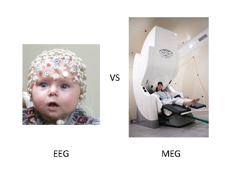
VS EEG MEG
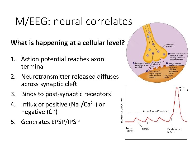
M/EEG: neural correlates What is happening at a cellular level? 1. Action potential reaches axon terminal 2. Neurotransmitter released diffuses across synaptic cleft 3. Binds to post-synaptic receptors 4. Influx of positive (Na+/Ca 2+) or negative (Cl-) 5. Generates EPSP/IPSP
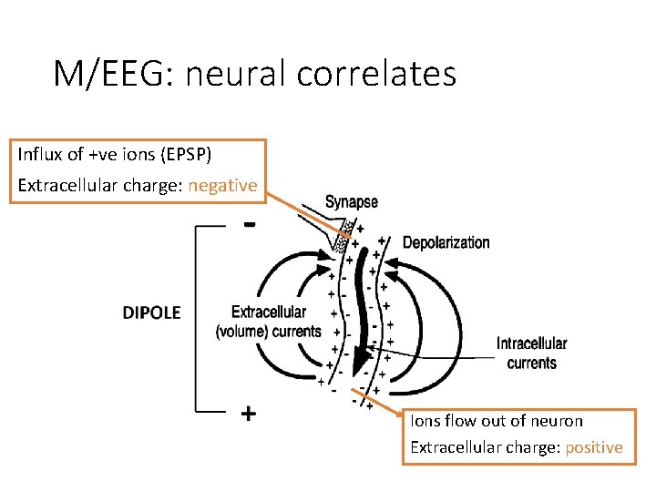
M/EEG: neural correlates Influx of +ve ions (EPSP) Extracellular charge: negative Ions flow out of neuron Extracellular charge: positive
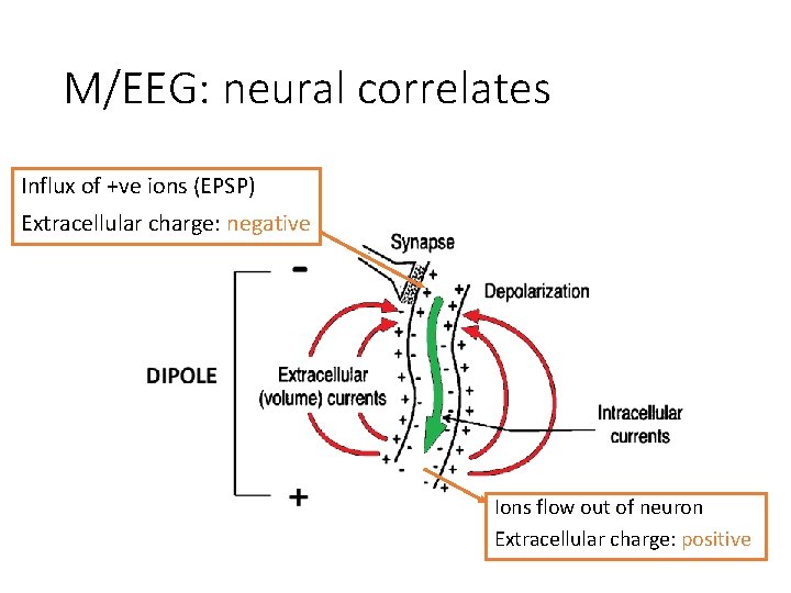
M/EEG: neural correlates Influx of +ve ions (EPSP) Extracellular charge: negative Ions flow out of neuron Extracellular charge: positive
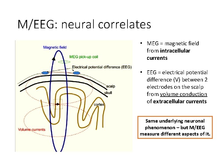
M/EEG: neural correlates • MEG = magnetic field from intracellular currents • EEG = electrical potential difference (V) between 2 electrodes on the scalp from volume conduction of extracellular currents Same underlying neuronal phenomenon – but M/EEG measure different aspects of it.
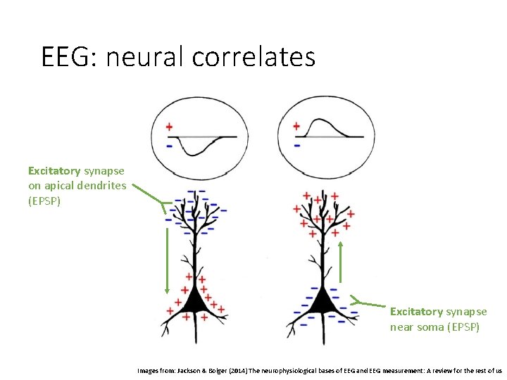
EEG: neural correlates Excitatory synapse on apical dendrites (EPSP) Excitatory synapse near soma (EPSP) Images from: Jackson & Bolger (2014) The neurophysiological bases of EEG and EEG measurement: A review for the rest of us
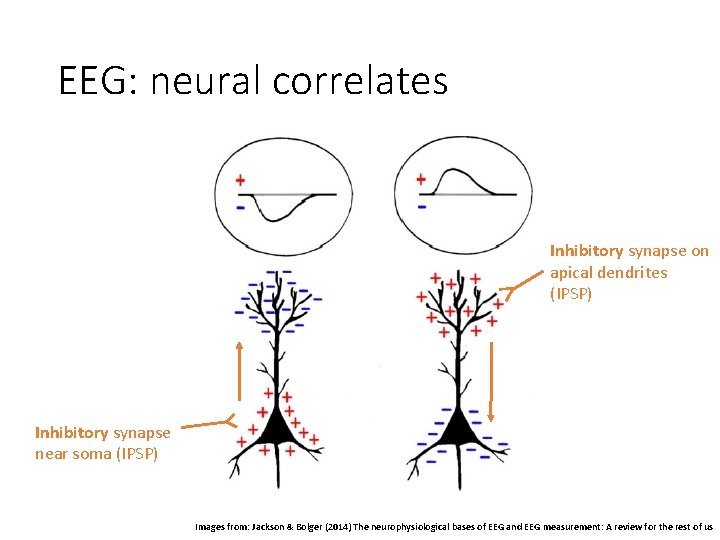
EEG: neural correlates Inhibitory synapse on apical dendrites (IPSP) Inhibitory synapse near soma (IPSP) Images from: Jackson & Bolger (2014) The neurophysiological bases of EEG and EEG measurement: A review for the rest of us
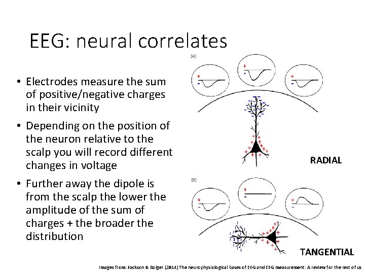
EEG: neural correlates • Electrodes measure the sum of positive/negative charges in their vicinity • Depending on the position of the neuron relative to the scalp you will record different changes in voltage • Further away the dipole is from the scalp the lower the amplitude of the sum of charges + the broader the distribution RADIAL TANGENTIAL Images from: Jackson & Bolger (2014) The neurophysiological bases of EEG and EEG measurement: A review for the rest of us
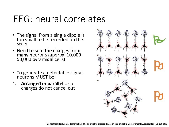
EEG: neural correlates • The signal from a single dipole is too small to be recorded on the scalp • Need to sum the charges from many neurons (approx. 10, 00050, 000 pyramidal cells) • To generate a detectable signal, neurons MUST be: 1. Arranged in parallel = so charges do not cancel out 2. Synchronously active = creates a large enough signal to measure + + + - - - Images from: Jackson & Bolger (2014) The neurophysiological bases of EEG and EEG measurement: A review for the rest of us
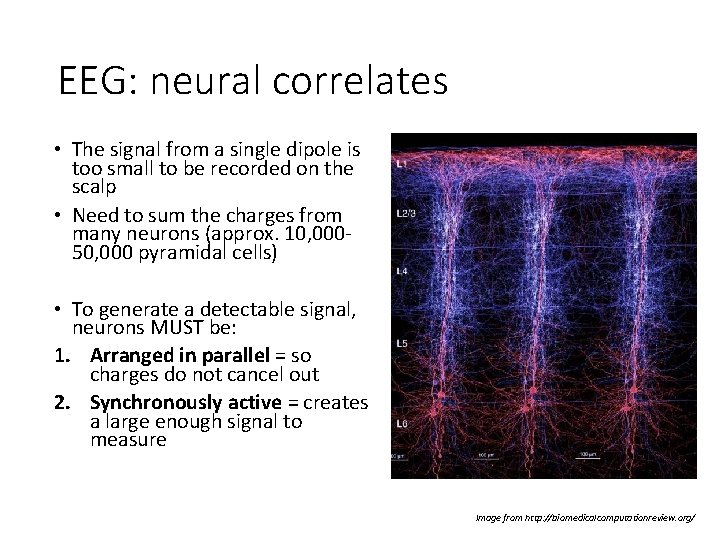
EEG: neural correlates • The signal from a single dipole is too small to be recorded on the scalp • Need to sum the charges from many neurons (approx. 10, 00050, 000 pyramidal cells) • To generate a detectable signal, neurons MUST be: 1. Arranged in parallel = so charges do not cancel out 2. Synchronously active = creates a large enough signal to measure Image from http: //biomedicalcomputationreview. org/
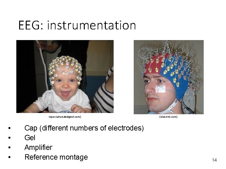
EEG: instrumentation (specialneedsdigest. com) • • Cap (different numbers of electrodes) Gel Amplifier Reference montage (biosemi. com) 14
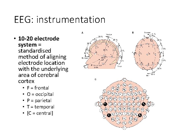
EEG: instrumentation • 10 -20 electrode system = standardised method of aligning electrode location with the underlying area of cerebral cortex • • • F = frontal O = occipital P = parietal T = temporal [C = central]
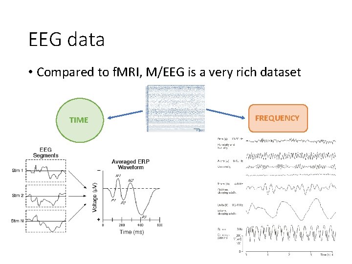
EEG data • Compared to f. MRI, M/EEG is a very rich dataset TIME FREQUENCY
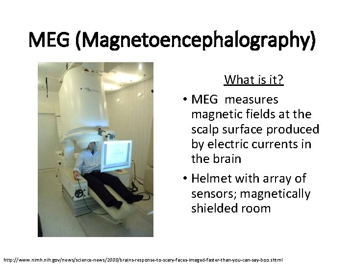
MEG (Magnetoencephalography) What is it? • MEG measures magnetic fields at the scalp surface produced by electric currents in the brain • Helmet with array of sensors; magnetically shielded room http: //www. nimh. nih. gov/news/science-news/2008/brains-response-to-scary-faces-imaged-faster-than-you-can-say-boo. shtml
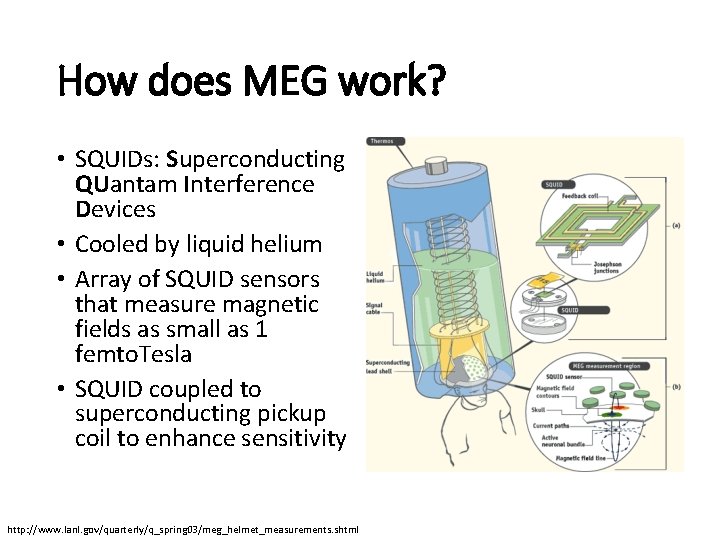
How does MEG work? • SQUIDs: Superconducting QUantam Interference Devices • Cooled by liquid helium • Array of SQUID sensors that measure magnetic fields as small as 1 femto. Tesla • SQUID coupled to superconducting pickup coil to enhance sensitivity http: //www. lanl. gov/quarterly/q_spring 03/meg_helmet_measurements. shtml
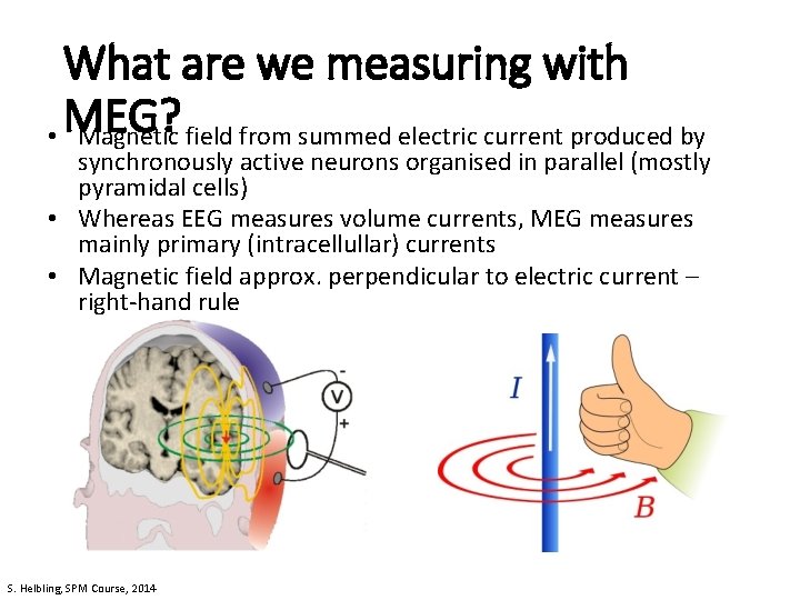
What are we measuring with • MEG? Magnetic field from summed electric current produced by synchronously active neurons organised in parallel (mostly pyramidal cells) • Whereas EEG measures volume currents, MEG measures mainly primary (intracellullar) currents • Magnetic field approx. perpendicular to electric current – right-hand rule S. Helbling, SPM Course, 2014
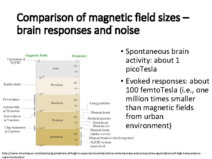
Comparison of magnetic field sizes – brain responses and noise • Spontaneous brain activity: about 1 pico. Tesla • Evoked responses: about 100 femto. Tesla (i. e. , one million times smaller than magnetic fields from urban environment) http: //www. intechopen. com/books/applications-of-high-tc-superconductivity/some-contemporary-and-prospective-applications-of-high-temperaturesuperconductors
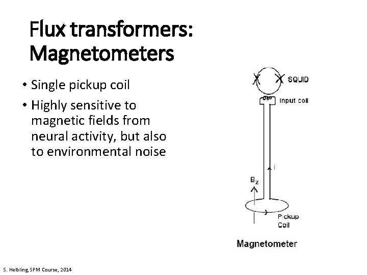
Flux transformers: Magnetometers • Single pickup coil • Highly sensitive to magnetic fields from neural activity, but also to environmental noise S. Helbling, SPM Course, 2014
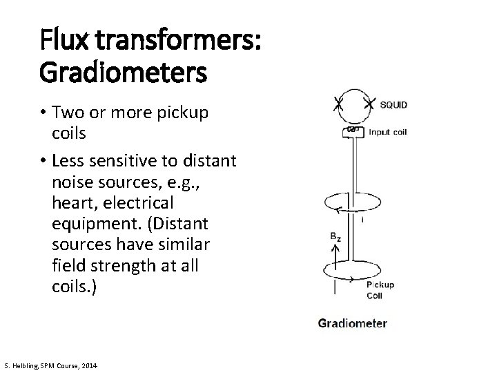
Flux transformers: Gradiometers • Two or more pickup coils • Less sensitive to distant noise sources, e. g. , heart, electrical equipment. (Distant sources have similar field strength at all coils. ) S. Helbling, SPM Course, 2014
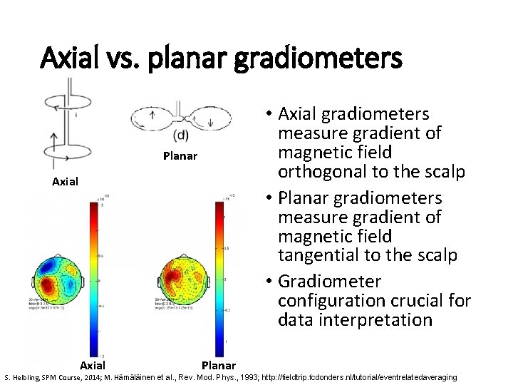
Axial vs. planar gradiometers • Axial gradiometers measure gradient of magnetic field orthogonal to the scalp • Planar gradiometers measure gradient of magnetic field tangential to the scalp • Gradiometer configuration crucial for data interpretation Planar Axial Planar S. Helbling, SPM Course, 2014; M. Hämäläinen et al. , Rev. Mod. Phys. , 1993; http: //fieldtrip. fcdonders. nl/tutorial/eventrelatedaveraging
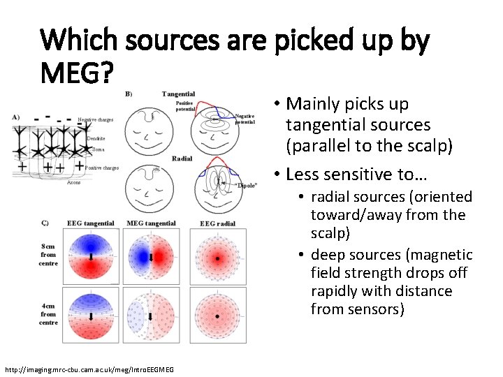
Which sources are picked up by MEG? • Mainly picks up tangential sources (parallel to the scalp) • Less sensitive to… • radial sources (oriented toward/away from the scalp) • deep sources (magnetic field strength drops off rapidly with distance from sensors) http: //imaging. mrc-cbu. cam. ac. uk/meg/Intro. EEGMEG
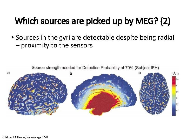
Which sources are picked up by MEG? (2) • Sources in the gyri are detectable despite being radial – proximity to the sensors Hillebrand & Barnes, Neuro. Image, 2002
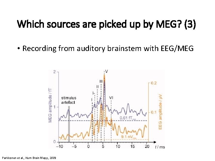
Which sources are picked up by MEG? (3) • Recording from auditory brainstem with EEG/MEG Parkkonen et al. , Hum Brain Mapp, 2009
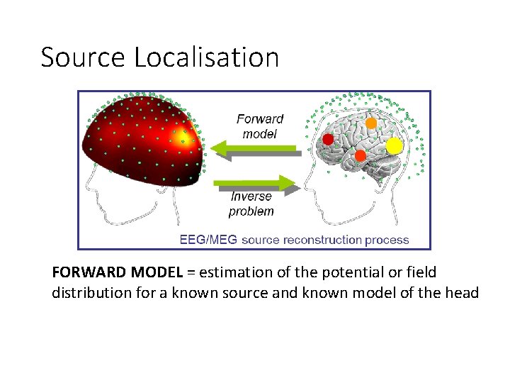
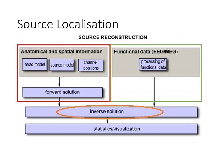
Source Localisation FORWARD MODEL = estimation of the potential or field distribution for a known source and known model of the head
Source Localisation
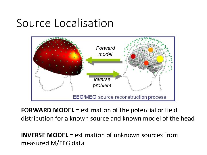
Source Localisation FORWARD MODEL = estimation of the potential or field distribution for a known source and known model of the head INVERSE MODEL = estimation of unknown sources from measured M/EEG data
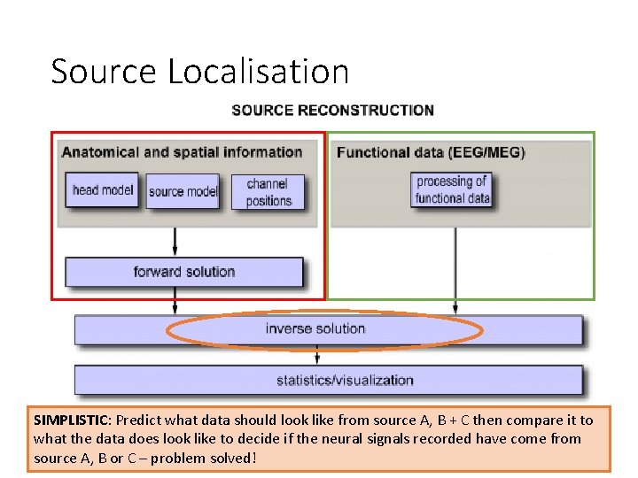
Source Localisation SIMPLISTIC: Predict what data should look like from source A, B + C then compare it to what the data does look like to decide if the neural signals recorded have come from source A, B or C – problem solved!
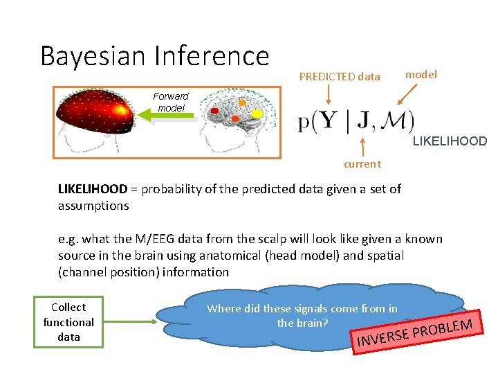
Bayesian Inference PREDICTED data model Forward model LIKELIHOOD current LIKELIHOOD = probability of the predicted data given a set of assumptions e. g. what the M/EEG data from the scalp will look like given a known source in the brain using anatomical (head model) and spatial (channel position) information Collect functional data Where did these signals come from in the brain? LEM B O R P E INVERS
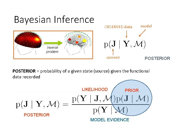
Bayesian Inference OBSERVED data model Inverse problem current POSTERIOR = probability of a given state (source) given the functional data recorded LIKELIHOOD PRIOR POSTERIOR MODEL EVIDENCE
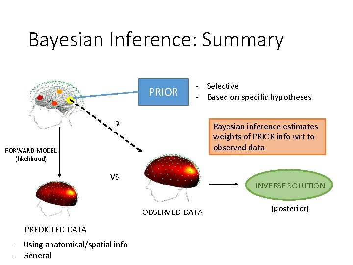
Bayesian Inference: Summary PRIOR - Selective - Based on specific hypotheses ? Bayesian inference estimates weights of PRIOR info wrt to observed data FORWARD MODEL (likelihood) VS INVERSE SOLUTION OBSERVED DATA PREDICTED DATA - Using anatomical/spatial info - General (posterior)
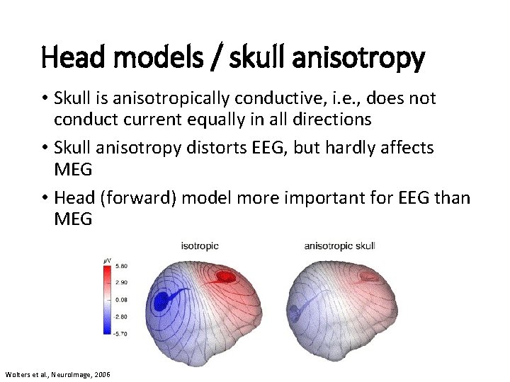
Head models / skull anisotropy • Skull is anisotropically conductive, i. e. , does not conduct current equally in all directions • Skull anisotropy distorts EEG, but hardly affects MEG • Head (forward) model more important for EEG than MEG Wolters et al. , Neuro. Image, 2006
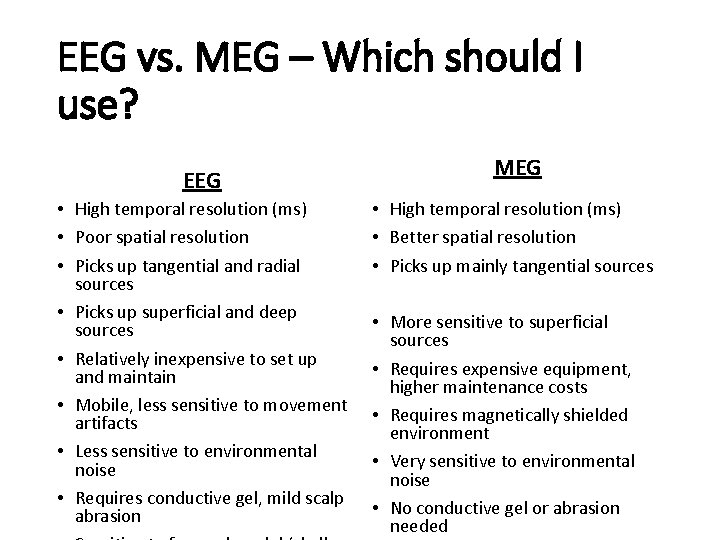
EEG vs. MEG – Which should I use? EEG • High temporal resolution (ms) • Poor spatial resolution • Picks up tangential and radial sources • Picks up superficial and deep sources • Relatively inexpensive to set up and maintain • Mobile, less sensitive to movement artifacts • Less sensitive to environmental noise • Requires conductive gel, mild scalp abrasion MEG • High temporal resolution (ms) • Better spatial resolution • Picks up mainly tangential sources • More sensitive to superficial sources • Requires expensive equipment, higher maintenance costs • Requires magnetically shielded environment • Very sensitive to environmental noise • No conductive gel or abrasion needed

Thanks to Gareth Barnes for all his help!
- Slides: 34