VCP511 TOPIC Treatment and management of wounds and


- Slides: 22
VCP-511, TOPIC: Treatment and management of wounds and abscess Dr Gyan Dev Singh, Assistant professor, VCC, BVC, Patna-14
Abscess • Abscesses are circumscribed collections of purulent material (pus) in a cavity • Corynebacterium, Pseudomonas, Streptococcus and Staphylococcus • Abscess consists of a wall, pyogenic membrane and pus (Liquor puris). • Pyogenic membrane controls spread of infection, and helps in phagocytosis and granulation tissue formation
• Pus contains necrosed tissue, dead bacteria, leukocytes and proteins of blood and tissues. • Pus cells mainly consist of polymorphonuclear leukocytes along with a few mononuclear cells. • Pus is alkaline in nature and yellow in colour. • Pus serum will not clot, since the fibrin of exudates is digested by the proteolytic enzymes of the leukocytes
Classification of abscess • Abscess may be classified as: • Acute Abscess (Hot abscess): Inflammatory symptoms are more active. • Chronic Abscess (Cold abscess): Inflammatory symptoms are less active. • Chronic abscess may be: • Hard with inspissated pus, or • Soft with liquid pus and thin abscess wall. • Superficial or deep abscess: based on location
Etiology of abscess • Pyogenic organisms like Staphylococci, Streptococci, Escherichia coli and Pseudomonas aeruginosa. • Specific organisms like Corynebacterium pyogenes, Actinomyces bovis etc. • Chemicals like mercuric chloride and Zinc chloride.
Common seats of abscess formation • Cattle: Yoke, udder and prominences • Horses: Shoulders, sub-maxillary and post pharyngeal lymph nodes. • Dogs: Anal region, and mammary glands.
ACUTE ABSCESS • Acute abscess forms in 3 to 5 days following infection. • In long duration abscess, the liquid part is absorbed and the solid part is left. This is called Inspissated Pus. Symptoms • Acute superficial abscess appears as a local painful swelling. • The dead tissues and dead inflammatory cells are continuously thrown into the cavity which leads to a gradual increase in the amount of pus. • Thus the abscess enlarges till it reaches the surface of skin or mucous membrane. • The center of abscess becomes soft (pointing) and later ruptures, discharging pus. • Local acute inflammatory symptoms without fever are observed in superficial abscess. • Deep abscess has no local symptoms, but fever and pain on manipulation of the part are evident.
CHRONIC ABSCESS (Cold abscess) • A chronic abscess develops slowly without any inflammatory symptoms. • It may be painless or slightly painful. • Primary chronic abscess usually occurs from repeated injuries and observed on the prominences of limbs and ribs due to bed sores. • Secondary chronic abscess develops in the course of various local affections. • Chronic abscess may be hard in consistency surrounded by fibrous tissue and containing small amount of pus or it may be soft and thin walled with comparatively larger amount of pus.
TREATMENT • maturation of abscess by using liniments, fomentations and mild blisters. • Drainage of pus • Tincture of Iodine soaked gauge packing (Gauze soaked with 0. 5% silver nitrate is best against most of the micro-organisms)
WOUND-CLASSIFICATION, SYMPTOMS, DIAGNOSIS AND TREATMENT
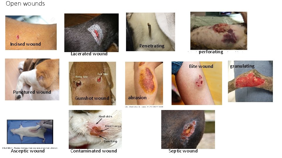
Open wounds Incised wound Penetrating perforating Lacerated wound Bite wound Punctured wound Asceptic wound Gunshot wound Contaminated wound abrasion Septic wound granulating
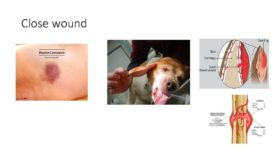
Close wound
SYMPTOMS OF WOUND • Localized pain and bleeding. • Gaping of the lips of wound. • Weakness, paralysis or a loss of function in a dependent portion. • Febrile disturbances in severe septic wound. • Neuritis extending along the course of the nerve involved in the wound.
FACTORS AFFECTING WOUND HEALING • LOCAL FACTORS: • Tissue vascularity • Infection • Topical medications • Lavage and dressings • Presence of foreign bodies • Obliteration of dead space • Ionizing radiation • Movement • Mutilation
Systemic factors • Advanced age • Nutrition • Protein • Glucose • Iron • Minerals like zinc, copper • Vitamins A and B complex • Vitamin C • Carbohydrates and fats
MEDICATION • Anti-inflammatory drugs • Chemotherapeutic agents • Anticoagulant drugs • Most NSAIDs lower resistance to infection and ultimately delay healing
SYSTEMIC DISEASES • malignancy, uncontrolled diabetes, renal and hepatic disturbances delay healing process. • Malignancy (by altering metabolism, producing chachexia, and minimizing inflammatory cell division. ) • Uremia • diabetes
MANAGEMENT OF WOUNDS • Contusions: are treated with cold and astringent applications to minimize extravasation. • Haematomas: when small get absorbed other wise they may have to be opened and treated. • Open wounds: surgical or aseptic wound, contaminated and septic wound or infected wounds.
Surgical or aseptic wounds • Surgeon should avoid drying of the tissue, excessive trauma and haemorrhage • Prophylaxis against tetanus. • Dependent drainage should be provided if haemotoma or seroma formation is expected. • Suture should be supported upto healing time 8 -14 days • Antibiotics as a therapeutic or prophylactic measure. • Local application of Fly repellents – hot summer months. • The patient and the affected injured part should be kept at rest.
Contaminated wound • A fresh wound gets contaminated when it is more than 4 -5 days old. • The principal therapeutic strategies of the open and contaminated wound are to convert it into a clean closed wound.
SEPTIC WOUND OR INFECTED WOUND • Debridement • Lavage • Wound drainage • Antimicrobial therapy • Sterile protective bandaging
Thank you