WOUNDS Wounds Tissue repair Scars Prepared by Dr

WOUNDS Wounds, Tissue repair & Scars Prepared by, Dr. Panchajani. R

Leaning objectives At the end of this topic, you should be able to, • Define“Wound” • Causes of wounds • Classification of wounds • Wound healing • Phases of wound healing • Types of wound healing • Factors affecting wound healing • Complications of wound healing • Management of wounds

Wounds Definition –Wound is a discontinuity or break in the surface epithelium. • A wound is a type of physical trauma whereby the integrity of the skin or of any tissue is compromised. • It is a separation or discontinuity of the skin, mucous membrane or tissue caused by physical, chemical or biological insult.

Wounds Aetiology The etiology of wounds can be classified as follows: - • Blunt injuries • Penetrating injuries • Surgical insult • Burn injuries

Wounds Blunt injuries • RTA • Falls • Assault • Sport injuries • Bite injuries [animal or human] Penetrating injuries • Stab wounds • Gunshot wounds Surgical wounds • Wounds caused by a surgical procedure Burn injuries • Thermal burn • Chemical burn • Electrical burn • Radiation burn

WOUND CLASSIFICATION Aim: Wound classification systems provide frameworks that: - • Aid diagnosis and stratification • Ensure uniformity of documentation • Offer prognostic information • Guide management

Types of wound classification Wounds can be classified as follows: - • According to the etiology • According to Rank-Wakefield classification system • According to the duration of the wound healing • According to the integrity of the skin • According to wound depth • According to morphological characteristics • According to degree of contamination • According to severity

Wound classification According to the etiology • Surgical wounds - These are wounds caused by surgical procedure • Penetrating wounds - Wounds caused by penetrating trauma , Stab injuries of abdomen is common. Look like an innocent injury but internal organs may be damaged. Pt. should be admitted & observed for at least 24 hrs. layer by layer exploration & repair is recommended. • Blunt wounds - Wounds caused by blunt trauma • Burn wounds - Wounds caused by burn injuries

Wound classification According to Rank-Wakefield classification system Tidy wounds – wounds inflicted by sharp instruments and contain no devitalized tissue. • They are usually single , incised, clean cut , healthy tissue &seldom associated with tissue loss. • Such wounds can be closed primarily with the repair of all structures may be possible. (primary healing) • Associated fractures are uncommon in tidy wounds • Examples: surgical incisions, cuts from glass and knife wounds

Wound classification Untidy wounds • These are wounds resulting from crushing, tearing avulsion, vascular injury or burns, and contain contaminated devitalized tissue & with tissue loss • They are usually multiple and irregular • Commonly associated with fractures • Such wounds can not be closed primarily and therefore should be allowed to heal by second intention • Contaminated wound with dead tissue requires debridement before repair can be carried out. • Multiple debridement are required after RTA which causes widespread muscle damage or even compartment syndrome.

Wound classification According to the duration of the wound healing Acute wounds • Acute wounds are wounds that usually heal in the anticipated time frame • Duration of the wound: immediately to few weeks • Examples are wounds acquired as a result of trauma or an operative procedure Chronic wounds • Wounds that fail to heal in the anticipated time frame and often reoccur • Duration of the wound ⇒ > 4 weeks to 3 months • Wounds occur as a result of an underlying condition such as extended pressure on the tissues, poor circulation, or even poor nutrition • Pressure ulcers, venous leg ulcers, and diabetic foot ulcers are examples

Wound classification According to the integrity of the skin Open wounds • Type of wounds in which the skin has been compromised and underlying tissues are exposed • Open wounds can be classified into a number of different types, according to the object that caused the wound • Examples include incised wounds, laceration, punctured wounds , penetrating , crushed etc

Open wounds Incised wounds – • caused by sharp objects • Has a sharp edge, Less contaminated • Primary suture is needed, as it gives a neat &clean scar. Lacerated wounds ; caused by blunt objects, RTA • Edges are jagged • Injury involves only the skin & subcutaneous tissue or sometimes deeper structures. • Due to blunt nature of instruments there is crushing of tissues, which may results in hematoma, bruising or necrosis of tissue. • Treated with in 6 hrs. of injury. • Treated by wound excision & primary suturing.

Caused by clean sharp edged objects such as knife, razor, glass splinder. Wound has a sharp edge, less contaminated.

LACERATED W-Caused by blunt objects like fall on a stone, road traffic accidents. Edges are jagged, injury involves only the skin and subcutaneous tissue or sometimes deeper structures.

PUNCTURED WOUND- It may look like an innocent injury with a small one or two cm long cut but internal organs may have been damaged.

Wound classification Closed wounds • Wounds in which the skin has not been compromised, but trauma to underlying structures has occurred • Closed wounds have fewer categories, but are just as dangerous as open wounds Examples of closed wounds are: • contusion, abrasion, haematoma
 –can be, • minor- soft tissue injury")
Closed wounds Contusions (known as a bruise) –can be, • minor- soft tissue injury without break in the skin • Major- when run over by a vehicle • caused by blunt force trauma that damages tissue under the skin • It produces discolouration of skin due to collection of blood underneath. Abrasion-epidermis of the skin is scrapped away exposing the dermis. • Painful as dermal nerve endings are exposed • Need cleaning & dressing Hematoma - (also called a blood tumor) – collection of blood following injury • caused by damage to a blood vessel that in turn causes blood to collect under the skin • Occur spontaneously in pts. with bleeding tendencies • Sites- subcutaneous, intramuscular, subperiosteal • Small hematoma get absorbed • Knee joint hematoma need to be aspirated, if not they can get infected

A contusion, commonly known as a bruise, is a type of hematoma of tissue in which capillaries and sometimes venules are damaged by trauma, allowing blood to seep, hemorrhage, or extravasate into the surrounding interstitial tissues.

An abrasion is a type of open wound that's caused by the skin rubbing against a rough surface. It may be called a scrape or a graze.

A Hematoma is a localized collection of blood outside the blood vessels, due to either disease or trauma including injury or surgery and may involve blood continuing to seep from broken capillaries.

Wound classification According to wound depth Superficial wounds • Only the epidermis is affected and has to be replaced. • A truly superficial wound does not bleed and heals within a few days. • Examples include most abrasions and blisters.

Wound classification Partial-thickness wounds • The epidermis and part of the dermis is affected • A partial-thickness wound does bleed • If left uncovered, a blood clot will cover the wound a scar will form • The missing tissue will then be replaced, followed by regeneration of the epidermis • A partial-thickness wound can take from several days to several weeks to heal, depending on the patient and the wound treatments chosen.

Wound classification Full-thickness wounds • A full-thickness wound involves the epidermis and the dermis • The underlying fatty tissue, bones, muscles, or tendons may also be damaged • If full-thickness wounds cannot be sutured, the healing process will create new tissue to fill the wound, followed by regeneration of the epidermis • The full-thickness wound takes longer time to heal than does a partial-thickness wound, sometimes as long as several months

Wound classification According to morphological characteristics Bruises / contusion • These are closed wounds • Caused by blunt trauma that damage the tissue under the skin without breaking the skin • Characterized by skin discoloration due to bleeding into the tissues • Blows to the chest, abdomen, or head with a blunt instrument can cause contusions

Wound classification Hematoma • These are also closed wounds caused by damage to a blood vessel that in turn causes blood to collect under the skin • Initially this is fluid, but it will clot within minutes or hours ⇒later after few days the hematoma will again liquefy → increased risk of secondary infection → pus formation

Wound classification Crush wounds or contused • Crush wounds are caused by a great or extreme amount of force applied over a long period of time • Caused by blunt trauma, run over by vehicle, wall collapse etc. . • These occur when a heavy object falls onto a person, splitting the skin and shattering or tearing underlying structures • These wounds are dangerous they cause severe haemorrhage, death of tissue& crushing of blood vessels • more prone to gas gangrene & tetanus • They are often accompanied by degloving injuries and compartment syndrome • Treatment includes good debridement & removal of all dead & necrotic tissues.

A crush injury is injury by an object that causes compression of the body. This form of injury is common following a natural disaster or after some form of trauma from a deliberate attack. Common concerns after an injury of this type are rhabdomyolysis and crush syndrome

Wound classification Abrasions • An abrasion is a shearing injury of the skin in which the surface is rubbed off • Most are superficial and will heal by epithelialization Lacerated wound • Caused by tearing of tissues • Wounds have irregular borders • Loss of tissue is limited to skin and s/c tissue

Wound classification Penetrated wound • Cause by sharp pointed objects like nails • Have relatively small opening • May be very deep • Infection/ foreign particles might have been carried deep in to wound opening is inadequate for drainage • eg: punctured wound on foot due to gathered nail Perforating wound • Have two opening one of entrance and other of exit • Eg. gunshot wounds

Wound classification • According to degree of contamination Clean wounds • No break in aseptic technique • Incision is made under sterile conditions • No inflammation is encountered • The respiratory tract, alimentary, genital or uninfected urinary tracts are not entered • Primary closure • No drain • Eg- Herniorrhaphy,

Wound classification Clean Contaminated wounds • Operative wounds in which the respiratory, alimentary, genital or urinary tract is entered under controlled conditions and without unusual contamination Contaminated wounds • Open, fresh or accidental wounds; operations with major breaks in sterile technique or gross spillage from the gastrointestinal tract; and incisions in which acute, non-purulent inflammation is encountered Dirty or Infected wounds • Old traumatic wounds with retained devitalized tissue and those that involve existing clinical infection

Wound classification According to severity Simple wounds • when skin only involved. • The integrity of the skin is traumatized without loss or destruction of tissue and without the presence of a foreign body in the wound Complex wounds • involves the underlying nerves, vessels, tendons. • Tissue is lost or destructed by means of a crush, burn, or foreign body in the wound

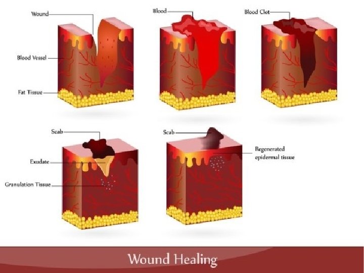
WOUND HEALING Definition • Wound healing, or wound repair, is the body's natural process of restoring normal function and structure after injury. • The entire wound healing process is a complex series of events that begins at the moment of injury and can continue for months to years.

wound healing

WOUND HEALING Phases of wound healing Three phases of wound healing include: • Inflammatory phase • Proliferative phase • Maturation and remodeling phase • Occassionally a haemostatic phase before the inflammatory phase

WOUND HEALING- PHASES Inflammatory phase – Immediately after wounding & last for 2 -3 days Early infla. - platelet enriched blood clot & blood vessels Late infla. - increased vascularity & increased poly morpho nuclear lymphocytes • Aim: to stop bleeding and to prevent further injury

WOUND HEALING Characterized by : • Clotting - haemostasis • Vasoconstriction and vasodilatation • Increased polymorphonuclear neutrophils • Increased Macrophages

Wound healing- Inflammatory phase • Vaso constriction & thrombus formation to limit the blood loss. • Platelets aggregation – platelet stick to the damaged endothelial lining of vessel releasing ADP which causes thrombocytic aggragates to fill the wound. • This clot acts to control active bleeding (haemostasis) • Platelet release several cytokines from their alpha granules, these atract inflammatory cells such as poly morphonuclear lymphocytes& macrophages. • Platelet and local injured site release vaso active amines ( histamine , serotonine , prosta glandin) which increase vascular permeability.

WOUND HEALING- INFLAMMATORY PHASE • vasoconstriction lasts 5 -10 minutes and is followed by vasodilatation which peaks at about 20 minutes post-wounding. • Macrophages remove devitalised tissue and micro organism & regulate fibroblastic activity in the proliferative phase. • The initial frame work for structural support of cells is provided by fibrin produced by fibrinogen

WOUND HEALING • Vasodilatation is the result of factors released by platelets and other cells • The main factor involved in causing vasodilatation is histamine • Histamine also causes ↑ vascular permeability→ entry of inflammatory cells like leukocytes into the wound site from the bloodstream

WOUND HEALING Increased polymorphonuclear neutrophils • Within an hour of wounding, PMNs arrive at the wound site and become the predominant cells in the wound for the first two days after the injury • These PMNs phagocytise debris and bacteria and also kill bacteria by releasing free radicals • They also cleanse the wound by secreting proteases that break down damaged tissue • PMNs usually undergo apoptosis once they have completed their tasks and are engulfed and degraded by macrophages

WOUND HEALING Increased Macrophages • Macrophages are essential to wound healing. • They replace PMNs as the predominant cells in the wound by two days after injury. • Attracted to the wound site as monocytes from blood vessels by growth factors released by platelets and other cells. • Once they are in the wound site, monocytes mature into macrophages.

WOUND HEALING • The macrophage's main role is to phagocytize bacteria and damaged tissue and they also debride damaged tissue by releasing proteases. • Macrophages also secrete a number of factors such as growth factors and other cytokines that attract cells involved in the proliferation stage of healing to the area

WOUND HEALING- PHASES Proliferative phase • After the inflammatory stage, the proliferative stage lasts from 3 day to about 3 weeks (or longer, depending on the severity of the wound) • Aim: repair of wounded tissue Characterized by • Fibroblastic activity • Angioneogenesis • Fibroplasia and granulation tissue formation • Re-epithelialization • Wound contraction

Proliferative phase • Fibroblastic activity with production of collagen & ground substance which helps in binding of collagen fibres.

WOUND HEALING Angioneogenesis • Angiogenesis is the process of new blood vessel formation and is necessary to support a healing wound environment • New blood vessels are formed by vascular endothelial cells • Growth of new blood vessels as capillary loops • Endothelial cells are attracted to the wound area chemo tactically by angiogenic factors released by platelets and macrophages. • Endothelial growth and proliferation is also directly stimulated by hypoxia, and presence of lactic acid in the wound.

WOUND HEALING Fibroplasia and granulation tissue formation • Fibroblasts begin accumulating in the wound site 2 -5 days after wounding and peaks at 1 -2 weeks post-wounding • Fibroblasts then deposit ECM into the wound bed, and later collagen and granulation tissue formation • Granulation tissue consists of new blood vessels, fibroblasts, inflammatory cells, endothelial cells, myofibroblasts, and extracellular matrix (ECM) • Fibroblast require vit. C to produce collagen • The wound tissue formed in the early part of this phase is granulation tissue. • In later part of this phase there is an increase in the tensile strength of the wound due to increased collagen & is of type III collagen.

WOUND HEALING Epithelialization • Epithelial cells migrate across the granulation tissue to form a barrier between the wound and the environment • Epithelialization occurs from the edge of the wound by a process of cell migration & cell multiplication brought about by marginal basal cells. thus with in 48 hrs. the entire wound is re-epithelialised • Basal keratinocytes from the wound edges and dermal appendages such as hair follicles, sweat glands and sebacious glands are the main cells responsible for the epithelialization phase of wound healing. • Epithelialization phase is usually complete within 7 -10 days • Wounds with skin loss skin appendages also helps in epithelialization. • Slowly surface cells get keratinised.

WOUND HEALING-PHASES Maturation and remodeling phase • The maturation phase of tissue repair begin when the levels of collagen production and degradation equalize. • Maturation of collagen – type 1 replacing type III until the ratio of 4: 1 is achieved • Re-allignment of collagen fibres along the lines. • The maturation phase can last for a year or longer, depending on the size of the wound and whether it was initially closed or left open

WOUND HEALING Wound contraction • Contraction is a key phase of wound healing • If contraction continues for too long, it can lead to disfigurement and loss of function • Decreased wound vascularity & wound contracture due to fibroblast & myofibroblastic activity • Contraction commences approximately a week after wounding, when fibroblasts have differentiated into myofibroblasts and can last for several weeks. • Myofibroblasts, which are similar to smooth muscle cells, are responsible for contraction • Wound contracture readily occurs in loose skin as in back & gluteal region, reduced over tibia, malleoli. • Cortico- steroids, Uv radiation, chemotherapy delays wound contracture

Wound healing Scar formation- Changes – • Fibroplasia & laying of increased collagen • Vascularity becomes less • Epithelialization continues • Ingrowth of lymphatics & nerve fibres • Remodelling of collagen with ciccatrisation resulting in a scar. •
")
WOUND HEALING- TYPES Types of wound healing • Healing by primary intention (Primary closure) • Healing by secondary intention (Secondary closure) • Healing by tertiary intention (Delayed primary closure)
 • Healing by primary intention (Primary")
WOUND HEALING Healing by primary intention (Primary closure) • Healing by primary intention (Primary closure) occurs when a wound is created aseptically with minimal tissue damage. • Occurs in clean incised wounds • Wound edges are opposed • Normal healing, minimal scar • Healing takes place by the approximation of tissue edges with suture, staples, wound sealant etc
 • Occurs in wounds that are")
WOUND HEALING Healing by secondary intention (Secondary closure) • Occurs in wounds that are already infected , discharging pus or wound with skin loss • Wound is usually left open and allowed to heal by granulation, epitheliazation and wound contraction. • Increased inflammation & proliferation • May be caused by infection, excessive trauma, tissue loss, or inability to re- approximate the tissue • It is a slow process, poor scar.
 • Wounds that are heavily")
WOUND HEALING Healing by tertiary intention (Delayed primary intention) • Wounds that are heavily contaminated and are likely to develop an infection if closed primarily, • may be left open for 3 -5 days, edges are later opposed when healing conditions favourable. • This allows the wound to be cleaned and allows the body’s natural defenses to decrease bacterial count • The wound can then be closed and allowed to heal, producing a wound with characteristics similar to primary closure

Abnormal healing • Some factors adversely affects healing • Delayed healing results in loss of function or poor cosmetic outcome • Aim is to achieve healing by primary intention & reduce the inflammation & proliferative responses

Factors affecting wound healing • Local factors affecting wound healing • Systemic factors affecting wound healing

Factors affecting wound healing Local factors affecting wound healing • • • • Site Structures involved Mechanism of wounding Contamination Loss of tissue Infection Surgical Technique Movement Hematoma formation Vascular insufficiency – poor blood supply Tissue ischemia- hypoxia Presence of foreign body Exposure to radiation Pressure Faulty technique of wound closure

Factors affecting wound healing Systemic factors affecting wound healing • Aging • Nutritional status – vit. & mineral deficiency • Diseases states • Uremia • Jaundice • Diabetes • Malignancies • Immunosuppression • Smoking • Drugs • Steroids • anti-neoplastics • NSAIDs

Complications of wound healing • • • Dehiscence – wound rupture along the incision Evisceration – to push outside through incision Hemorrhage Adhesions Infection Herniation Fistula formation Sinus formation Suture complications Hypertrophic scar Keloids Malignant changes

MANAGEMENT OF WOUNDS Surgical toilet with: - • Primary closure • Delayed primary closure • Skin grafting • Flaps • Wound dressing • Skin grafting
")
Management of wounds • Examine the whole patient according to acute trauma life support(ATLS) principles • Consider site, possible structures damaged, movement & sensation • Monitoring tempt. , pulse, resp. • Bleeding wound elevated, pressure & packing applied • prophylaxis against tetanus- TT

Wound treatment • Cleaning& dressing • Suturing • Debridement & excision in aberation, RTA, explosion • Repair of all damaged structures • Repair of nerves & arteries under magnification • Skin repair by flap or graft

Wound closure / suturing 1. primary suturing – • Suturing with in few hours following injury(with in 6 hrs. ) • Done in incised or cut wound with sharp objects • Minimal injury to structures , no infection Precautions while suturing- • Foreign body present , removed • Associated injury to blood vessels, nerves, tendons repaired • Prevention of tetanus by TT

WOUND CLOSURE 2. Wound excision & primary suturing of skin • Indications- Wound edges are jagged( lacerated) Contaminated with organism or foreign body Crushed or devitalized tissues • Wound is explored , devitalized tissues or foreign bodies if present removed. • Irrigated with antiseptic solution • Lacerated wound is converted in to incised & then sutured Precautions- • done with in 6 hrs. • Tetanus & gas gangrene prophylaxis taken. • Repair of tendons & vessels done

Wound closure 3. wound excision & delayed primary suturing • Indicated in lacerated wounds with major crush injuries • Primary suturing with in 6 hrs. is not done because of , oedema of the part, increased tissue tension, hematoma, contamination with bacteria. • Excision of all dead tissue is done • Wound is irrigated with saline & left open without suturing & dressing is applied • Wound is re-examined 4 -6 days later • If there is no infection or non viable tissues wound is sutured.

Wound closure 4. wound with skin loss Complications of skin loss • Secondary infection • Underlying structures tendons, nerves are in danger • Diabetic pts. can develop septicaemia • Deformity & disability can occur later Tt. –Debridement • Done under G. A • Assess the extent of injury • Control bleeding • Excision of devitalised tissue • Saline irrigation • Skin grafting with proper asepis • Blood transfusion

MANAGEMENT OF ACUTE WOUND • • • Cleaning Exploration & diagnosis Debridement Repair of structures Skin cover if required

SKIN GRAFTING • Used to reconstruct wounds • Sites- thigh, in children- buttock • Full thickness G- whole dermis with underlying fat trimmed away • Split thickness G- taking all the epidermis together with some dermis leaving the remaining dermis behind to heal the donor site. • Composite SG- skin, fat, cartilage • Nerve graft • Tendon graft

Skin grafting • The graft adheres to & heal a wound is an unnatural process& normal healing is altered by the presence of graft. • The survival of the graft depends on how fast the graft derives a new blood supply from the wound on which it is placed. • Nutrition is derived by diffusion through the fibrin layer formed between it & wound bed • After 48 -72 hrs. fine capillary network grows in to the graft& anastamosis with the native vasculature of the graft. • Graft are hairless, donot sweat because these structures are not transferred. • Factors inhibit this process are hematoma, seroma, bacterial exudate between graft and wound bed.

Compartment syndrome • Occur in closed lower limb injuries • Severe pain on movement of the affected compartment muscles • Sensory disturbances& absence of pulses distally , color changes, inflammatory signs, oedema • Fasciotomy is the treatment

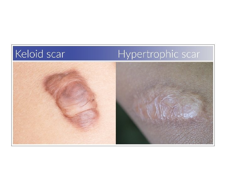
Hypertrophied scar& Keloid • Scars – occur in the maturation phase • The immature scar become mature over a period lasting a year or more. • It is pink , hard, raised & itchy • Collagen matures, scar becomes acellular as the fibroblast & blood vessels reduce, scar becomes pale, softer, flattens • Scar types- atrophic, hypertrophic, keloid

Scars Atrophic- • pale, flat, stretched in appearance • Easily traumatised as dermis &epidermis are thinned. • Excision & resuturing may improve the scar

HYPERTROPHIED SCAR • A hypertrophic scar is a cutaneous condition characterized by deposits of excessive amounts of collagen which gives rise to a raised scar, but not to the degree observed with keloids. Like keloids, they form most often at the sites of pimples, body piercings, cuts and burns. They often contain nerves and blood vessels. They generally develop after thermal or traumatic injury that involves the deep layers of the dermis and express high levels of TGF-β.

Hypertrophic scar- • Excessive scar tissue that doesnot extend beyond the boundary of the original incision or wound. • It results from prolonged inflammatory phase of wound healing • There is hypertrophy of mature fibroblast • Blood vessels are minimum. • Hypertrophied scar improve spontaneously

Scars- KELOID Keloid • Excessive scar tissue that extent beyond the boundary of original incision or wound. • It is associated with elevated growth factors, deeply pigmented skin, inherited tendency & certain areas of the body • Proliferation of immature fibroblasts with immature blood vessels • Very common in black • Tendency to local recurrence after excision. • It takes a shape of butterfly over sternum(common site) Tt- collagen bundles are absent in keloid so difficult to treat. • Pressure • Excision & steroid injection • Excision & post operative radiation • Laser therapy • Vitamin E

Keloids are a type of raised scar. They occur where the skin has healed after an injury. They can grow to be much larger than the original injury that caused the scar. Anything that can cause a scar can cause a keloid. This includes being burned, cut, or having severe acne

REFERENCE • • • Bailey and Love’s short practice of Surgery Manipal manual of Surgery SRB’s Manual of surgery Text book of Surgery- ASI Clinical Surgery, Das

Thank you, Dr. Panchajani. R Associate professor, Dept. of Surgery, SKHMC

- Slides: 83