
BURNS 30 yo patient transfered to your burn

BURNS
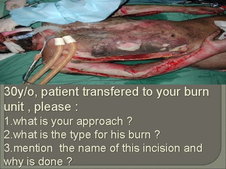
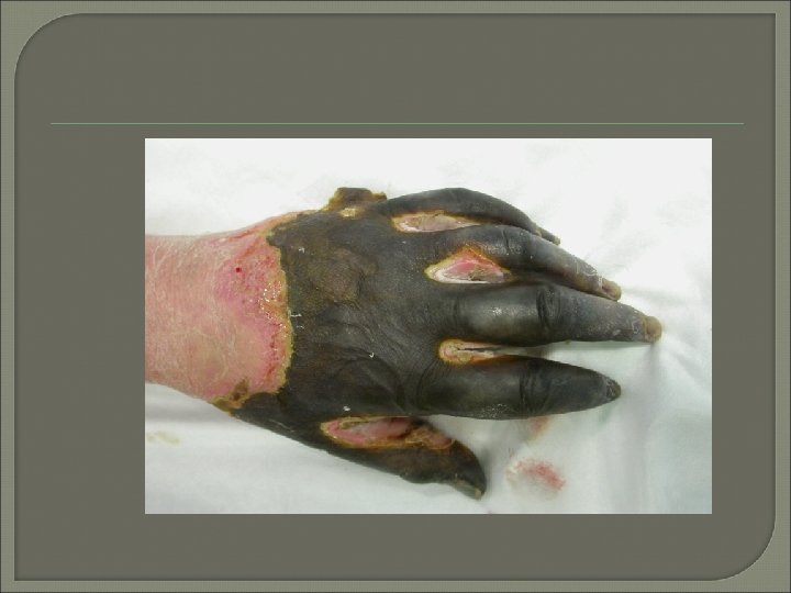
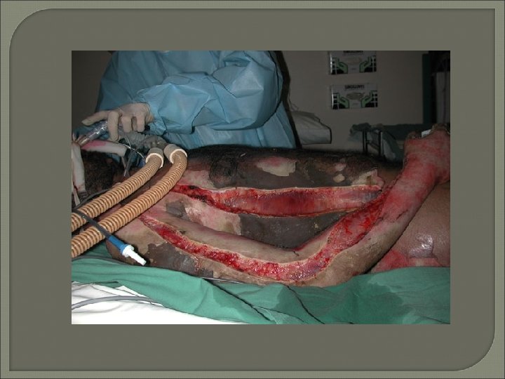
30 y/o, patient transfered to your burn unit , please : 1. what is your approach ? 2. what is the type for his burn ? 3. mention the name of this incision and why is done ?
Epidemiology � 0. 5 -1% of UK population suffer burn /year. � 10% of these require admission � Of these admissions, 10% are life threatening � 45% of US admissions for the scald burn are in the children < 5 years of age
TYPES OF BURNS �Flame burns; fires �Scold burns; hot liquids �Electrical burns; lightening injuries �Chemical burns; acids & alkali Radiation

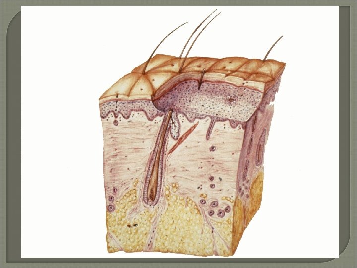
Burn wound depth �Superficial (first degree) �Partial thickness (second degree) superficial partial deep partial �Full thickness (third degree)

Superficial (epidermal) Burn �Redness �Painful �Dry �Spontaneous scarless healing within 7 days
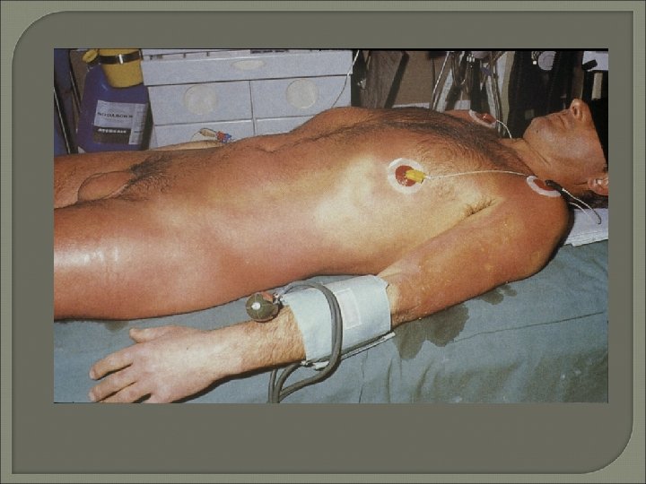
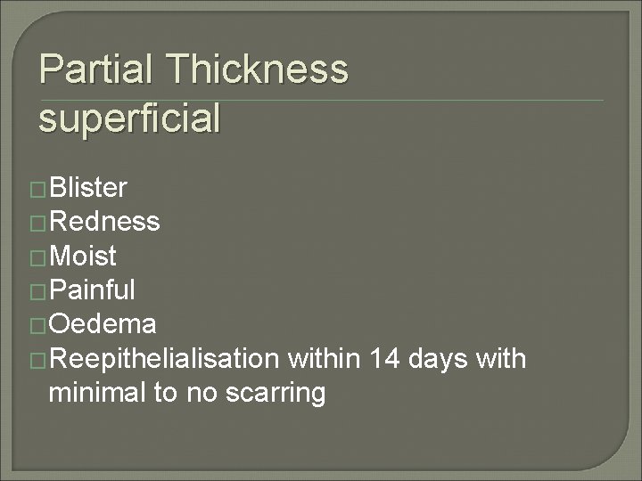
Partial Thickness superficial �Blister �Redness �Moist �Painful �Oedema �Reepithelialisation within 14 days with minimal to no scarring
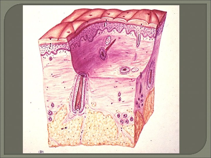
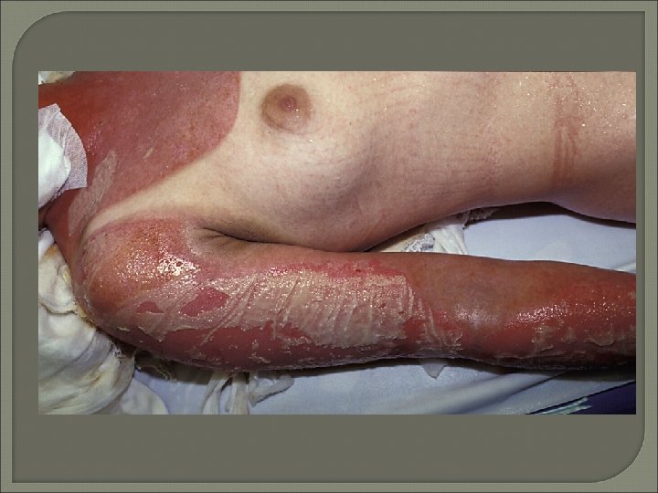
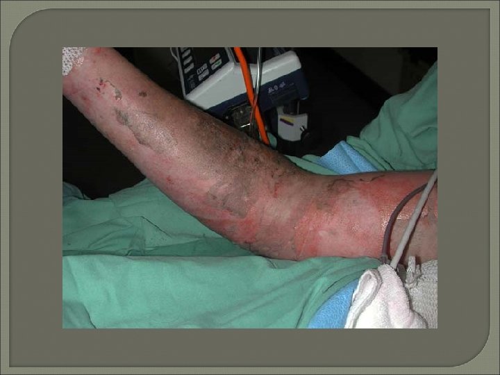
Partial thickness deep �Grey-white �Dry �Little to no pain �Reepithelialization with copious scarring over weeks (extensive collagen deposition)
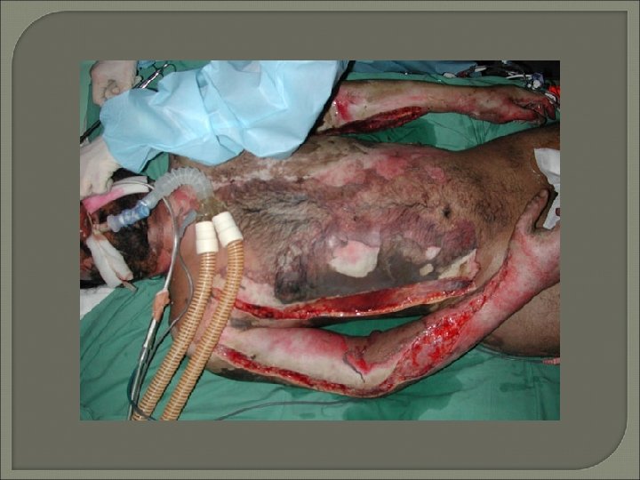
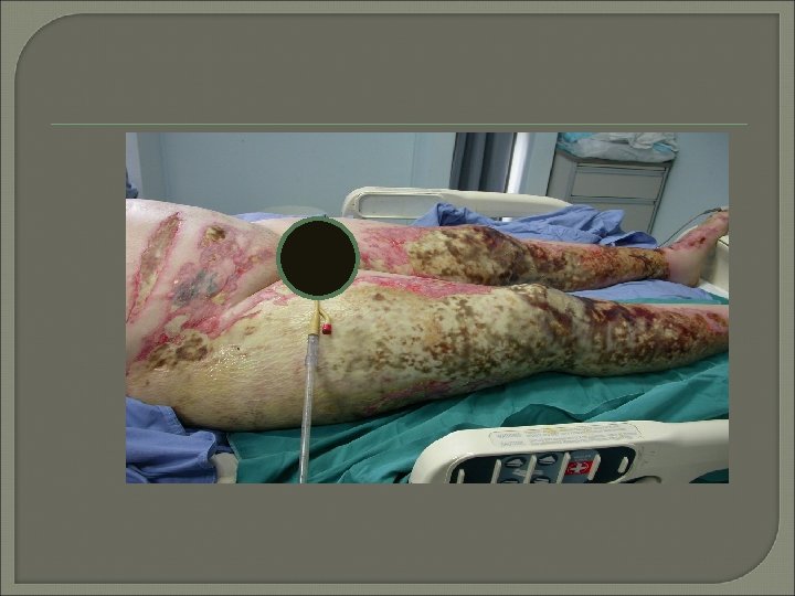
Full thickness �Dry �No pain �Clay like colour, white, charred �Leathery �No spontaneous wound closure possible (except very small wounds)

Inhalational injury �Fires �Closed space �Singed nasal hair �Carbonaceous material in nose & mouth
Inhalational injury �Direct thermal injury (upper airway) �Chemical injury ; products of combustion (lower airway) �CO poisoning ; systemic effect
CO poisoning �Toxic symptoms > 20%, death at 60% �CO-Hb > 5% is indicative of inhalational injury but not severity � 200 – 250 × greater affinity for Hb than oxygen �Treatment ; 100% O 2, hyperbaric oxygen
pathophysiology • Fluid Shift Period of inflammatory response– Vessels adjacent to burn injury dilate → ↑ capillary permeability– Continuous leak of plasma from intravascular space into interstitial space. Associated imbalances of fluids, electrolytes and Lasts 24 -36 hours
Fluid remobilization -Capillary leak stop and fluid shifts back into the circulation – Restores fluid balance and renal perfusion and Increased urine formation -Imbalances Hyponatremia, Hypokalemia
SYSTEMIC CHANGES �Cardiac– Decreased cardiac output � Pulmonary– Respiratory insufficiency as a secondary process– Can progress to respiratory failure � Gastrointestinal– Decreased or absent motility (may need NG tube)– Curling’s ulcer formation

Metabolic � Hypermetabolic state Increased oxygen and calorie requirements �Increase in core body temperature �Immunologic– Loss of protective barrier– Increased risk of infection– Suppression of humoral and cell-mediated responses
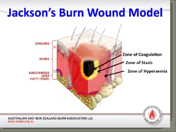
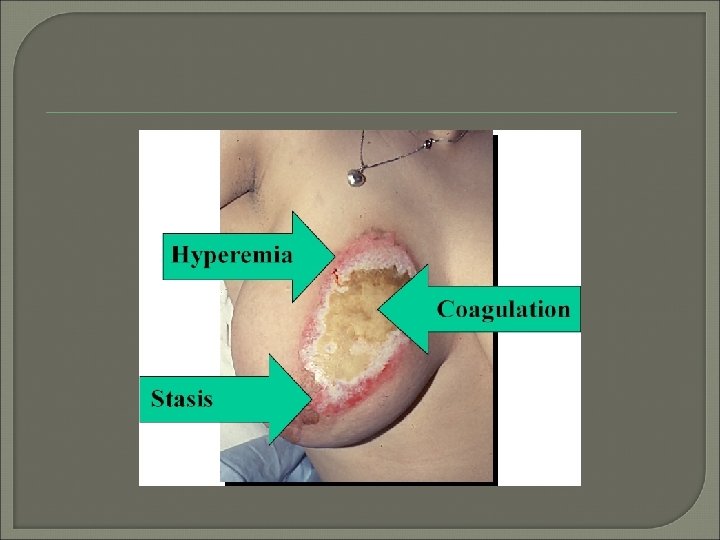
Jackson’s Theory of Thermal Wounds � Zone of coagulation ; Area in a burn nearest the heat source that suffers the most damage � Zone of stasis ; Area surrounding zone of coagulation characterized by decreased blood flow. � Zone of Hyperemia Peripheral area around burn that has an increased blood flow
Organization of Burn Care

First Aid scene � Extinguish flame, switch off power source, remove chemical soaked clothes, etc… � Cool burn wound � Dilute acid- alkali
First Aid Scene 1. A B C, Like all other trauma patients, should be evaluated systematically 2. Intubation ? ? 3. Look for other injuries 4. Assess burn wound
�Cool burn wound: - reduce direct thermal trauma & stabilize mast cells, reducing release of histamine and other inflammatory mediators - pain relief - running water (15 degree C. ) - worth considering for up to 2 hours

Intubation : �Suspicion of inhalational injury �Severe facial & neck burn “ easy early intubation will become difficult and impossible later on”

Assessment of burn wound �Remove all clothes/ maintain warm temp. �Check the back �Estimate % BSA burn �Estimate depth �Recognize need for escharotomy

Estimation of % TBSA burn
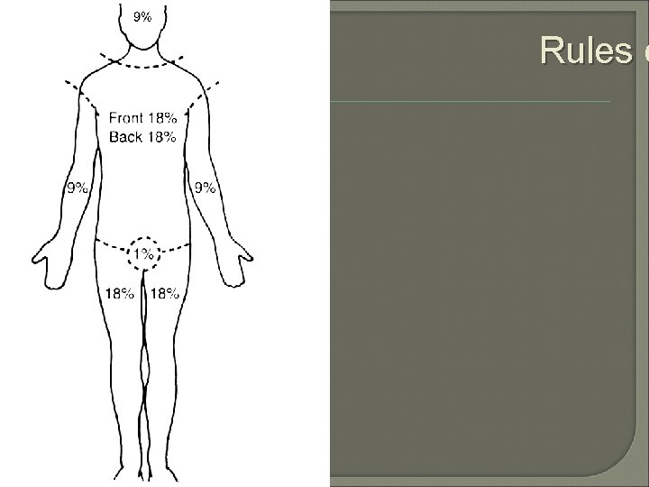
Rules o
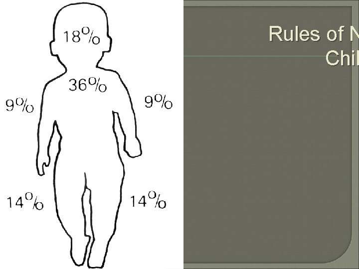
Rules of N Chil

Lund and Browder Chart

The Rule of Nines Isn’t Always the Rule � The Rule of Nines provides reasonable estimates of body surface area for patients ranging from 10 to 80 kg. � For obese patients weighing more than 80 kg, a rule of fives is proposed: � 5% body surface area for each arm � 5 x 4 or 20% for each leg � 10 x 5 or 50% for the trunk �and 2% for the head
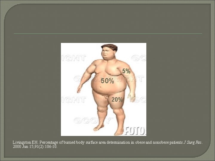
5% 50% 20% Livingston EH. Percentage of burned body surface area determination in obese and nonobese patients J Surg Res. 2000 Jun 15; 91(2): 106 -10.
Limbs: Signs of Circulatory Obstruction � Loss of distal circulation �pallor �coolness �absent pulse �loss capillary refill �decreased oxygen saturation � Pain on passive extension � Deep pain at rest
Escharotomy �Chest: �Limb: To allow respiratory movement To restore circulation in limb with excess swelling under rigid eschar
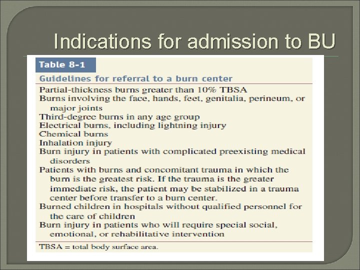
Indications for admission to BU

Management of Major Burns Initial patient management 1. Intravenous access established & I. V. fluids 2. Folley’s catheter 3. Analgesia 4. Tetanus vaccine 5. Patient must be kept warm 6. Dressing

Intravenous fluids Parkland formula : � 4 ml R. L. × %TBSA × Wt (kg) half over 8 hrs half over 16 hrs

Other formula • Modified Brook LR 2 m. L/kg percent burn • Galveston LR 5000 ml/m² BSA burned/day plus 2000 ml/m² BSA total/day (also used in TEN)
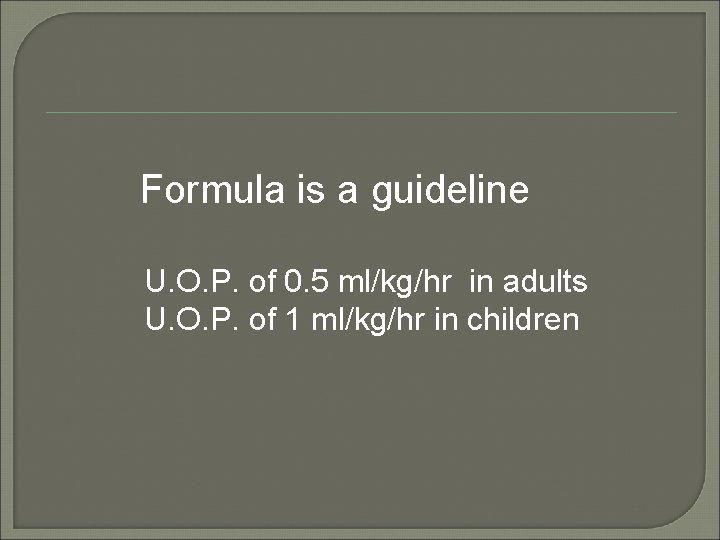
Formula is a guideline U. O. P. of 0. 5 ml/kg/hr in adults U. O. P. of 1 ml/kg/hr in children
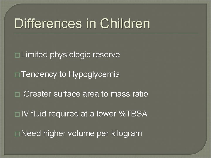
Differences in Children � Limited physiologic reserve � Tendency to Hypoglycemia � Greater surface area to mass ratio � IV fluid required at a lower %TBSA � Need higher volume per kilogram
Monitoring Resuscitation �Urinary output �Heart rate �Blood pressure �Central invasive haemodynamic �Electrolytes �Blood gases �p. H (<7. 39 - lactic acidosis ? )
OUTCOME PROGNOSIS Sum of Age in years Plus Area of burn in % TBSA < 80 80 -100 >100 good life threatening bad

INFECTION �Infection is the leading cause of death in burn patients. �Risk factors : 1. Burns > 30% TBSA 2. Deep partial / full thickness burns 3. Age: under 16 or over 60 years 4. Preexisting disease; DM, malnutrition, . .

Surgical Therapy Sharp Debridement (tangential, epifascial) Split Thickness Skin Grafts Alternative Wound Coverage � � �

PRINCIPLES OF BURN RECONSTRUCTION �Direct closure �Adjacent tissue transfer �Skin grafts; FTSG vs STSG �Cultured epithelial autograts �Flaps �Tissue expansion
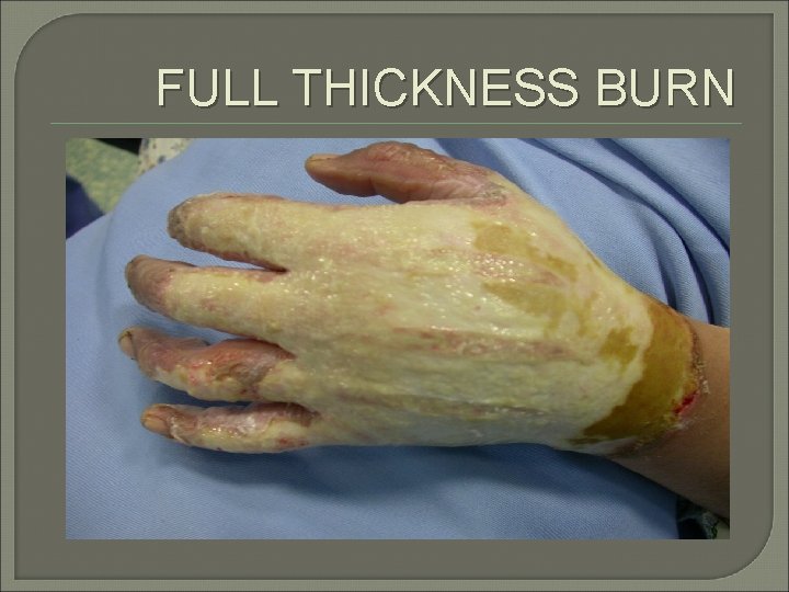
FULL THICKNESS BURN
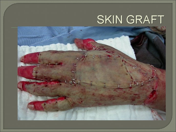
SKIN GRAFT

SIX MONTHS LATER
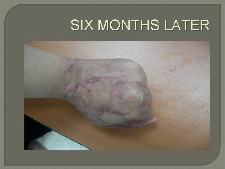
SIX MONTHS LATER
- Slides: 59