Treatment Localized disease Radical nephrectomy Metstatic disease Radiation


- Slides: 12

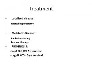
Treatment • Localized disease: Radical nephrectomy. • Metstatic disease: Radiation therapy. Immunotherapy • PROGNOSIS: stage 1 80 -100% 5 yrs survival stage. II 60% 5 yrs survival.

Urethelial tumor of the renal pelvis • 4% of urethelial tumors. Male-Female ratio 4 -1. High incidence of multicentric. • Etiology: Risk factor: smoking industrial dye, solvent, analgesic such as phenacetin, aspirin, caffeine, acetaminophen. . Pathology: Majority are Transitional cell carcinoma. Rarely squamous cell carcinoma, or adenocarcinoma.

Pathology of TCC. Metastasis: Regional LNs, Lung, bone. Staging: TNM: Ta. Tis: confined to mucosa. T 1 Invasion of lamina propria. T 2 Invasion of muscularis. T 3 a Invasion of deep muscles. T 3 b Extension into fat or renal parechyma. T 4 Spread to adjacent organs. N+ LNs Metastasis. M+ Distant metastasis.

Clinical Findings: • Symptoms&Signs: Gross Hematuria. Flank pain. Flank mass(Hydronephrosis). Weight loss anorexia. • Laboratory: Hematuria. Urine cytology(voided urine or ureteric catheter).

X-ray • I. V. U.

Retrograde Pyelogram:

Ureteropyeloscopy: Ultrasonography: C. T.

Treatment: Localized Tumor: -Nephroureterectomy. -Conservative : open or endoscopic excision + instillation of immuno-0 r chemotherapeutic Single kidney. Bilateral tumors. Metastatic Tumor: Chemotherapy.

Adrenal gland: Benign Tumors: -Adenoma. Malignant: - Neuroblastoma.

Neuroblastoma of Adrenal Gland -Origin: Neural crest. -Age: 1 st 2 ½ yrs. -Poor prognosis. -Hereditary. -Rarely bilateral. Clinical Findings: Symptoms: Abdominal mass (parent). Symptoms related to metastases (failure to thrive, Fever, malaise, bone pain, constipation, diarrhea).

Signs: -Palpable, visible abdominal mass. -In metastatic patient: enlarged nodular liver, mass in bone, ocular protrusion. - Hypertension. Laboratory Findings: -Anemia. -Increase level of serum epinephrine , nor epinephrine, and urinary VMA. X-ray Findings: U. S, I. V. P, CT, Angiography.

Treatment: Localized Tumor: -Tumor Excision followed by radiotherapy to the tumor bed -Very large tumor : Radiotherapy followed by excision. Metastatic Tumor: Chemotherapy.
 Metstatic
 What is a mixed radical
 Unit 6 radical functions homework 4 rational exponents
 Radical section
 Prion disease symptoms
 Disease control phase
 Shahzad ahmad md
 Maple syrup urine disease treatment
 Heart disease symptoms
 Reliability centered maintenance
 How to tell if a lone pair is delocalized
 Localized convective lifting
 Section 18.2 cloud formation
