The Small Intestine Pages 469 470 and 480


- Slides: 12

The Small Intestine Pages 469 -470 and 480 -484

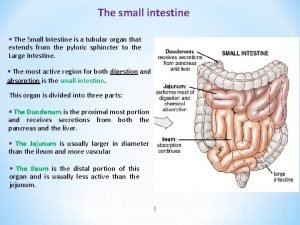
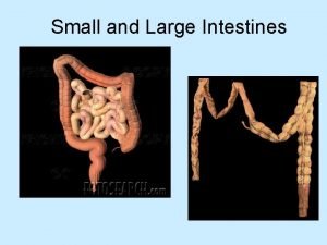
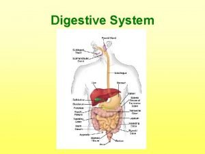
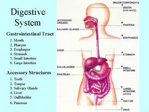
Anatomical Subdivisions � From the stomach to the large intestine: � Duodenum ◦ Attached to the stomach via the pyloric sphincter � Jejunum � Ileum ◦ Meets the large intestine at the ileocecal valve © 2015 Pearson Education, Inc.

Chemical Digestion of nutrients into the blood � Begins in the small intestine via enzymes from: ◦ Intestinal cells ◦ Pancreas �Pancreatic ducts carry enzymes to the duodenum ◦ Bile, formed by the liver, enters the duodenum via the bile duct �The pancreatic and bile ducts come together to form a joint duct that releases into the duodenum – the hepatopancreatic ampulla © 2015 Pearson Education, Inc.

Figure 14. 6 The duodenum of the small intestine and related organs. Bile duct and sphincter Accessory pancreatic duct Pancreas Gallbladder Jejunum Duodenal papilla Hepatopancreatic ampulla and sphincter Main pancreatic duct and sphincter Duodenum

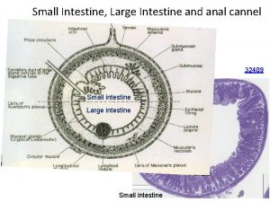
Food absorption and surface area � Three structural modifications increase surface area for food absorption: 1. Villi—fingerlike projections formed by the mucosa � House a capillary bed and lacteal 2. Microvilli—tiny projections off of the villi (create a brush border appearance) 3. Circular folds (plicae circulares)—deep folds of mucosa and submucosa © 2015 Pearson Education, Inc.

Figure 14. 7 a Structural modifications of the small intestine. Blood vessels serving the small intestine Muscle layers Villi (a) Small intestine Lumen Circular folds (plicae circulares)

Figure 14. 7 b Structural modifications of the small intestine. Absorptive cells Villus Blood capillaries Lymphoid tissue Muscularis mucosae Lymphatic vessel Submucosa (b) Villi

Digestion in the Small Intestine : What comprises intestinal juice? � Protein and some carbohydrate breakdown started in the stomach ◦ Fats begin in the intestine � Enzymes are released by the microvilli ◦ “brush-border enzymes” �Break down larger sugars into simple sugars �finish protein digestion � Protective mucus is secreted � Pancreatic juice and bile

Chemicals in the Small Intestine � Pancreatic Juice: pancreatic enzymes are essential and act specifically on organic molecules: ◦ ◦ ◦ Amylase : starch A collection of protein enzymes including trypsin Lipase: fats Nucleases: nucleic acids Bicarbonate keeps the p. H slightly alkaline �Neutralizes the chyme upon entry to the small int. � Bile: breaks down fats; aids in absorption of fats and fat-soluble vitamins (K, D, E, A)

Regulation of Digestion � Neural and hormonal regulation control: ◦ Pace of digestion ◦ Secretion of enzymes and hormones � The presences of chyme stimulates hormone release by the mucosa ◦ These hormones stimulate the release of bile and pancreatic juice

Absorption � Water and most end products (except fats) are absorbed into the blood via active transport ◦ from here they travel to the liver via the hepatic portal vein � Fats are absorbed through diffusion � What remains at the ileum: (the end) ◦ Water ◦ Undigestible foods ◦ Lots of bacteria (which cannot enter the blood) �Peyer’s Patches (clusters of lymph tissue) help prevent this

Peyer’s Patches
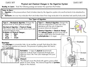
 Esophagus stomach small intestine large intestine
 480+480
 Printed pages vs web pages
 Main function of intestine
 Small intestine extends from
 Small intestine gangrene
 Calot's triangle boundaries
 Parts of the large intestine
 Another name for small intestine
 Git layers
 13.2 structures of the digestive system
 Is the stomach a physical or chemical change
 Blood supply small bowel
