Sizes of Some Neural Do not need to


- Slides: 22

Sizes of Some Neural Do not need to know specific size values

The Nervous System Is Composed of Cells • Types of cells in the nervous system – Neurons, or nerve cells • are the most important part of the nervous system • There are many types of neurons – Glial cells provide support for neurons • Microglia surround and break down any debris • Astrocytes regulate adjacent capillaries to control blood flow • Oligodendrocytes form myelin wrapping on axons in CNS • Schwann cells form myelin wrapping on axons in PNS • Neuron doctrine states that: – The brain is composed of independent cells. – Information is transmitted from cell to cell across synapses. • Chemical synapses • Electrical synapses

Figure 2. 7 Glial Cells (A) Star-shaped astrocytes detect neural activity and regulate adjacent capillaries to control blood flow, supplying neurons with more energy when they are active. (B) Tiny microglial cells surround and break down any debris that forms, especially after damage to the brain. (C) Unmyelinated axons are embedded in the troughs of glial cells. The light-colored circular shapes in the photograph are unmyelinated axons surrounded by the cytoplasm (blue) of a glial cell (the large dark area is the glial nucleus). (D) Extensions of oligodendrocytes form myelin wrapping (blue) on axons (yellow). The colorized electron micrograph of a myelinated axon (lower right) shows the many layers of the myelin sheath. The longitudinal micrograph of an axon (lower left) shows a node of Ranvier, the gap between adjacent myelinated segments. (E) Processes from astrocytes (blue) surround and insulate synapses and directly modify synaptic activity.

The Principal Components of Neurons Which neurons can collect the most information? a. Bipolar b. Monopolar c. Multipolar d. Unipolar

Neuron Classification • By Structure into three groups – Unipolar: dendrite and axon emerging from same process. – Bipolar: axon and single dendrite on opposite ends of the soma. – Multipolar: more than two dendrites: • Golgi I: neurons with long-projecting axonal processes; examples are pyramidal cells, Purkinje cells, and anterior horn cells. • Golgi II: neurons whose axonal process projects locally; the best example is the granule cell. • By Function into three groups, which is all but useless – Sensory Neurons – Interneurons – Motor Neurons • Most neurons are named as they are discovered which is not classification and results in a very long list • There currently is no useful classification system for neurons

Histology Techniques for Nervous System Box • Shape and size of neurons: • Golgi's method is a silver staining technique that is used to visualize nervous tissue under light microscopy. • • used by Spanish neuroanatomist Santiago Ramón y Cajal stains a limited number of cells at random in their entirety • Counting Cells: • The Nissl method refers to staining of the cell body, and in particular endoplasmic reticulum. • Expression of cellular products: • Autoradiography: shows the distribution of radioactive chemicals in tissues. • Immumohistochemistry: can detect a protein in tissue: • • • An antibody binds to the protein. Chemical treatments make the antibody visible. Reveals cells that have a common protein • Tracing Interconnections • Tract tracers • • Anterograde labeling reveals the axonal targets of cell bodies in a particular region. Retrograde labeling reveals the cell bodies of axons terminating in a region.

Box 2. 1 Visualizing the Cells of the Brain

Nineteenth-Century Drawing of Neurons The great Spanish neuroanatomist Santiago Ramón y Cajal created detailed renderings of the cells of the nervous system, such as this drawing of mammalian brain neurons. Another example of neuron diversity from the work of Cajal whom realized the importance of this diversity for function of brain circuits. Diversity increases the capacity to process information and adapt to new information i. e. new experiences.

The Great Diversity of Neurons come in a bewildering variety of shapes and sizes. These examples, drawn to scale, are taken from the nervous systems of various species.

Diversity of Neurons • Thousands of different types of neurons – “No two cells are the same. ” (Sanders, p 22, 2011) – Improves information processing – Flexible processing to assess unexpected things • Circuit diagram of sensory neocortex – Yuste lab composite diagram of circuits in the sensory neocortex – http: //blogs. cuit. columbia. edu/rmy 5/ • Classifying neurons based on shape, connections and neurophysiology • The Allen Institute for Brain Science – Characterizing and cataloguing the wide variety of cells that constitute the brain • What does this have to do with brain circuits?

Synapses • Neurons are interconnected through synapses – Individual neurons do not have specific behavioral or cognitive functions – neurons connected into a circuit have function • The neuronal cell body and dendrites receive information across synapses. • Dendrites have a branched arborization pattern to facilitate contacts. • Information is transmitted from the presynaptic neuron to the postsynaptic neuron. (Mostly) – There are chemical signals from postsynaptic neuron to presynaptic neuron called “retrograde” which is some sort of feedback signal

Figure 2. 5 Synapses (A) Axon terminals typically form synapses on the cell body or dendrites of a neuron. On dendrites, synapses may form on dendritic spines or on the shaft of a dendrite. (B) Information flows through a synapse from the presynaptic membrane across a gap called the synaptic cleft to the postsynaptic membrane. (C) This photomicrograph shows a synapse with some structures color coded.

Electron Micrograph of Chemical Synapse mitochondria Figure 5. 3, Bear, 2001 Active Zone

First electron micrograph showing synapse published in 1956 Wyckoff, R. W. G. and Young, J. Z. (1956) The motor neuron surface. Proc. R. Soc. London Biol. Sci 144, 440– 450

Brain Circuits • How can we best understand localization of brain function? – study of brain circuits with new technologies – a functional description of the brain and how circuits generate behavior – “brain’s ability to generate coherent thoughts derives from the spatiotemporal orchestration of neuronal activity” (Hebb, 1949) – cell assemblies • transiently active ensembles of neurons • underlie numerous operations of the brain • formation and disbanding of cell assemblies and temporal evolution of cell assembly sequences are not well understood • Human Connectome Project: plans to produce a map of the anatomical connections of the human brain

Figure 3. 18 The knee-jerk reflex. A neural chain is a simple series of neurons. The knee-jerk reflex is a circuit for the stretch reflex, consisting of Sensory neuron

Figure 3. 19 Two representations of neural circuitry. This simple representation shows the input part of the visual system. (B) This more realistic representation illustrates convergence and divergence. The visual system is a circuit with other features: Convergence—many cells send signals to one cell Divergence—one cell send signals to many cells Units are arranged in parallel, and have lateral interaction across units.

The Arrangement of Cells within the Cerebellum Basket cell Golgi cell

Layers of the Cerebral Cortex

Cortical Tracts Connect Cortical Regions • Human Connectome Project: plans to produce a map of the anatomical connections of the human brain

Basal Ganglia Anatomy Not On Exam The basal ganglia, important in motor control, include four nuclei: The caudate nucleus, the putamen, and the globus pallidus under the cerebral cortex The substantia nigra, in the midbrain

The Limbic System The limbic system includes structures important for learning and memory: Amygdala–emotional regulation and perception of odor Hippocampus and fornix–learning Cingulate gyrus–attention Olfactory bulb–sense of smell Anatomy Not On Exam
 Men's shirt sizes are determined by their neck sizes
 Sudden and violent but brief; fitful; intermittent
 I need some fire
 Fill in the gaps with speak say or tell
 Sometimes you win some sometimes you lose some
 Sometimes you win some
 Pears countable or uncountable
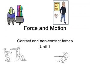
 What are some contact forces and some noncontact forces
 Fire and ice diamante poem
 Some say the world will end in fire some say in ice
 Some trust in horses
 Love is not all rhyme scheme
 Facts admitted need not be proved
 Anosognosia
 Facts that need not be proved
 A workman not ashamed
 The inspector need not be a big man
 Good people do not need laws plato
 Inspector calls mind map
 Key issue 4 why are some human actions not sustainable
 Key issue 4 why are some actions not sustainable
 Some things are caught not taught
 How do you calculate mechanical advantage


