Quantitative of protein Methods of Quantitative of protein


- Slides: 15
Quantitative of protein
Methods of Quantitative of protein • Method 1: protein assay based on dye binding assay • Method 2: protein assay based on alkaline copper
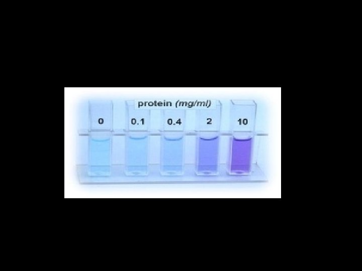
Method 1: protein assay based on dye binding assay
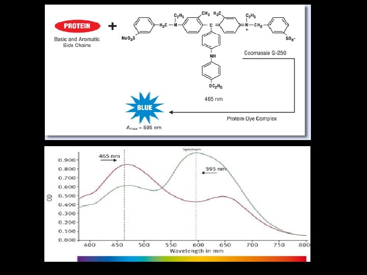
• BRADFORD METHOD • Use of coomassie G-250 dye in a colorimetric reagent for the detection and quantitation of total protein. • In the acidic environment of the reagent, protein binds to the coomassie dye. • This results in a spectral shift from the reddish/brown form of the dye (absorbance maximum at 465 nm) to the blue form of the dye (absorbance maximum at 610 nm).
• The difference between the two forms of the dye is greatest at 595 nm, so that is the optimal wavelength to measure the blue color from the coomassie dye-protein complex • Development of color in coomassie dye-based (Bradford) protein assays has been associated with the presence of certain basic amino acids (primarily arginine, lysine and histidine) in the protein.
• Free amino acids, peptides and low molecular weight proteins do not produce color with coomassie dye reagents. (unbound) forms are green or red.
• Advantages: This assay is quick, and the reagent is not affected by the presence of reducing agents, like DTT • Disadvantages: Basic conditions and detergents, such as SDS, can interfere with the dye’s ability to bind to the protein
Method 2: protein assay based on alkaline copper Biuret Test
• Principle: • Under alkaline conditions substances containing two or more peptide bonds form a purple complex with copper salts in the reagent.
• Biuret Reagent contains: • Hydrated Copper sulphate – this provides the Cu (II) ions which form the chelate complex. Cu (II) ions give the reagent its characteristic blue color. • Potassium hydroxide does not participate in the reaction but provides the alkaline medium. • Potassium sodium tartrate (KNa. C 4 H 4 O 6· 4 H 2 O) stabilizes the chelate complex
• The Biuret reaction can be used to measure the concentration of proteins because peptide bonds occur with the same frequency per amino acid in the peptide. • The intensity of the color, and hence the absorption at 540 nm, is directly proportional to the protein concentration, according to the Beer-Lambert law.
tubes D. W Protein standard 1 0. 5 0 0 2 0. 4 0. 1 0. 4 3 0. 2 0. 8 4 0. 2 0. 3 1. 2 5 0. 1 0. 4 1. 6 6 0 0. 5 2 Unknown 0 0 1. 2. 3. 4. Unknown 0. 5 Add 1 ml of biuret reagent to each tube and mix Incubation 30 min Read at 540 nm Draw the standard curve and measure the unknown Conc
Reference range • Reference range for total proteins is 66. 6 to 81. 4 g/L