Primary glomerulonephritides GN II Proliferative GN Miroslav Merta
- Slides: 20
Primary glomerulonephritides (GN) II Proliferative GN Miroslav Merta Klinika nefrologie 1. LF a VFN
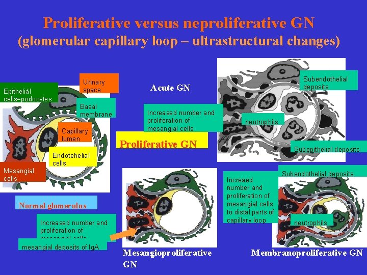
Proliferative versus neproliferative GN (glomerular capillary loop – ultrastructural changes) Epitheliál cells=podocytes Urinary space Basal membrane Capillary lumen Mesangial cells Subendothelial deposits Acute GN Increased number and proliferation of mesangial cells neutrophils Proliferative GN Subepithelial deposits Endotehelial cells Increaed number and proliferation of mesangial cells to distal parts of capillary loop Normal glomerulus Increased number and proliferation of mesangial cells mesangial deposits of Ig. A Mesangioproliferative GN Subendothelial deposits neutrophils Membranoproliferative GN
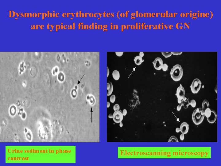
Dysmorphic erythrocytes (of glomerular origine) are typical finding in proliferative GN Urine sediment in phase contrast Electroscanning microscopy
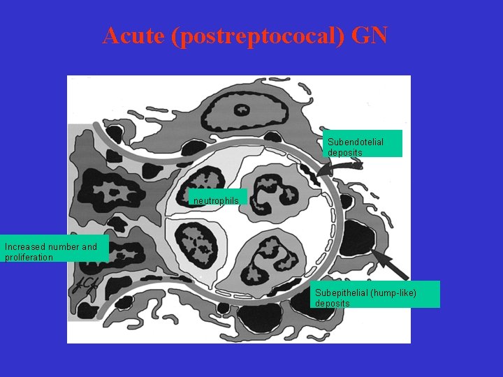
Acute (postreptococal) GN Subendotelial deposits neutrophils Increased number and proliferation Subepithelial (hump-like) deposits
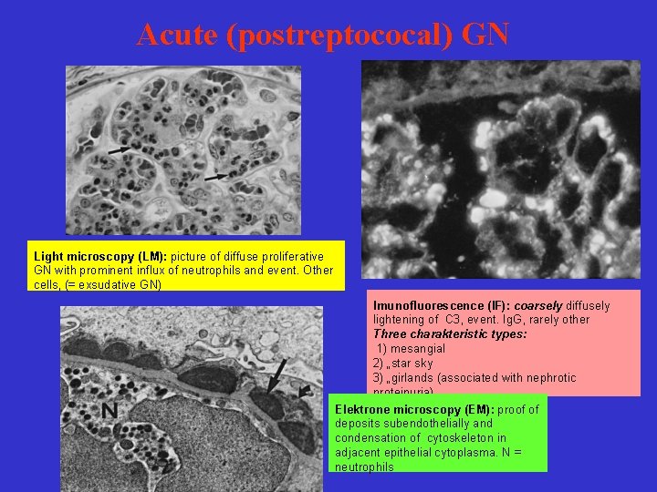
Acute (postreptococal) GN Light microscopy (LM): picture of diffuse proliferative GN with prominent influx of neutrophils and event. Other cells, (= exsudative GN) Imunofluorescence (IF): coarsely diffusely lightening of C 3, event. Ig. G, rarely other Three charakteristic types: 1) mesangial 2) „star sky 3) „girlands (associated with nephrotic proteinuria) Elektrone microscopy (EM): proof of deposits subendothelially and condensation of cytoskeleton in adjacent epithelial cytoplasma. N = neutrophils
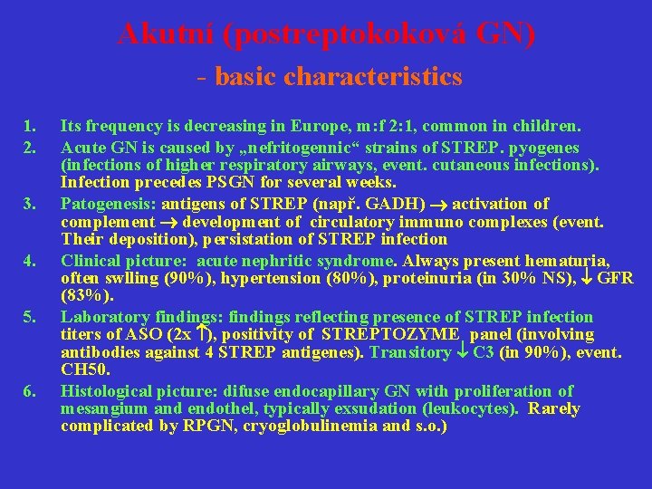
Akutní (postreptokoková GN) - basic characteristics 1. 2. 3. 4. 5. 6. Its frequency is decreasing in Europe, m: f 2: 1, common in children. Acute GN is caused by „nefritogennic“ strains of STREP. pyogenes (infections of higher respiratory airways, event. cutaneous infections). Infection precedes PSGN for several weeks. Patogenesis: antigens of STREP (např. GADH) activation of complement development of circulatory immuno complexes (event. Their deposition), persistation of STREP infection Clinical picture: acute nephritic syndrome. Always present hematuria, often swlling (90%), hypertension (80%), proteinuria (in 30% NS), GFR (83%). Laboratory findings: findings reflecting presence of STREP infection titers of ASO (2 x ), positivity of STREPTOZYME panel (involving antibodies against 4 STREP antigenes). Transitory C 3 (in 90%), event. CH 50. Histological picture: difuse endocapillary GN with proliferation of mesangium and endothel, typically exsudation (leukocytes). Rarely complicated by RPGN, cryoglobulinemia and s. o. )
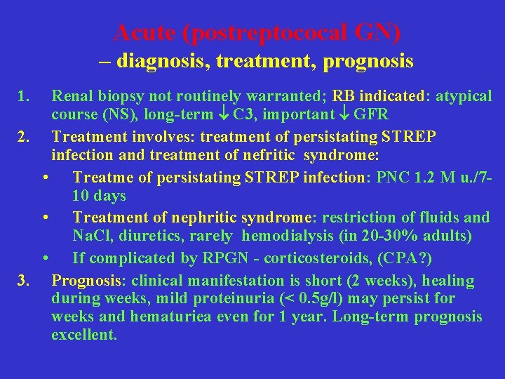
Acute (postreptococal GN) – diagnosis, treatment, prognosis 1. Renal biopsy not routinely warranted; RB indicated: atypical course (NS), long-term C 3, important GFR 2. Treatment involves: treatment of persistating STREP infection and treatment of nefritic syndrome: • Treatme of persistating STREP infection: PNC 1. 2 M u. /710 days • Treatment of nephritic syndrome: restriction of fluids and Na. Cl, diuretics, rarely hemodialysis (in 20 -30% adults) • If complicated by RPGN - corticosteroids, (CPA? ) 3. Prognosis: clinical manifestation is short (2 weeks), healing during weeks, mild proteinuria (< 0. 5 g/l) may persist for weeks and hematuriea even for 1 year. Long-term prognosis excellent.
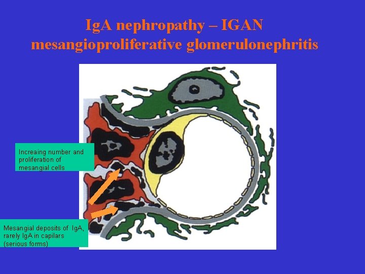
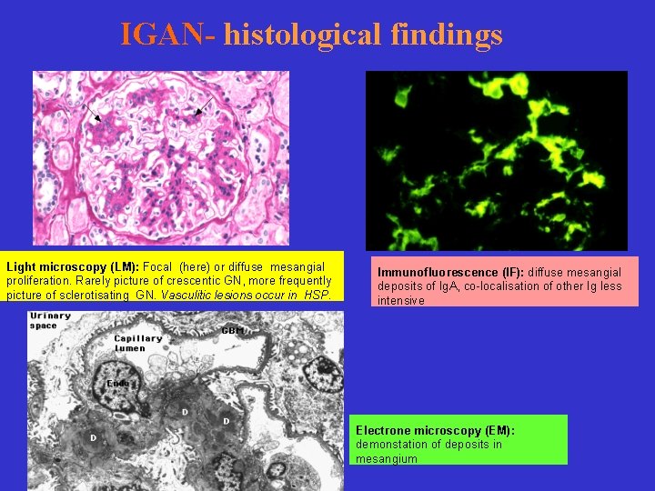
Ig. A nephropathy – IGAN mesangioproliferative glomerulonephritis Increaing number and proliferation of mesangial cells Mesangial deposits of Ig. A, rarely Ig. A in capilars (serious forms)
IGAN- histological findings Light microscopy (LM): Focal (here) or diffuse mesangial proliferation. Rarely picture of crescentic GN, more frequently picture of sclerotisating GN. Vasculitic lesions occur in HSP. Immunofluorescence (IF): diffuse mesangial deposits of Ig. A, co-localisation of other Ig less intensive Electrone microscopy (EM): demonstation of deposits in mesangium
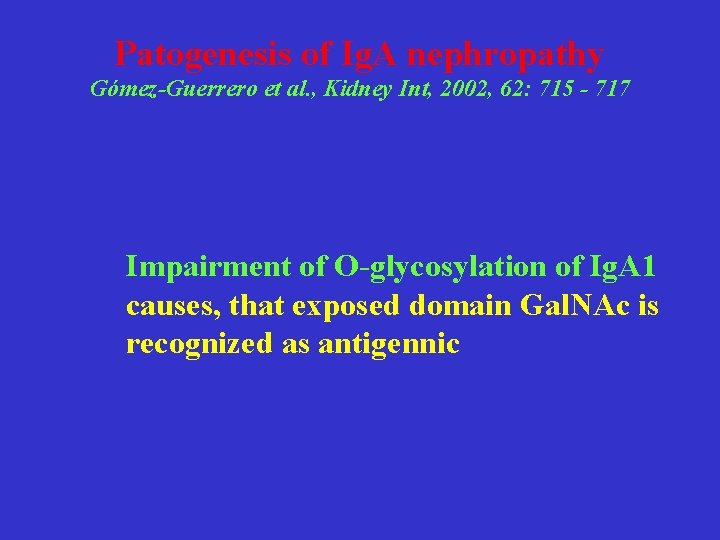
Patogenesis of Ig. A nephropathy Gómez-Guerrero et al. , Kidney Int, 2002, 62: 715 - 717 Impairment of O-glycosylation of Ig. A 1 causes, that exposed domain Gal. NAc is recognized as antigennic
Ig. A nephropathy - basic characteristics 1. Commonest GN in Europe (20 -40% of all primary GN) 2. Typical clinical picture – asymptomatic microskopic hematuria či episodes of parainfectious macroskopic hematuria 3. Natural course of the disease is not benign – at least in 20% patients ESRD develops within 20 years 4. Histology – mesangial deposits of Ig. A demonstrated by IF
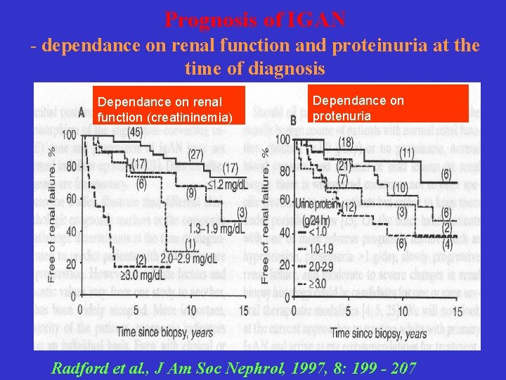
Prognosis of Ig. AN – negative prognostic factors a. klinical hypertension proteinuria (> 1 g/24 h) decrease of GFR at the time of diagnosis b. histological glomerulosclerosis interstitial fibrosis vascular sclerosis
Prognosis of IGAN - dependance on renal function and proteinuria at the time of diagnosis Dependance on renal function (creatininemia) Dependance on protenuria Radford et al. , J Am Soc Nephrol, 1997, 8: 199 - 207
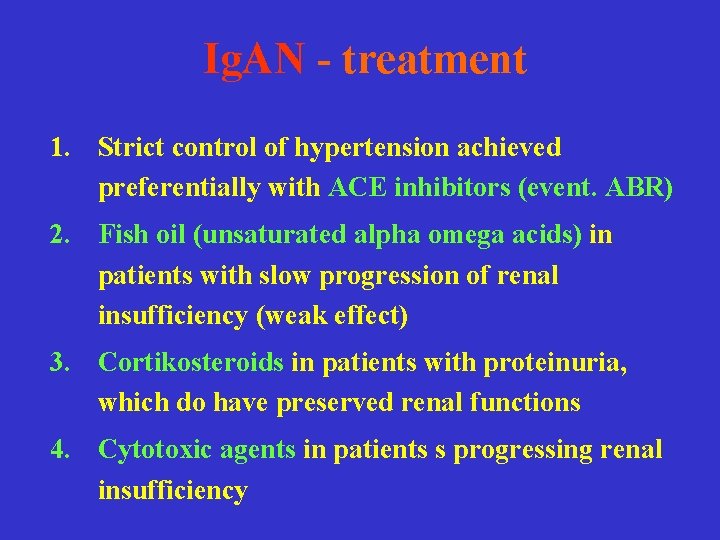
Ig. AN - treatment 1. Strict control of hypertension achieved preferentially with ACE inhibitors (event. ABR) 2. Fish oil (unsaturated alpha omega acids) in patients with slow progression of renal insufficiency (weak effect) 3. Cortikosteroids in patients with proteinuria, which do have preserved renal functions 4. Cytotoxic agents in patients s progressing renal insufficiency
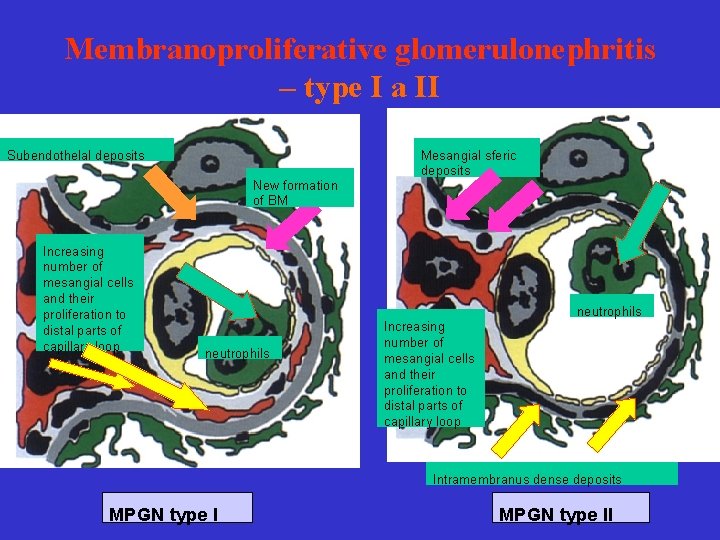
Membranoproliferative glomerulonephritis – type I a II Subendothelal deposits New formation of BM Increasing number of mesangial cells and their proliferation to distal parts of capillary loop Mesangial sferic deposits neutrophils Increasing number of mesangial cells and their proliferation to distal parts of capillary loop Intramembranus dense deposits MPGN type II
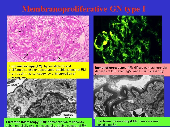
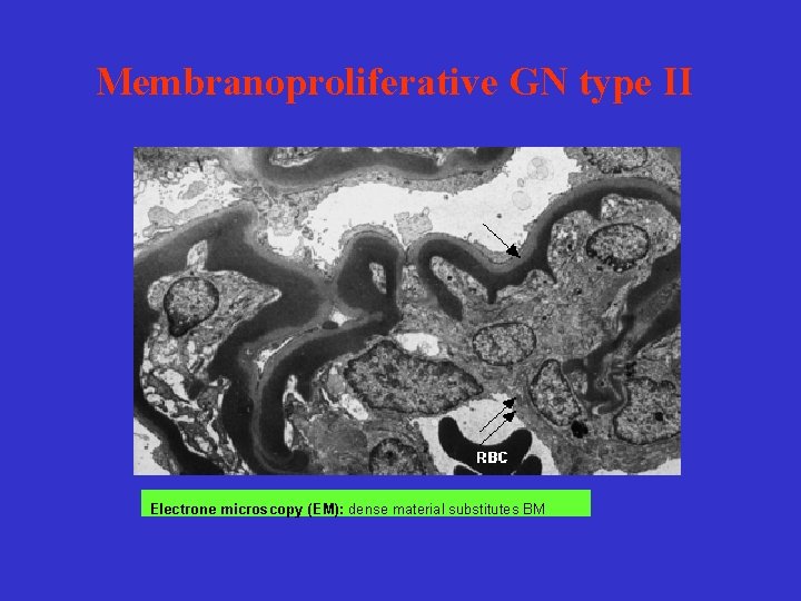
Membranoproliferative GN type I Light microscopy (LM): hypercelullarity and proliferation, , lobular appearance, double contour of BM (tram track) – as consequence of interposition of mesangium), Electrone microscopy (EM): demonstration of deposits subendothelially and a mesangially, double contour of BM, Immunofluorescence (IF): diffuse periferal granular deposits of Ig. G, event. Ig. M, and C 3 (in type II only C 3) Electrone microscopy (EM): dense material substitutes BM
Membranoproliferatie GN (MPGN) Type I a. Idiopatic b. Secondary – infection (visceral abscesses, endocarditis, infection of atrioventricular shunts, malaria) - systemic diseases ( SLE – LN type III-IV) - paraproteinemias ( LCDD, cryoglobulinemia) - thrombotic microangiopathy ( HUS/TTP, APS) Type II – disease of dense deposits
Idiopatic MPGN type I - basic characteristics 1. Relatively rare disease in developed countries 2. It is found in less aged subjects 3. Clinical findings at the time of dg: usually NS and microskopic hematuria 4. Slowly progressing disease – during 10 -y interval 50% patients do progress into ESRD 5. Treatment in adults is controversial (corticosteroids, CPA, anticoagulation)
Membranoproliferative GN type II Electrone microscopy (EM): dense material substitutes BM
Idiopatic MPGN type II - basic characteristics 1. Very rare disease 2. Clinically: presence of nephritic factor with hypocomplementemia 3. More pronounced nephritic features and more agressive course of diasease 4. Efficient treatment unknown