Plenary case discussion 1 Fracture assessment and decision
- Slides: 21
Plenary case discussion 1 Fracture assessment and decision making
Objectives • Describe how to approach a fracture patient • Apply a descriptive fracture classification system • Describe the mechanical and biological demands of different fractures • Use the fracture planning assessment scheme (FPAS) • Mechanical, biological, clinical (MBC)
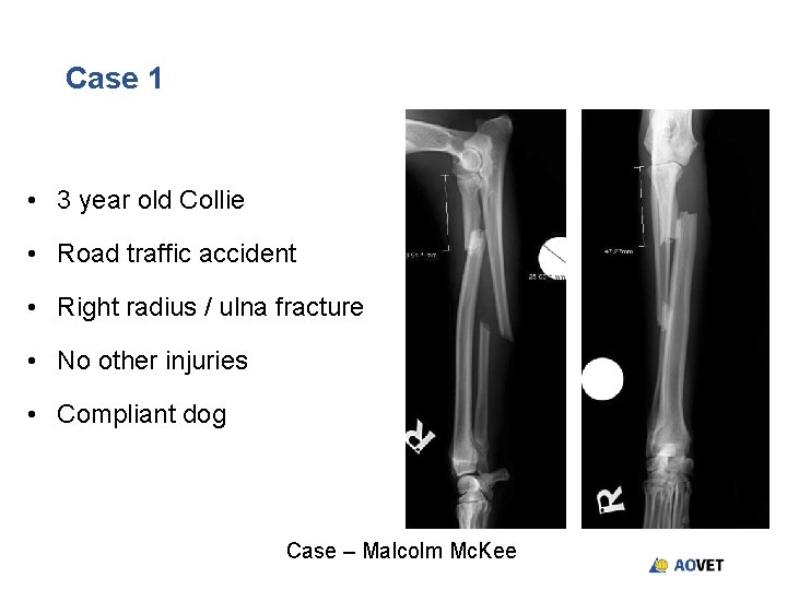
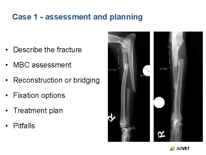
Case 1 • 3 year old Collie • Road traffic accident • Right radius / ulna fracture • No other injuries • Compliant dog Case – Malcolm Mc. Kee
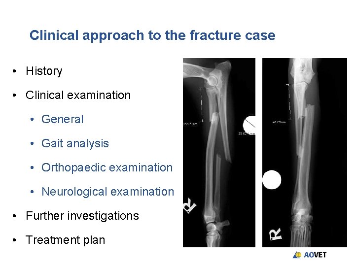
Clinical approach to the fracture case • History • Clinical examination • General • Gait analysis • Orthopaedic examination • Neurological examination • Further investigations • Treatment plan
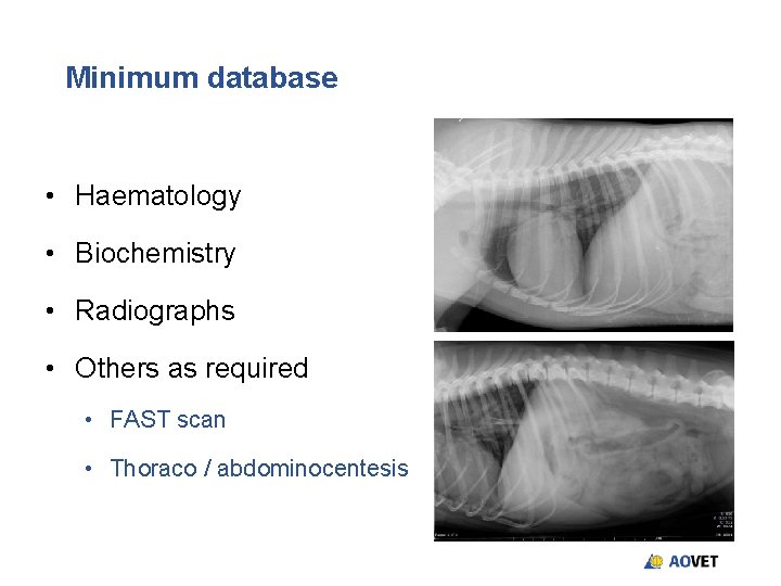
Minimum database • Haematology • Biochemistry • Radiographs • Others as required • FAST scan • Thoraco / abdominocentesis
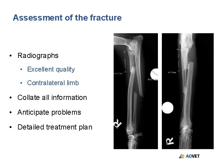
Assessment of the fracture • Radiographs • Excellent quality • Contralateral limb • Collate all information • Anticipate problems • Detailed treatment plan
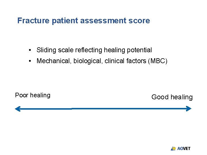
Fracture patient assessment score • Sliding scale reflecting healing potential • Mechanical, biological, clinical factors (MBC) Poor healing Good healing
Key Questions • Absolute or relative stabililty • Should the fracture be reconstructed • Mechanical advantage versus biological disadvantage • What are the forces acting on the fracture • What fixation is most appropriate for this fracture • What is my plan A, B and C • What are the potential challenges
Case 1 - assessment and planning • Describe the fracture • MBC assessment • Reconstruction or bridging • Fixation options • Treatment plan • Pitfalls
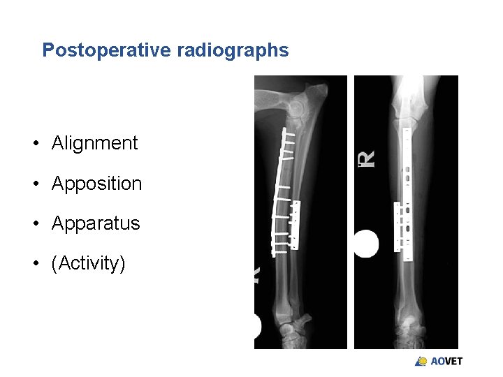
Postoperative radiographs • Alignment • Apposition • Apparatus • (Activity)
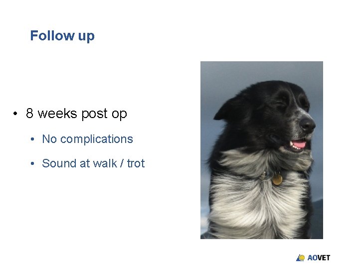
Follow up • 8 weeks post op • No complications • Sound at walk / trot
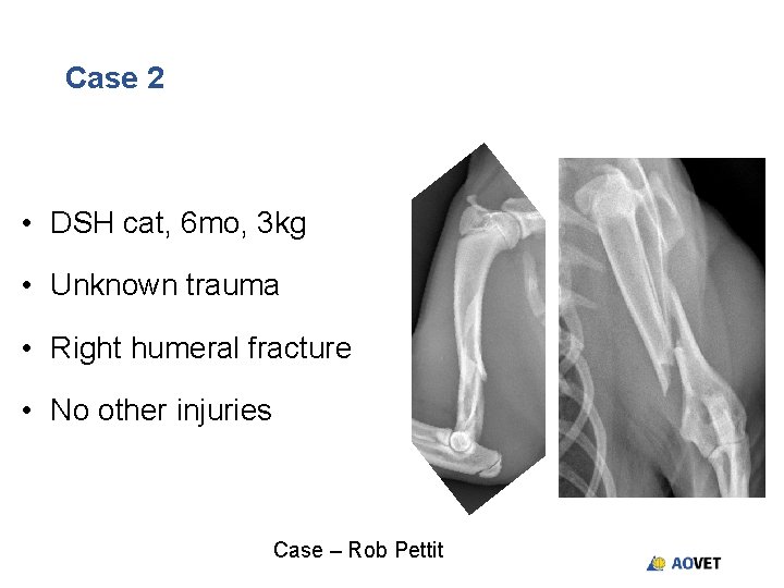
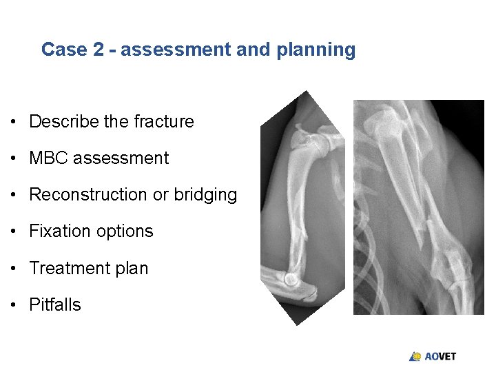
Case 2 • DSH cat, 6 mo, 3 kg • Unknown trauma • Right humeral fracture • No other injuries Case – Rob Pettit
Case 2 - assessment and planning • Describe the fracture • MBC assessment • Reconstruction or bridging • Fixation options • Treatment plan • Pitfalls
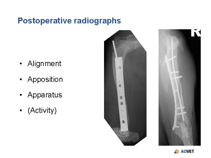
Postoperative radiographs • Alignment • Apposition • Apparatus • (Activity)
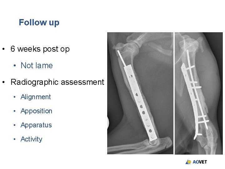
Follow up • 6 weeks post op • Not lame • Radiographic assessment • Alignment • Apposition • Apparatus • Activity
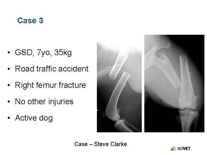
Case 3 • GSD, 7 yo, 35 kg • Road traffic accident • Right femur fracture • No other injuries • Active dog Case – Steve Clarke
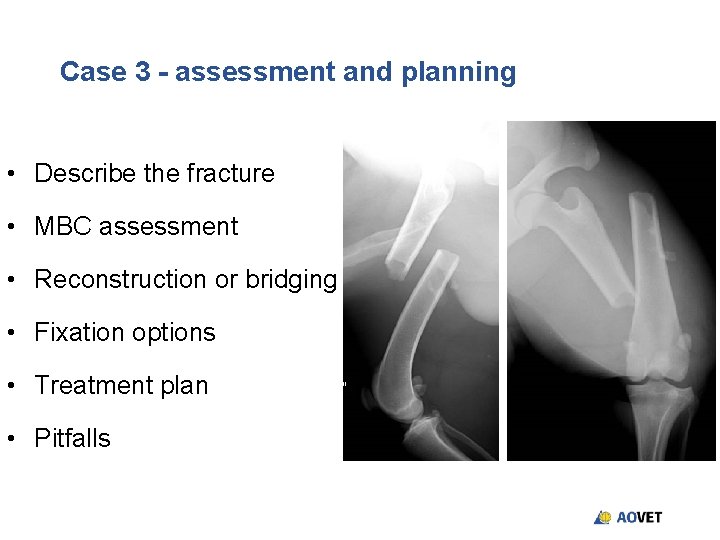
Case 3 - assessment and planning • Describe the fracture • MBC assessment • Reconstruction or bridging • Fixation options • Treatment plan • Pitfalls
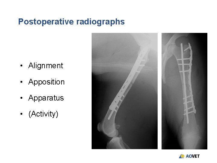
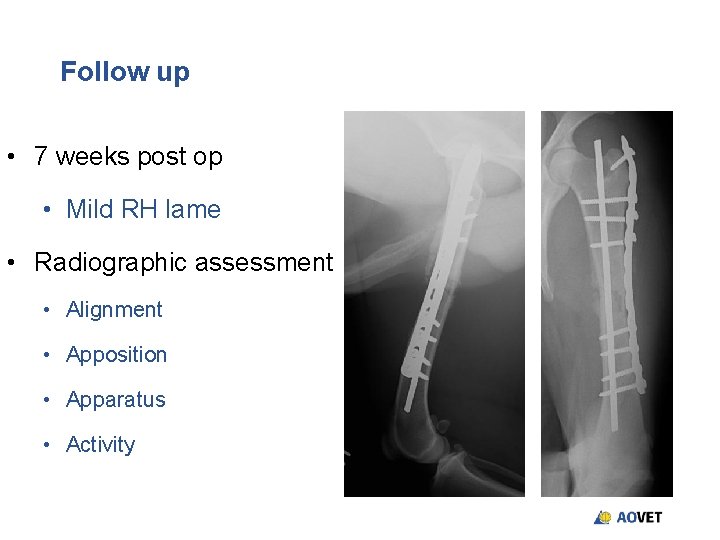
Postoperative radiographs • Alignment • Apposition • Apparatus • (Activity)
Follow up • 7 weeks post op • Mild RH lame • Radiographic assessment • Alignment • Apposition • Apparatus • Activity
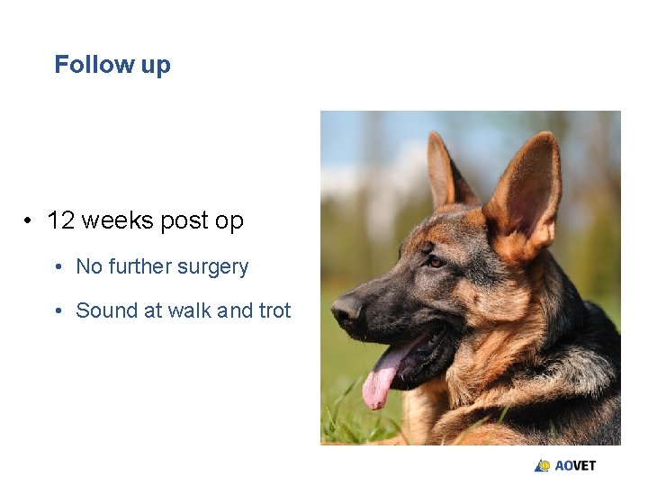
Follow up • 12 weeks post op • No further surgery • Sound at walk and trot
Summary • Summarise how you would approach a fracture case • Write down the key learning points • What will you do differently in the future