Perch Dissection Some pictures from 2001 Agri Teach

- Slides: 20
Perch Dissection Some pictures from © 2001 Agri. Teach. com (92601 ms)
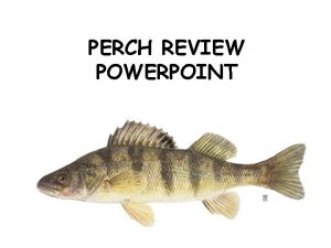
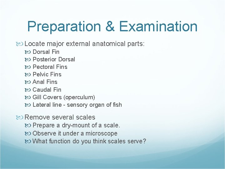
Preparation & Examination Locate major external anatomical parts: Dorsal Fin Posterior Dorsal Pectoral Fins Pelvic Fins Anal Fins Caudal Fin Gill Covers (operculum) Lateral line - sensory organ of fish Remove several scales Prepare a dry-mount of a scale. Observe it under a microscope What function do you think scales serve?
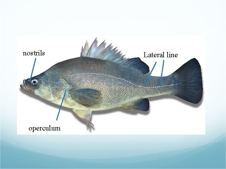
nostrils operculum Lateral line
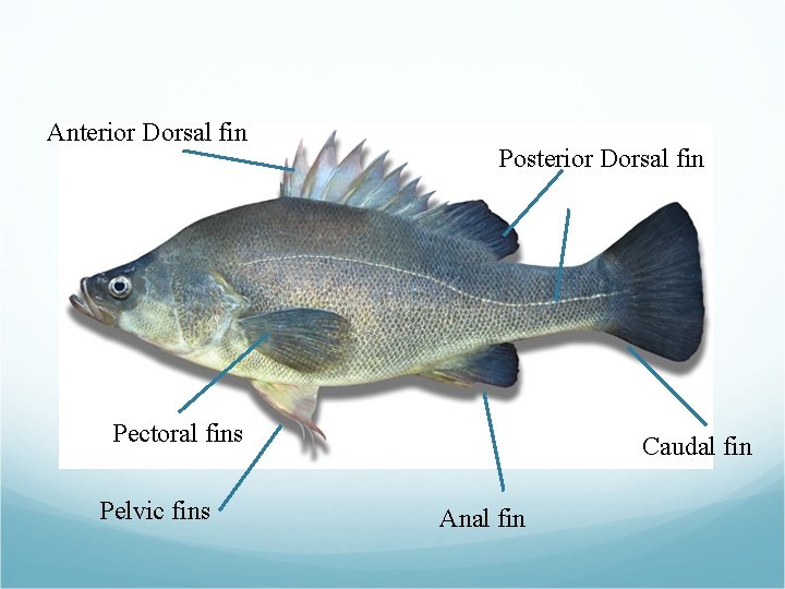
Anterior Dorsal fin Posterior Dorsal fin Pectoral fins Pelvic fins Caudal fin Anal fin
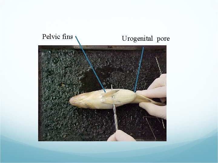
Pelvic fins Urogenital pore
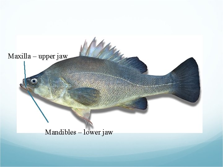
Maxilla – upper jaw Mandibles – lower jaw
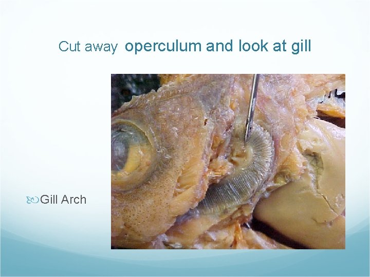
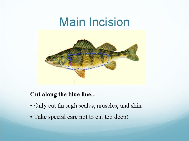
Dissection Remove operculum with scissors Observe gill anatomy Rakers - white, comb-like arches Filaments - Red fingerlike projections With a scalpel, remove a section of the lateral line What function does this organ serve? Do you notice a concentration of nerve endings? Begin the main incision Open the abdomen (below the gill) carefully with a scalpel Cut with a scissors: remove a oval-shaped piece of skin (only skin) running from underneath the gills, to the anus, up to the lateral line, along the lateral line, to the gill, down to where you started the incision. Remove flap of skin (see diagram on next slide)
Cut away operculum and look at gill Gill Arch
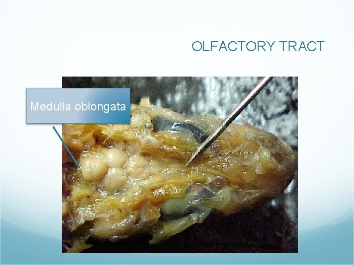
OLFACTORY TRACT Medulla oblongata
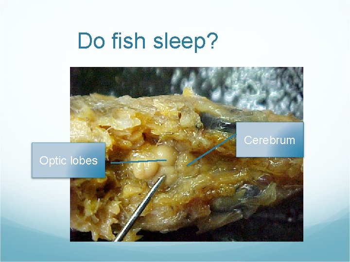
Do fish sleep? Cerebrum Optic lobes
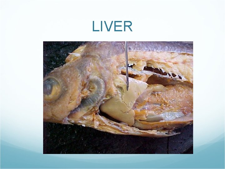
LIVER
Main Incision Cut along the blue line. . . • Only cut through scales, muscles, and skin • Take special care not to cut too deep!
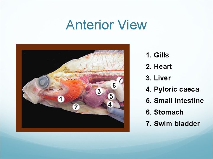
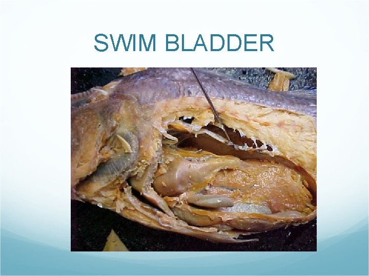
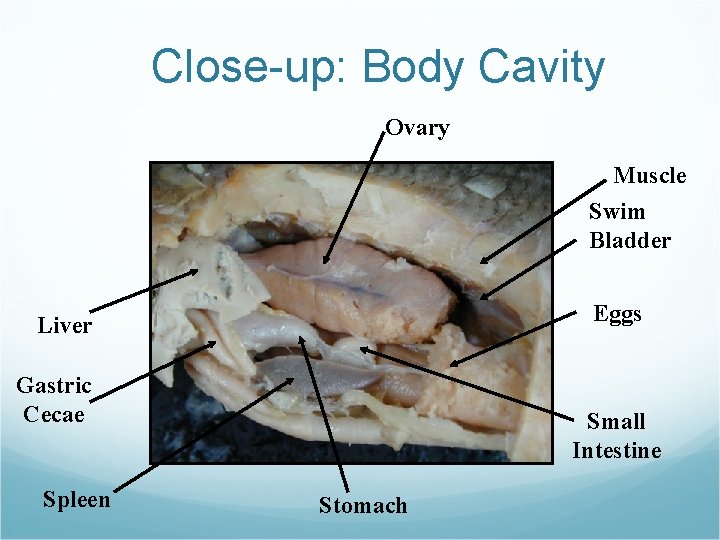
Anterior View 1. Gills 2. Heart 3. Liver 4. Pyloric caeca 5. Small intestine 6. Stomach 7. Swim bladder
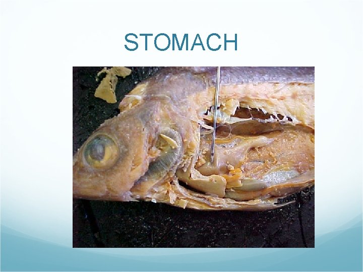
STOMACH
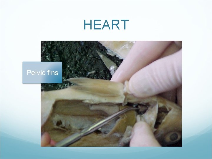
HEART Pelvic fins
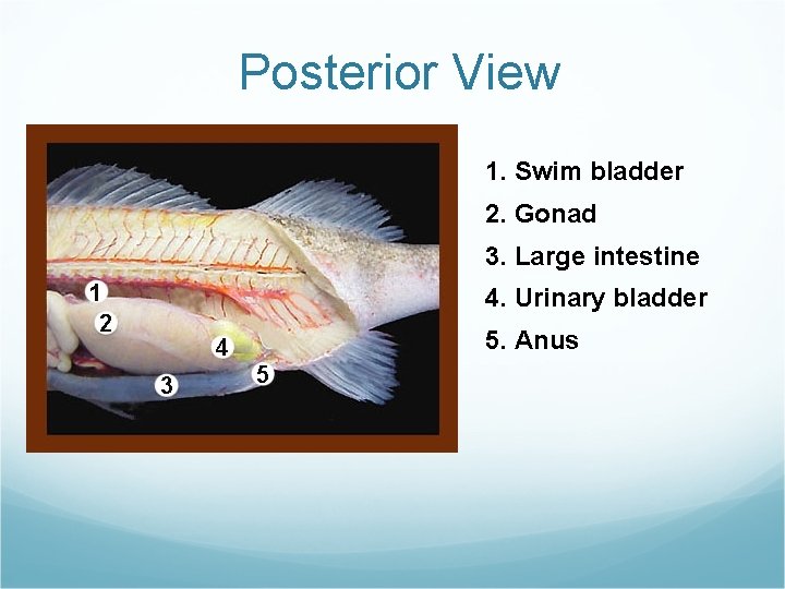
Posterior View 1. Swim bladder 2. Gonad 3. Large intestine 4. Urinary bladder 5. Anus
SWIM BLADDER
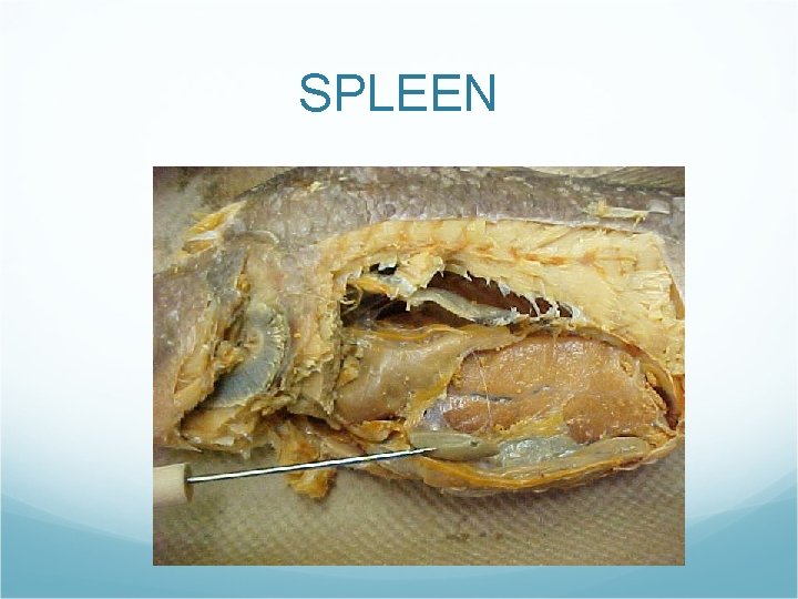
SPLEEN
Close-up: Body Cavity Ovary Muscle Swim Bladder Eggs Liver Gastric Cecae Spleen Small Intestine Stomach
Other sources used to put presentation together: Pictures from Mr. Tippett’s Biology Class at http: //www. flushing. k 12. mi. us/srhigh/tippettl/biology/perch/index. html Lab modified from Massengale’s Biology Place found at http: //sps. k 12. ar. us/massengale/perch_dissection 2. htm