PARATHYROID GLANDS They are 4 glands present at


- Slides: 20


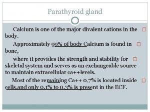
PARATHYROID GLANDS � They are 4 glands present at the back of thyroid glands. � Each measures 4 mm in diameter & their combined weight = 120 mg. � They secrete parathyroid hormone (parathromone) which is essential for life. Microscopically, they contain 2 types of cells: 1. Chief cells : they are the most abundant & they secrete pathormone. 2. Oxyphil cells : they are little & may represent inactive chief cells

PARATHYROID HORMONE (PTH) = PARATHORMONE Source : � Parathyroid glands. Nature : � PP formed of 84 AA.

Functions: � The prime function of PTH is to keep Ca++ level (9 – 11 mg%). � Normally, the plasma inorganic phosphate is inversely related to Ca++ concentration & the product Ca++ × Po 4 - = constant (solubility product). � The function of parathormone is to ↑ plasma Ca++ & ↓ plasma PO 4 - thus maintain the solubility product constant. Parathyroid hormone (PTH) raises the lowered Ca++ level through acting on: 1 - On the intestine : A. ↑ Ca++ absorption - This action is mediated by active vitamin D. B. ↑ phosphate & Mg++ absorption

2 - On the bone: • ↑ Ca++ mobilization from bone by activating osteoclasts (bone destroying cells) → release of Ca++ & phosphate into the blood stream. 3 - On the kidney: • ↑ Ca++ & Mg++ reabsorption. • ↑ phosphate excretion 4 - ↓ Ca++ excretion in milk to maintain its blood levels high. Mode of action of PTH 1. Activation of adenyl cyclase enzyme →↑ C-AMP in the target cells 2. ↑ intracellular Ca++ content into the cells by enhancing the permeability of bone cells to Ca++. N. B. : β adrenergic receptors were discovered in the parathyroid gland, this may explains hypercalacemia found in patients with pheochromocytoma

DISORDERS OF THE PARATHYROID GLAND 1 - Hypo-para-thyroidism Cause : � Accidental damage or removal of the parathyroid gland during thyroid surgery. � Hypoparathyroidism is characterized by hypocalcemia due to decrease ionized Ca++. � hypocalcemia is associated with increased neuromuscular excitability due increased membrane permeability to Na+. Leading to tetany.

� Is a disease characterized by increased neuromuscular excitability caused by reduction of blood levels of ionized Ca++. Cause : 1. Hypoparathyroidism 2. Renal failure due to phosphate retention. 3. Alkalaemia due to precipitation of ionized Ca++. 4. Decreased Ca++ absorption from the intestine due to : I. Low dietary Ca++ intake. II. Vitamin D deficiency. III. steatorrhea (fatty diarrhea) which ↓ Ca++ absorption

Manifestations: - Manifestations of tetany depends on the degree of Ca++ lowering: I- Manifest tetany - Occurs if Ca++ is markedly ↓ i. e. < 7 mg% II- Latent tetany - Ca++ isn’t marked ly ↓ ( )7&9 mg% Manifestations: 1 - In adults, carpopedal spasm: a) In the hands, carpal spasm: - Flexion of the wrist & metacarpophalangeal joint. - Extension of the interphalangeal joint. - Adduction of the thumb. b) In the feet, pedal spasm: - Dorsiflexaion of the ankle & plantar flexion of the toes. 2 - In children : may be convulsions 3 - In infants : may be laryngeal spasm - No carpopedal spasm except if the person is exposed to stress. - The pateint may feel numbness & heat flushings


Diagnosis Provocative tests of latent tetany: - They are a group of tests designed to diagnosis latent tetany: 1 - Chvosteck’s test: - Tapping of the facial nerve → twitching of epsi-lateral facial ms. - Due to increased excitability 2 - Erb’s sign : - Stimulation of the motor nerve by sub-threshold galvanic current → ms. Contraction. - Due to increased excitability. 3 - trousseau’s test: - Is done by inflating the sphygmomanometer cuff around the arm to a pressure more than the systolic BP for 2 minutes: - Ischemia of the nerve trunk →↑ their excitability & re-enforces the effects of hypocalcemia → carpal spasm.

Treatement of tetany 1. Intravenous Ca++ gluconate stops immediately the spasm. 2. Diet rich in Ca++ & vitamin D. 3. Acidifying salts e. g. ammonium Cl- (↑) Ca++ solubility in GT) 4. Dihydro-tachysterol: has similar effects to parathormone but doesn’t produce antibodies like exogenous parathormone.

CALCITONIN HORMONE (= THYRO-CALCITONIN) Calci= calcium, tonin = lowering Nature : • Polypeptide hormone formed of 32 Aa. Source : • Parafollicular C cells of the thyroid gland. Control of release: 1. Rise of serum Ca++, the major stimulus - ↑ serum Ca++ by 1 mg% →↑ cacitonin release about 10 times. 2. Ingestion of food : - ingestion of food →↑ calcitonin release. - this action is mediated by GIT hromones specially gastrin

inactivation • It is degradated by the kidney • Its half life is less than 1 hours Actions • Although its role in human is uncertain, calcitonin seems to be the hypocalcaemic hormone of the body & exerts this effects as follows: The major effect of calcitonon administration is rapid fall in plasma Ca++, this action is more marked in the young than adults

1 - On the intestine: • ↓ Ca++ absorption & Po 4 - 2 - On the bone : • It inhibits osteoclastic activity →↓ bone resorption & mobilization of Ca++ from bone into the blood 3 - On the kidney: • ↑ urinary excretion of Ca++ &Po 4 - • Inhibits renal α 1 -hydroxylase enzyme which activate vit D. 4 - It act as physiological antagonist to parathormone as regards Ca++, and its has the same effect as regards phosphate, but it has no effect as regards magnesium

OTHER HORMONES AFFECTING BONE & CALCIUM METABOLISM • Although parathromone and calcitonin are the major calcium regulating hormones, a number of other hormones are known to have an important influence on the bone and mineral metabolims. • These include vitamin D, estrogens androgens, glucocortcoids, thyroid hormones, and growth hormone. • Bone remodeling is a process which continues througout life, long after epiphyseal fusion and cessation of linear growth of bone. • Remodeling consists of bone formation and bone resorption I. Osteoblasts : are the primary cells concerned with synthesis of new bone. II. Osteoclasts : function to resorb bone

1 - VITAMIN D • Vitamin D have both dietary & endogenous precursors : I. Vitamin D 2 (ergo-calciferol) formed in plants II. Vitamin D 3 (chole-calciferol) formed in the skin by the ultraviolet rays (UVR) Actions: 1 - On the intestine : - it stimulates absorption of both Ca++ & phosphate. 2 - On the kidney: - it stimulates re-absorption of both Ca++ & phosphate. 3 - On the bone: - it provides Ca++ & phosphate needed for bone formation. - it promotes differentiation of monocyte precursors to monocytes & macrophages.

2 - ESTROGENS & ANDROGENS • Have a role in childhood & puberty. • These hormones favours bone formation over resorption. • In the female estrogen protect the skeleton from development of oesteoporosis. 3 - GLUCOCORTICOIDS I. At physiological levels they are essential for skeletal growth. II. At high level they have deleterious effect on Ca++ homeostasis.

4 - THYROID HORMONES I. At physiological levels they are essential for skeletal growth. . II. At high level e. g. in hyperthyroidism they cause bone resorption. III. Also, in hypothyroidism bone growth is retarded. 5 - GROWTH HORMONE I. Has strong stimulatory effect on bone growth dependent on somatomedins. II. III. It increase intestinal Ca++ absorption through vit D It increase also renal phosphate reabsorption
 Antigentest åre
 Nerve supply of thyroid gland
 Thyrooid
 Thyroid and parathyroid glands histology
 Pseudostratified columnar epithelium in trachea
 Janos lobe
 Parathyroid cell
 Calcitonin vitamin d
 Parathyroid gland chief cell
 Pth function
 Parathyroid hormone pathway
 Shrsies
 Parathyroid
 Pharyngeal pouches definition
 Parathyroid hormone and calcitonin
 Present perfect present simple present continuous
 Do and does use
 Present perfect simple and present continuous
 Present simple present continuous present perfect
 Present perfect simple and continuous romana
 Present simple present continuous and present perfect

