Organogenesis1 Somitogenesis and derivatives of somites M A
- Slides: 21
Organogenesis(1). Somitogenesis and derivatives of somites M. A. Kai-Kai
Learning Objectives Understand process of somitogenesis segmental pattern of somitomeres and somites along the neural tube. Review the adult derivatives of each of the three morphological subdivisions of the somites dermatome, sclerotome and myotome. Understanding of patterns of osteogenesis from the sclerotome. Understanding the process of myogenesis. Examples of congenital malformations in development of the skeleton.
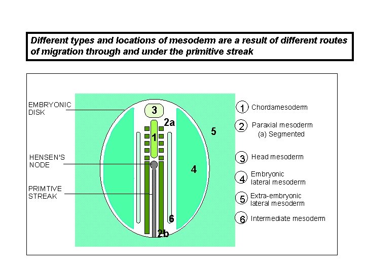
FORMATION OF THE MAMMALIAN GASTRULA - 7 Different types and locations of mesoderm are a result of different routes of migration through and under the primitive streak Dorsal view of embryonic disc Rostral EMBRYONIC DISK 3 2 a 5 1 HENSEN'S NODE 4 PRIMTIVE STREAK 6 2 b Caudal 1 Chordamesoderm 2 Paraxial mesoderm (a) Segmented (b) Unsegmented 3 Head mesoderm 4 Embryonic lateral mesoderm 5 Extra-embryonic lateral mesoderm 6 Intermediate mesoderm
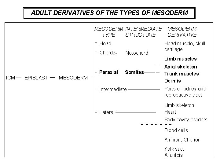
ADULT DERIVATIVES OF THE TYPES OF MESODERM INTERMEDIATE TYPE STRUCTURE Head Chorda- Notochord MESODERM DERIVATIVE Head muscle, skull cartilage Limb muscles Paraxial ICM EPIBLAST Somites MESODERM Intermediate Lateral Axial skeleton Trunk muscles Dermis Parts of kidney and reproductive tract Limb skeleton Heart Body cavity dividers Blood cells Amnion, Chorion Yolk sac, Allantois
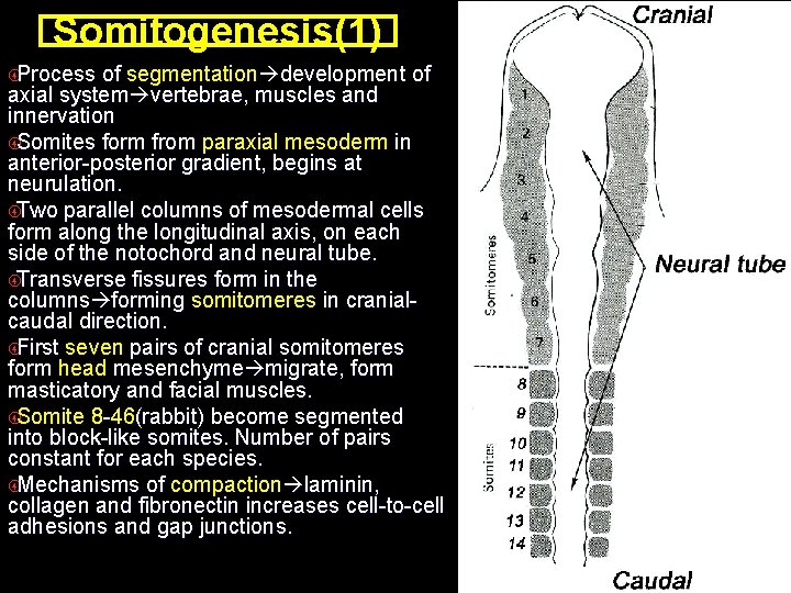
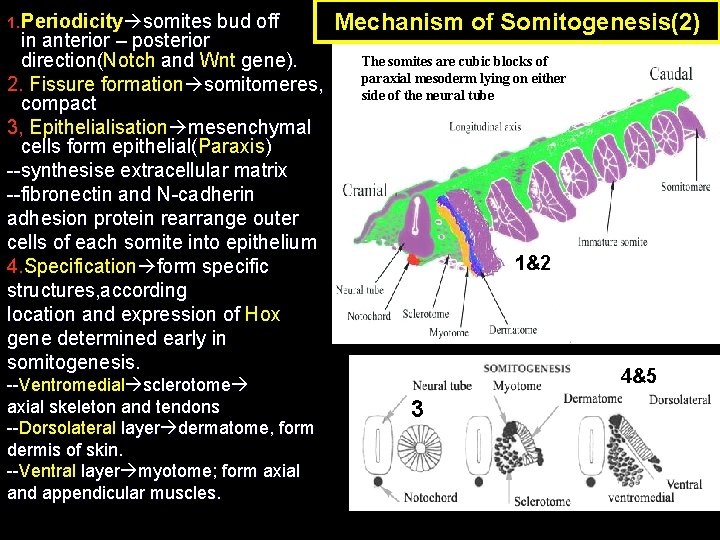
Somitogenesis(1) Process of segmentation development of axial system vertebrae, muscles and innervation Somites form from paraxial mesoderm in anterior-posterior gradient, begins at neurulation. Two parallel columns of mesodermal cells form along the longitudinal axis, on each side of the notochord and neural tube. Transverse fissures form in the columns forming somitomeres in cranialcaudal direction. First seven pairs of cranial somitomeres form head mesenchyme migrate, form masticatory and facial muscles. Somite 8 -46(rabbit) become segmented into block-like somites. Number of pairs constant for each species. Mechanisms of compaction laminin, collagen and fibronectin increases cell-to-cell adhesions and gap junctions.
1. Periodicity somites bud off in anterior – posterior direction(Notch and Wnt gene). direction( 2. Fissure formation somitomeres, compact 3, Epithelialisation mesenchymal cells form epithelial(Paraxis ) epithelial( --synthesise extracellular matrix --fibronectin and N-cadherin adhesion protein rearrange outer cells of each somite into epithelium 4. Specification form specific 4. structures, according location and expression of Hox gene determined early in somitogenesis. --Ventromedial sclerotome -axial skeleton and tendons --Dorsolateral layer dermatome, form -dermis of skin. --Ventral layer myotome; form axial -and appendicular muscles. Mechanism of Somitogenesis(2) The somites are cubic blocks of paraxial mesoderm lying on either side of the neural tube 1&2 4&5 3
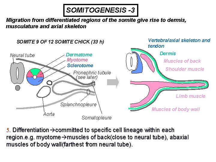
SOMITOGENESIS -3 Migration from differentiated regions of the somite give rise to dermis, musculature and axial skeleton SOMITE 9 OF 12 SOMITE CHICK (33 h) Dermatome Myotome Sclerotome Neural tube Pronephric tubule (see later) Vertebra/axial skeleton and tendon Dermis Muscles of back Shoulder muscle Limb muscle Splanchnopleure Aorta Muscles of body wall Somatopleure 5. Differentiation committed to specific cell lineage within each region. e. g. myotome muscles of back(close to neural tube), abaxial muscles of body wall(farthest from neural tube).
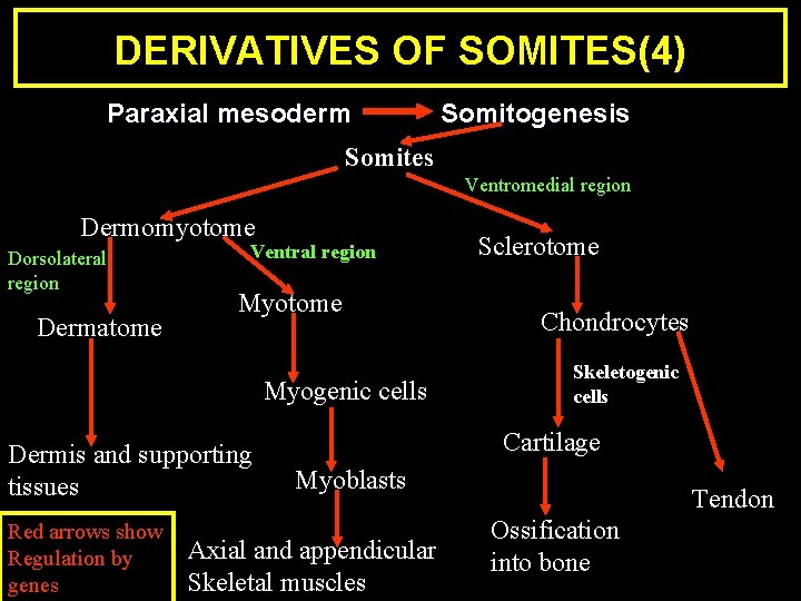
DERIVATIVES OF SOMITES(4) Paraxial mesoderm Somitogenesis Somites Ventromedial region Dermomyotome Dorsolateral region Dermatome Ventral region Myotome Myogenic cells Dermis and supporting tissues Red arrows show Regulation by genes Sclerotome Chondrocytes Skeletogenic cells Cartilage Myoblasts Axial and appendicular Skeletal muscles Tendon Ossification into bone
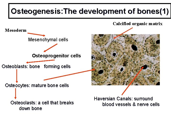
Osteogenesis: The development of bones(1) Calcified organic matrix Mesoderm Mesenchymal cells Osteoprogenitor cells Osteoblasts: bone forming cells Osteocytes: mature bone cells Osteoclasts: a cell that breaks down bone Haversian Canals: surround blood vessels & nerve cells
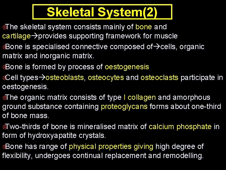
Skeletal System(2) The skeletal system consists mainly of bone and cartilage provides supporting framework for muscle Bone is specialised connective composed of cells, organic matrix and inorganic matrix. Bone is formed by process of oestogenesis Cell types osteoblasts, osteocytes and osteoclasts participate in oestogenesis. The organic matrix consists of type I collagen and amorphous ground substance containing proteoglycans forms about one-third of bone mass. Two-thirds of bone is mineralised matrix of calcium phosphate in form of hydroxyapatite crystals. Bone has range of physical properties giving high degree of flexibility, undergoes continual replacement and remodelling.
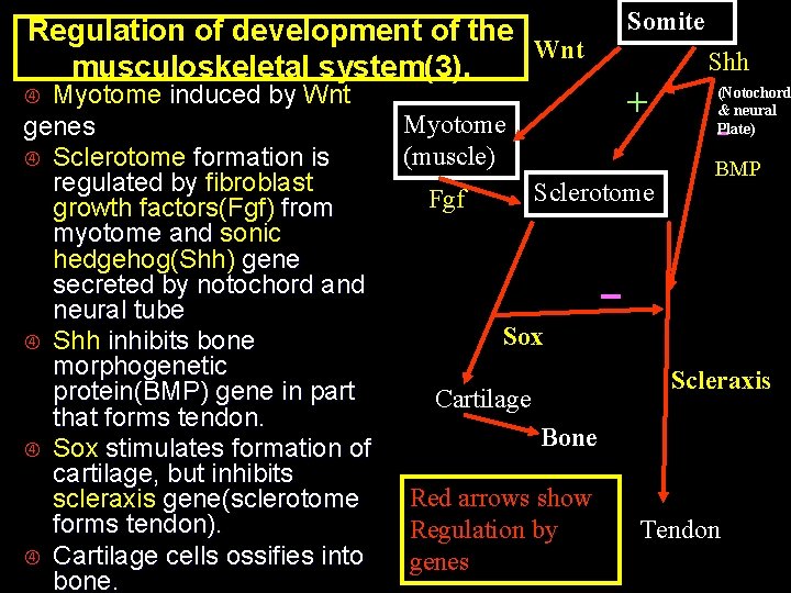
Regulation of development of the Wnt musculoskeletal system(3). Myotome induced by Wnt genes Sclerotome formation is regulated by fibroblast growth factors(Fgf) from myotome and sonic hedgehog(Shh) gene secreted by notochord and neural tube Shh inhibits bone morphogenetic protein(BMP) gene in part that forms tendon. Sox stimulates formation of cartilage, but inhibits scleraxis gene(sclerotome forms tendon). Cartilage cells ossifies into bone. Myotome (muscle) Somite Shh + Sclerotome Fgf (Notochord & neural Plate) - BMP Sox Scleraxis Cartilage Bone Red arrows show Regulation by genes Tendon
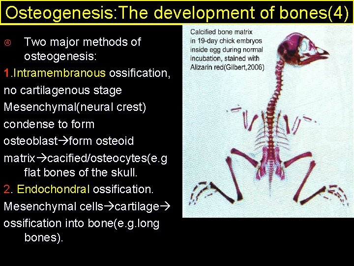
Osteogenesis: The development of bones(4) Two major methods of osteogenesis: 1. Intramembranous ossification, no cartilagenous stage Mesenchymal(neural crest) condense to form osteoblast form osteoid matrix cacified/osteocytes(e. g flat bones of the skull. 2. Endochondral ossification. Mesenchymal cells cartilage ossification into bone(e. g. long bones).
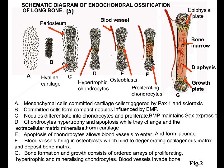
(5) Fig. 2
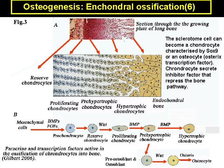
Osteogenesis: Enchondral ossification(6) Fig. 3 The sclerotome cell can become a chondrocyte characterised by Sox 9 or an osteocyte (osterix transcription factor). Chrondrocyte secrete inhibitor factor that repress the bone pathway. Pre-osteoblast & Osteoblast
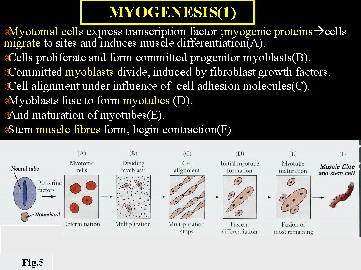
MYOGENESIS(1) Myotomal cells express transcription factor ; myogenic proteins cells migrate to sites and induces muscle differentiation(A). Cells proliferate and form committed progenitor myoblasts(B). Committed myoblasts divide, induced by fibroblast growth factors. Cell alignment under influence of cell adhesion molecules(C). Myoblasts fuse to form myotubes (D). And maturation of myotubes(E). Stem muscle fibres form, begin contraction(F) Fig. 5 (Gilbert 2006)
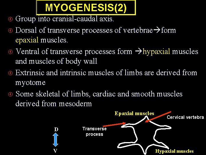
MYOGENESIS(2) Group into cranial-caudal axis. Dorsal of transverse processes of vertebrae form epaxial muscles. Ventral of transverse processes form hypaxial muscles and muscles of body wall Extrinsic and intrinsic muscles of limbs are derived from myotome Some skeletal of limbs, cardiac and smooth muscles derived from mesoderm Epaxial muscles D V Cervical vertebra Transverse process Hypaxial muscles
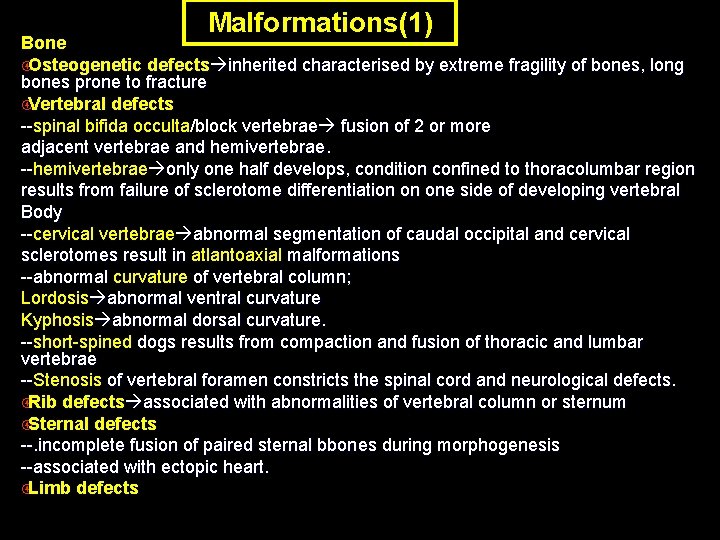
Malformations(1) Bone Osteogenetic defects inherited characterised by extreme fragility of bones, long bones prone to fracture Vertebral defects --spinal bifida occulta/block vertebrae fusion of 2 or more -adjacent vertebrae and hemivertebrae. --hemivertebrae only one half develops, condition confined to thoracolumbar region -results from failure of sclerotome differentiation on one side of developing vertebral Body --cervical vertebrae abnormal segmentation of caudal occipital and cervical -sclerotomes result in atlantoaxial malformations --abnormal curvature of vertebral column; Lordosis abnormal ventral curvature Kyphosis abnormal dorsal curvature. --short-spined dogs results from compaction and fusion of thoracic and lumbar -vertebrae --Stenosis of vertebral foramen constricts the spinal cord and neurological defects. - Rib defects associated with abnormalities of vertebral column or sternum Sternal defects --. incomplete fusion of paired sternal bbones during morphogenesis --associated with ectopic heart. Limb defects
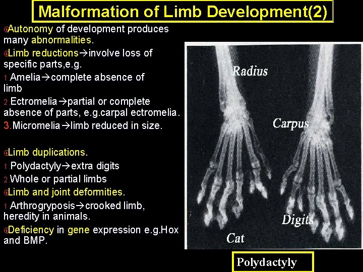
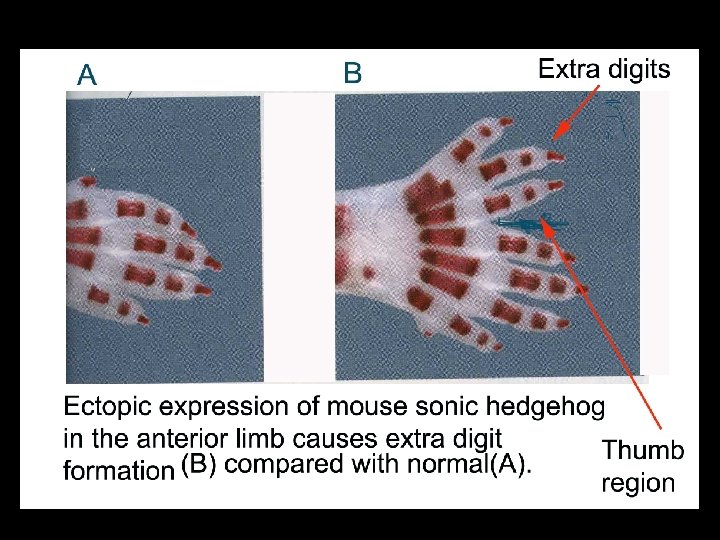
Malformation of Limb Development(2) Autonomy of development produces many abnormalities. Limb reductions involve loss of specific parts, e. g. 1. Amelia complete absence of limb 2. Ectromelia partial or complete absence of parts, e. g. carpal ectromelia. 3. Micromelia limb reduced in size. Limb duplications. 1. Polydactyly extra digits 2. Whole or partial limbs Limb and joint deformities. 1. Arthrogryposis crooked limb, heredity in animals. Deficiency in gene expression e. g. Hox and BMP. Polydactyly
Somitogenesis Summary is the process of segmentation of the paraxial mesoderm in anterior-posterior gradient, begins at neurulation. The first seven pairs of cranial somitomeres form head mesenchyme, migrate to specific regions and form masticatory and facial muscles. Somite 8 -46(rabbit) become segmented into block-like somites. Number of pairs constant for each species. Each somite has three morphological regions. Ventromedial sclerotome chondrocytes form axial skeleton and syndetome from within sclerotome form tendons. The dorsolateral layer dermatome, form dermis of skin. Ventral layer myotome; form axial and appendicular muscles. The molecular mechanisms regulating osteogenesis and myogenesis are transcription factors, growth factors, the Hox gene, Shh, BMP, and Wnt genes. Few examples of common congenital malformations in development of the axial skeleton and in limb development
References Carlson, B. M. , Foundations of Embryology (6 th. Edition) 1996. Mc. Graw-Hill inc. London. Page 393 - 424 1. 2. Gilbert, S. F. , Developmental Biology (8 th. Edition) 2006. Sinauer Associates Inc. Sunderland, Massachuetts. USA. Page 505 -527 Mc. Geady, T. A. , Quinn, P. J. , Fitzpatrick, E. S. , & Rayan, M. T. , (2006). Veterinary Embryology. Page 184 -203 3. Noden, D. M. , De. La. Hunta, A. , The Embryology of Domestic Animals. 1985, Williams & Wilkins. London. Page 196 -206 4.