Neurons Signals Synapses Chapter 37 I Neuron Structure


 • If change is")

 3 Rising phase")


 – Neurotransmitter is released by exocytosis into the synaptic cleft – Neurotransmitter binds")

- Slides: 22
Neurons, Signals, Synapses Chapter 37
I. Neuron Structure Dendrites • Dendrites receive signals from other neurons • Cell body contains Cell body nucleus and organelles Axon • Axon transmits signals to other neurons or muscles Fig. 37. 5 Motor neuron
Presynaptic neuron Figure 37. 2 Stimulus Cell body Axon Synapse Synaptic terminals Postsynaptic neuron Neurotransmitter
• Synapse – junction of a nerve cell with another cell • Relative terms depending on cell location in network: – Pre-synaptic cell – cell sending signal across synapse – Post-synaptic cell – cell receiving signal
Figure 37. 3 80 m Real neurons Green = dendrites Cyan = neuron cell bodies Red = support cells (glia) Cell bodies of neurons
II. Resting potential • Bioflix: How Neurons Work
II. Resting potential • Resting potential = charge difference across a cell membrane of ALL Body Cells • Develops because of Na+ & K+ movement • Ion movement through 3 membrane proteins: – Na+-K+ pump – Passive K+ channels – Passive Na+ channels
Figure 37. 6 Key Na K Sodiumpotassium pump Potassium channel OUTSIDE OF CELL Sodium channel INSIDE OF CELL
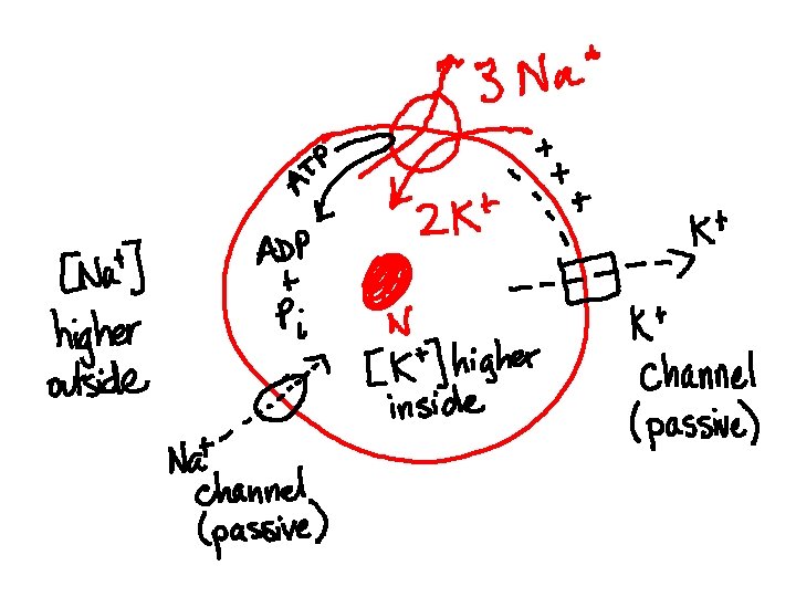
Description of ion movement • Na+-K+ pump: pumps 3 Na+ out & 2 K+ in to the cell • Result: – [Na+] = 15 m. M inside – [K+] = 140 m. M inside 150 m. M outside 5 m. M outside
• Passive K+ channel: always open – K+ leaves cell – Cl- and Protein- are stuck inside cell • Result: – (-) charge now greater inside cell – (-) charge attracts (+) charge on outside, electrical gradient limits K+ efflux (outflow)
III. Action potential • Bioflix: Action Potential
III. Action Potential • Depends on voltage-gated Na+ & K+ channels – Passive channels that are closed at rest – Open in response to a change in voltage across cell membrane – Like an electrically operated gate
Description of ion movement • Stimulus changes membrane potential (voltage) • If change is large enough that voltage exceeds a threshold, voltage-gated Na+ channels open • Na+ flows into cell • Change in potential CLOSES voltage-gated Na+ channels and OPENS voltage-gated K+ channels • K+ leaves cell
• Na+-K+ pump re-establishes gradients of Na+ and K+ • Action potential spreads because Na+ diffuses along the inside of the cell membrane, changing voltage & opening the next batch of voltagegated Na+ channels
Figure 37. 11 Key Na K 50 Membrane potential (m. V) 3 Rising phase of the action potential OUTSIDE OF CELL 100 Sodium channel Potassium channel Action potential 3 0 50 2 Depolarization 4 Falling phase of the action potential Threshold 2 1 4 5 Resting potential Time INSIDE OF CELL Inactivation loop 1 Resting state 5 Undershoot 1
IV. Synapse • Bioflix: How Synapses Work
A real synapse Synaptic terminals of pre-synaptic neurons 5 m Postsynaptic neuron Figure 37. 16
Figure 37. 15 Presynaptic cell Postsynaptic cell Axon Synaptic vesicle containing neurotransmitter 1 Postsynaptic membrane Synaptic cleft Presynaptic membrane 3 K Ca 2 2 Voltage-gated Ca 2 channel Ligand-gated ion channels 4 Na
At chemical synapse • Electrical signal crosses the synaptic cleft = gap between neurons • How? – A. p. reaches end of neuron – Voltage changes in the neuron membrane – Voltage-gated Ca 2+ channels open – Ca 2+ binds to vesicles containing neurotransmitter – Vesicles fuse with neuron membrane
(continued) – Neurotransmitter is released by exocytosis into the synaptic cleft – Neurotransmitter binds to receptors on next neuron – Receptors are ligand-gated Na+ channels = Na+ channels that open when the right molecule (ligand) attaches – Neurotransmitter detaches from receptors and is degraded by enzymes to stop signal
What happens next? • The postsynaptic cell responds if the postsymatic potential reaches threshold • Postsynaptic cells could be : – Nerve cells – Muscle cells – Glandular cells