Neuron The neuron Neurons The functional and structural


 Astrocytes: Play and important role in homeostasis in the CNS.")

 Microglia Oligodentrocyte Astrocyte (fibrous)")


- Slides: 38
Neuron
The neuron
Neurons • The functional and structural unit of the nervous system • Specialized to conduct information from one part of the body to another • There are many, many different types of neurons but most have certain structural and functional characteristics in common: - Cell body (soma) - One or more specialized, slender processes (axons/dendrites) - An input region (dendrites/soma) - A conducting component (axon) - A secretory (output) region (axon terminal)
Part of axon
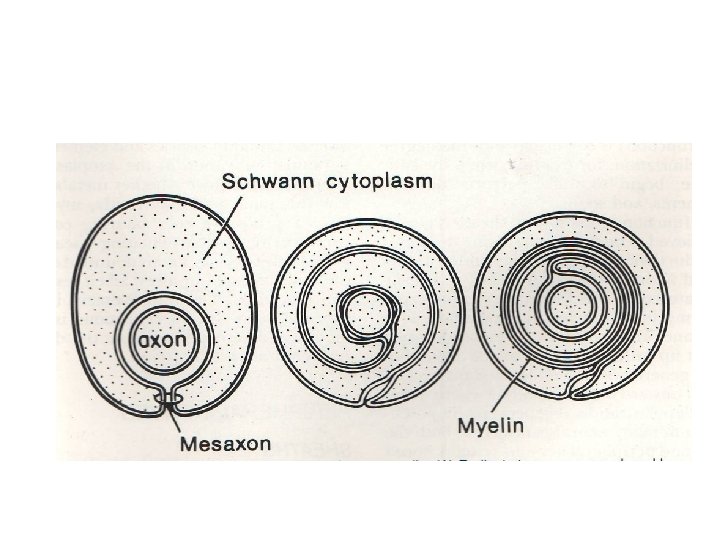
The myelin sheath
Types of neurons Neurons can be classified on their structure, size, position of cell body, length and mode of branching, etc. Golgi type-I neurons: large cells with long neurons. The cell bodies are generally in the CNS while the axons may traverse down the length of the spinal cord or to the peripheral nerves. Golgi type-II neurons: usually smaller cells with shorter axons. Such neurons are present in the cerebral and cerebellar cortex and the retina. Projections neurons: These are for long distance communication. Local circuit neurons: These are for short distance communication.
Unipolar neurons: These are rare in vertebrates except in early embryonic stages. A single processes arises from the cell body that has both dendritic and axonal properties. Bipolar neurons: A process projects from end of a fusiform cell body. These occur in the vestibular and cochlear ganglia and in the nasal olfactory epithelium. Pseudounipolar neurons: These arise as bipolar neurons but with time the two processes shift around the perikaryon and unite into one. Later it lengthens and divides forming the letter T. One acts as a dendrite and other as axon. Multipolar neurons: Most vertebrate neurons are multipolar. There are many different kinds of these neurons primarily in the central nervous system.
Types of neurons
Multipolar neurons of the brain
The neuroglia (neuroglial cells) Astrocytes: Play and important role in homeostasis in the CNS. They clear the extra-cellular spaces of K⁺ ions, glutamate and αaminobutyric acid. They also store glycogen as an energy reserve. Astrocytes are typically star shaped cells. They have numerous highly branched processes. Protoplasmic astrocytes that occur in the gray matter. Fibrous astrocytes have been found in the white matter. Their processes are long and thin and branch less frequently. Oligodendrocytes: They have fewer cellular processes which do not branch much. The cell body is small and the cytoplasm is very dense. Interfascicular oligodendrocytes occur in the white matter and make and maintain the myelin. Satellite oligodendrocytes are closely associated with the cell bodies in the gray matter. Their exact role is not known.
Microglia: These are small cells scattered throughout the CNS. They are smaller than oligodendrocytes and are darker. They have small dense nucleus and scant cytoplasm. The cells have short processes with minute spines. Generally thought to have developed through the macrophage line of cells. They are phagocytic in nature. Schwann cells: Ependyma cells:
The neuroglia Astrocyte (protoplasmic) Microglia Oligodentrocyte Astrocyte (fibrous)
Neuroglia • • – 1. Outnumber neurons by about 10 to 1 (the guy on the right had an inordinate amount of them). 6 types of supporting cells 4 are found in the CNS: Astrocytes • • • Star-shaped, abundant, and versatile Guide the migration of developing neurons Act as K+ and NT buffers Involved in the formation of the blood brain barrier Function in nutrient transfer
Neuroglia 2. Microglia • • Specialized immune cells that act as the macrophages of the CNS Why is it important for the CNS to have its own army of immune cells? 3. Ependymal Cells • • Low columnar epithelial-esque cells that line the ventricles of the brain and the central canal of the spinal cord Some are ciliated which facilitates the movement of cerebrospinal fluid
Neuroglia 4. Oligodendrocytes • Produce the myelin sheath which provides the electrical insulation for certain neurons in the CNS
Neuroglia • 2 types of glia in the PNS 1. Satellite cells • • Surround clusters of neuronal cell bodies in the PNS Unknown function 2. Schwann cells • • Form myelin sheaths around the larger nerve fibers in the PNS. Vital to neuronal regeneration
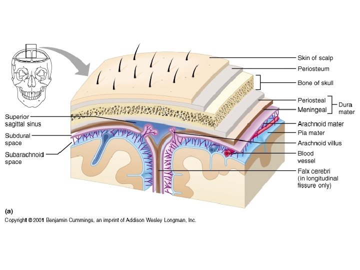
Protection • What is the major protection for the brain? • There also 3 connective tissue membranes called the meninges: • Cover and protect the CNS • Protect blood vessels • Contain cerebrospinal fluid • The 3 meninges from superficial to deep: • Dura mater • Arachnoid mater • Pia mater
Skin Galea Aponeurotica Connective Tissue Bone Dura Mater Arachnoid mater
Dura Mater • Tough and leathery. • Most superficial. • 2 layers: – Periosteal attached to the skull – Meningeal true external covering, extends downward and surrounds spinal cord • In several locations, the inner dura mater extends in to the cranial cavity, forming a sheet that dips inward and then returns. These dural folds provide additional support for the brain. Dural sinuses may be found btwn the 2 layers of a dural fold.
Arachnoid and Pia Mater • Arachnoid: – Loose spider-web of connective tissue. – Beneath it is the subarachnoid space – filled with blood vessels and CSF • Pia – Deepest and most delicate – Covers the brain tissue – Follows its every ridge and groove • What do you call an inflammation of the meninges?
The meninges of the brain
The meninges of the brain
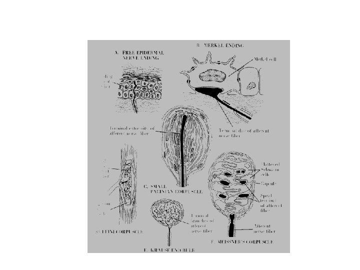
Sensory nerve terminations Several types are known. Free nerve endings Expanded tip endings: Merkels receptors Encapsulated nerve endings: Pacinian corpuscles, Meissner corpuscles, end bulb of Krause, Ruffini corpuscles. Free nerve endings: These are slender unmyelinated axonal branches with tapering tips. They are the most widely distributed receptors in the body. Free nerve endings mediate pain (noniceptors), heat and cold sensations (thermoreceptors) and inputs from tactile hairs (mechanoreceptors). Widely distributed in the epidermis and cornea of eyes. Merkels receptors: These are slow adapting mechanoreceptors. They consist of s swollen nerve ending associated with the Merkel cell. Signal prolonged touch or pressure on the skin. Generally found in the dermis on the lips and distal regions of limbs.
Epithelial nerve endings
Ruffini corpuscles: More widely distributed in the dermis. Consist of numerous axonal branches terminating in a tiny bulb-like expansion. Tension along the axis of the corpuscle is most effective. Serve as low threshold stretch receptors of the skin. They signal direction and magnitude of the forces acting on them. Pacinian corpuscle: This is the largest of the receptors usually visible to the naked eye. It is an ovoid structure up to 1 mm in length. The myelin of the axon is lost up on entering the capsule. Schwann cells become flattened and these in turn are surrounded by concentric lamellae forming the thick capsule. The lamellae consist of thin flat cells and successive lamellae are separated by a gel-like substance. Usually found in the dermis especially under the finger tips. These corpuscles respond to rapid changes in pressure and not to sustained pressure. Often called high frequency mechanoreceptors.
Pacinian corpuscle
Meissner corpuscle: This is a pear-shaped or elliptical structure. It is smaller and occurs in more superficial region of the skin. More common in regions of the skin that are more sensitive. Such as palms and soles, tips of fingers and toes and lips. The nerve enters at the broader end of the corpuscle and spirals upwards between the cells and layers. It is fast adapting mechanoreceptors but is tuned to low frequency mechanical stimuli.
Meissner’s corpuscle
Degeneration of nerve fibre
Regenerating nerve fibres
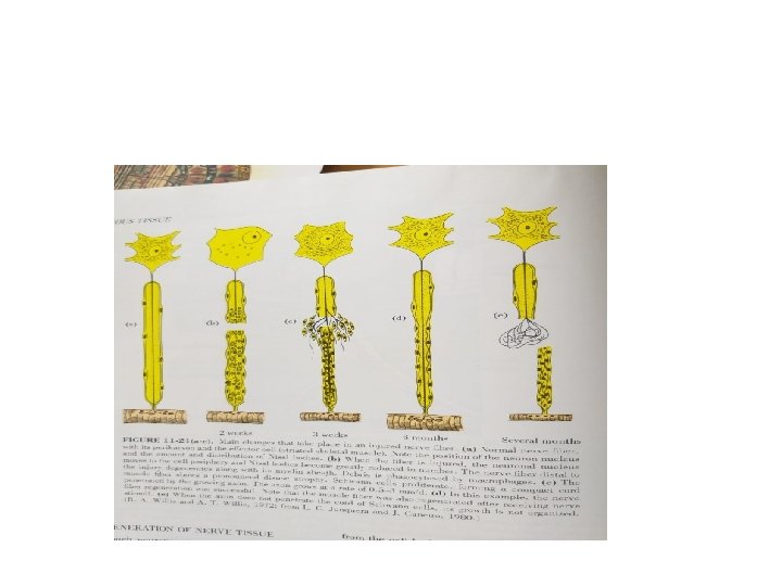
Wallerian Degeneration • When the fiber is cut, segment of the fiber separated from cell body undergoes degeneration. This is called Wallerian degeneration. • Some degeneration of the fiber occur in retrograde direction, but only for an internodal segment or two. • Changes also occur in cell-body showing disorganization of Nissl bodies, ( the process called Chromatolysis), migration of nucleus to more peripheral location.
Degeneration Contd • The amount of Axoplasm separated from cell-body by the injury has a direct effect on how severely the cellbody reacts to the injury. • If axon is cut far from the cellbody, separating only a small amount of axoplasm, changes in cell body are not so severe. . • If even after severe injury with loss of substantial axoplasm resulting in minimal reactions in the cell body , it is postulated that axoplasm of collateral branches sustains the cell body. • When injury is close to the cellbody, with extensive loss of Axoplasm, death of cell occurs. • When the fiber is cut, the muscle innervated by the neuron undergoes atrophy. • The peak of chromatolysis reaction is menifest about two weeks after the injury.
Regeneration of fibre • During this time macrophages enter the schwann cell sheath and remove the fragments of axon and myelin. • At the same time schwann cells multiply and form a cord of cells along the route of damaged fibre. • The axonal sprouts begin to grow from the cutend of the proximal stump. • A successful follows the rout to the effector which was reestablished by cord of schwann cells. • Other nerve sproutswithout appropriate guidance from Schwann cell regress.
Regeneration contd. • The successful sprout grows at a rate of 3 mm per day and ultimately reestablishes contact with the muscle. • After success reestablishment of the contact with the nerve, the muscle returns to its healthy state. • By this time the Nissl bodies and nucleus position return to normal original state. • If the axonal sprout does not reestablish contact with cord of Schwann cells, it grows in a disorganised manner and muscle stays atrophic.