Neuronal Ceroid Lipofuscinosis in Two Cats MARK CHALKLEY


 What is LSD anyway? • Ly. Sergic acid Diethylamide •")


 Lysosomal Function")


- Slides: 24
Neuronal Ceroid. Lipofuscinosis in Two Cats MARK CHALKLEY – 3 rd YEAR ANATOMIC PATHOLOGY RESIDENT
History & Signalment • Similar history and signalment for both case #1 and #2: - Both between 1 or 2 -years old male DSH cats - Submitted to animal shelters as strays with no previous history - Prolonged, progressive history of visual dysfunction (running into walls, difficulty tracking moving objects) and neurological deficits (falling, frantic running, behavior abnormalities) • Both euthanized and submitted for necropsy
Case #1 Control
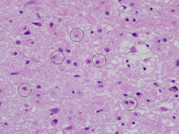
Cerebral Cortex
Normal Case #2
H&E
H&E LFB/ H&E AF PAS
Immunofluorescence • Normal and abnormal
H&E GFAP
Normal Case #2
Lesion Distribution Anatomic Location Lipopigment/Ceroid Neuronal/ Cell Loss Gliosis 1 2 1 2 Cerebral cortex +++ ++ Thalamus + ++ + + Hypothalamus + + + Basal Nuclei + ++ + + Hippocampus +++ + + ++ + Cerebellum ++ + Spinal Cord + ++ + + Retina + + ++ - - Extra-CNS tissues - - - * + = mild, ++ = moderate, +++ = severe; 1 = Case No. 1; 2 = Case No. 2
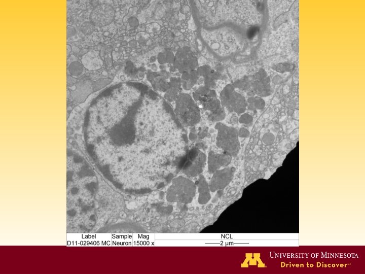
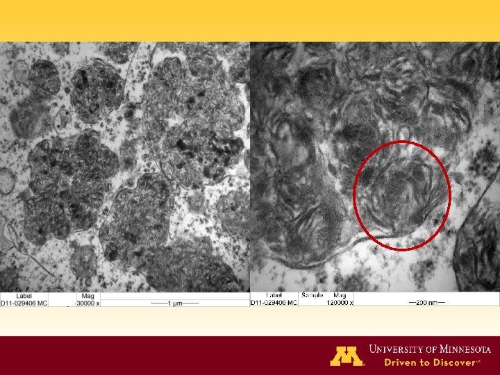
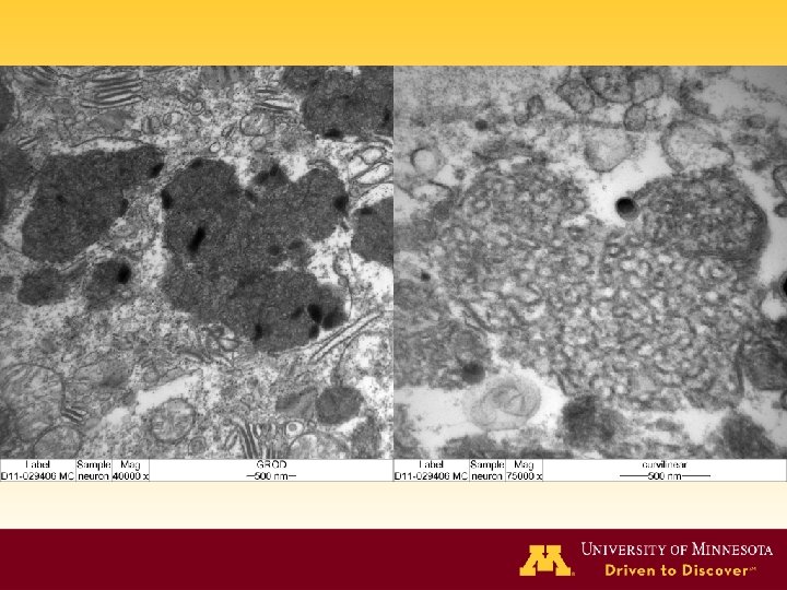
Disease Cause Organs affected 1. Ceroid. Lipofuscinoses Unknown or lysosomal protein defects Predominantly cerebrum, cerebellum & retina in cats but can be in many tissues 2. Gangliosidoses Lysosomal enzyme deficiency CNS, PNS, liver, kidney, lymph n. , cardiac Purkinje cells 3. Glucocerebrosidoses 4. Mannosidosis (glycoproteinosis) 5. Mucopolysaccharidoses (MPS VII in cats) Glucocerebrosidase deficiency Lysosomal α or β mannosidase or α-L-fucosidase deficiency Various enzyme deficiencies CNS, macrophages (esp liver, LNs) Many cells (neurons, epithelium, endothelium, mesenchyme) Predominantly bone and other connective tissues, neurons Storage material Special stains Subunit c of mitochondrial ATPase, amyloid-β, precursor protein, SAPs Magneta with PAS LFB+ Sudan black+ Autofluorescent Glycosphingolipids PAS+ LFB+ Sudan black+ Glucocerebrosides PAS+ in macs PAS – in neurons LFB+ Carbohydrate (saccharides with mannose residues) PAS+, diastase resistant, Variably alcian blue+ Glucoaminoglycan Variable PAS+ LFB+ Sudan black+ Autofluorescent Lectin histochemistry EM Variable to nil reactivity Curvilinear bodies Fingerprint profiles Granular osmophilic deposits O-linked Membranous cytoplasmic bodies and/or zebra bodies O-linked Twisted, branching tubules or zebra bodies in neurons N-linked Membrane bound vesicles, with floccular material & fine membrane stacks N-linked Empty vesicles, or floccular material /zebra bodies
Ceroid-lipofuscinosis Gangliosidoses Sphingolipidoses & Mucopolysaccharidoses Mannosidoses
Diagnosis Brain, spinal cord, eyes: Degenerative lysosomal storage encephalomyelopathy and retinopathy, widespread, marked, chronic, with neuronal loss and astrogliosis - Consistent with neuronal ceroid-lipofuscinosis (NCL)
Lysosomal Storage Diseases (LSD) What is LSD anyway? • Ly. Sergic acid Diethylamide • Lumpy Skin Disease • Lichenoid-p. Soriasiform Dermatoses • Louisiana School for the Deaf
NCL in Animals and Humans • • • Four main variants – infantile, late-infantile, juvenile, adult 160 mutations in 10 human genes (CLN 1 -10) Lead to defects in 8 known proteins Gene defects also recognized in dogs, sheep and cattle Similar distribution and character of lesions & material Main storage material in animals and humans - subunit c of mitochondrial ATP synthase (SCMAS) or sphingolipid activator proteins A and D (SAPs) • A correlation between the age of clinical onset of disease, ultrastructure of the storage material and the variants of NCL has not been established in animals
Characteristic Lipofuscin vs Ceroid Circumstance of deposition Aging Pathological Location Lysosome Positive Protein Heterogenous mix, amyloid-β protein Subunit c of mitochondrial ATPase, amyloid-β, precursor protein, saposins Lipids Triglycerides, FFA, dolichols Phosphorylated dolichols, phospholipids, neutral lipids Carbohydrates Dolichol-linked oligosaccharides Metals Fe, Cu, Al, Zn, Ca, Mn Predominantly Fe Sudan Black Positive Luxol fast blue Positive PAS Positive Concanavalin A Positive Agglutinin Negative Positive Granular osmophilic deposits Positive Fingerprint profiles Negative Positive Curvilinear bodies Negative Positive Autofluorescence Storage components Histochemistry (including lectins) Ultrastructure *Table adapted from SS Seehafer, DA Pearce, Neurology of Aging 27 (2006), p 580.
Mechanisms of Ceroid Oxidation Accumulation Autophagy Seehafer S & Pearce D (2007) Lysosomal Function
Acknowledgements • ACVP/STP coalition fellowship • Pfizer & Dr Peter Schmidt • University of Minnesota – - Dr Anibal Armien & Dr Gerry O’Sullivan - Electron microscopy laboratory
Questions?