NERVOUS SYSTEM NERVOUS SYSTEM 2 Main Divisions Central


 – sensory nerves from the skin and")
 – 90%")
 – most numerous – form webs that surround")
 – toward brain and")


 insulation • – speeds up transmission (Schwann or")
- Slides: 24
NERVOUS SYSTEM
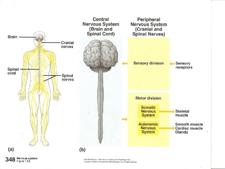
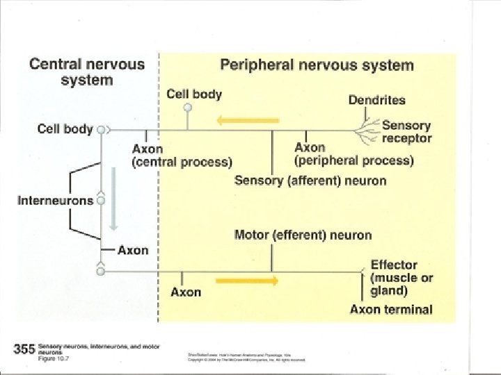
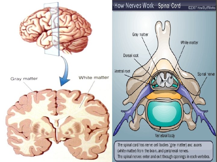
NERVOUS SYSTEM 2 Main Divisions • Central NS – brain and spinal cord • Peripheral NS – all other nerves (cranial, spinal) CNS – median structure; all sensory impulses arrive there, all motor impulses begin there PNS -- nerves that lie outside (peripheral to) the CNS – convey all sensations and motor impulses to and from the CNS. -cranial nerves – originate from the brain and brainstem -spinal nerves – originate from the spinal cord • both give rise to smaller branches.
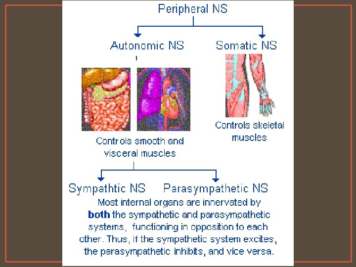
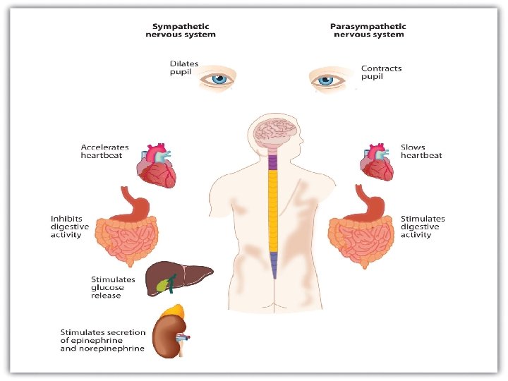
PERIPHERAL NS 2 Subdivisions Somatic System (voluntary) – sensory nerves from the skin and sensory organs (ear, eye, nose) and motor nerves to skeletal muscles Autonomic System (involuntary) – controls blood vessels, gland • further subdivided: Sympathetic – times of stress (injury, excitement, physical activity) Parasympathetic – conditions of normal organ functioning
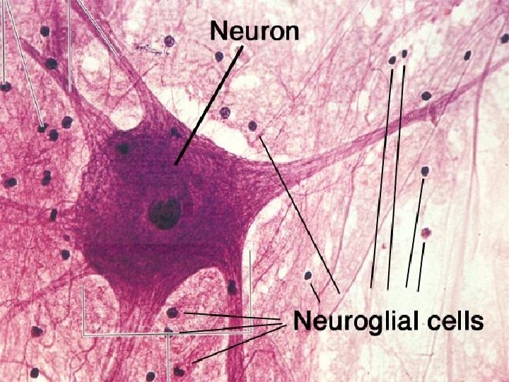
NERVOUS TISSUE 2 Types of Nervous Tissue : Neuroglia – (glial cells) – 90% of the CNS -Small part of the PNS. -Provide structural framework for the neurons. -Non-conducting. • Neurons – Structural and functional unit of the N. S. even though they are outnumbered by glial cells - Perform the functions of the N. S. – sense changes, integrate information, make a motor response.
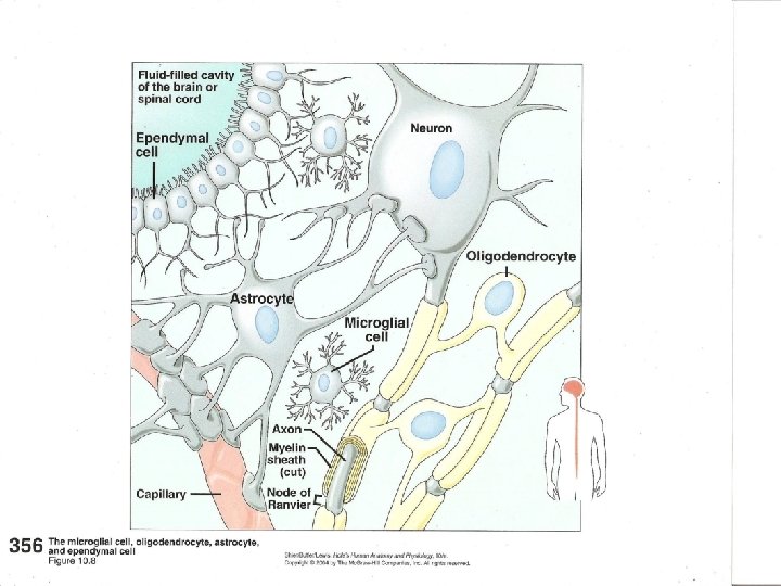
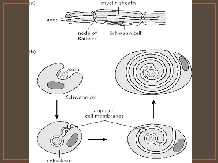
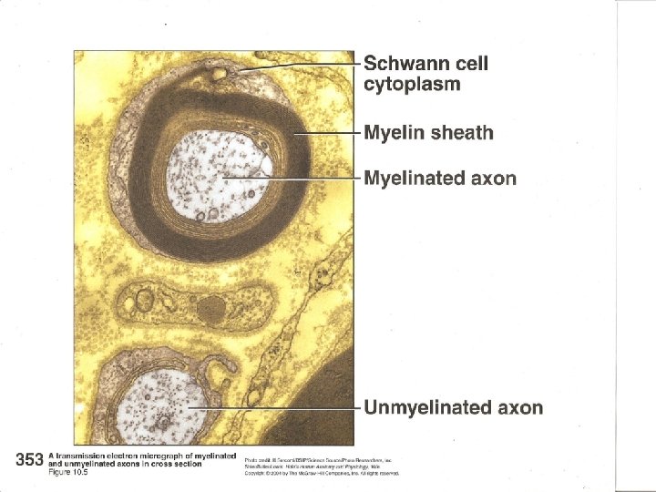
TYPES OF GLIAL CELLS -Astrocytes (CNS) – most numerous – form webs that surround capillaries, allowing small molecules (O 2 and CO 2) to pass through, keep others out. -Oligodendrocytes (oligodendroglia) (CNS) – help hold nerve fibers together – produce a myelin sheath, a protective covering -Microglia (CNS) – engulf and destroy microbes and debris in inflamed/damaged tissue -Ependymal (CNS) – line cavities of CNS – form and circulate CSF -Schwann cells (PNS) – form myelin sheath/neurolemma around peripheral nerves. Have high lipid content
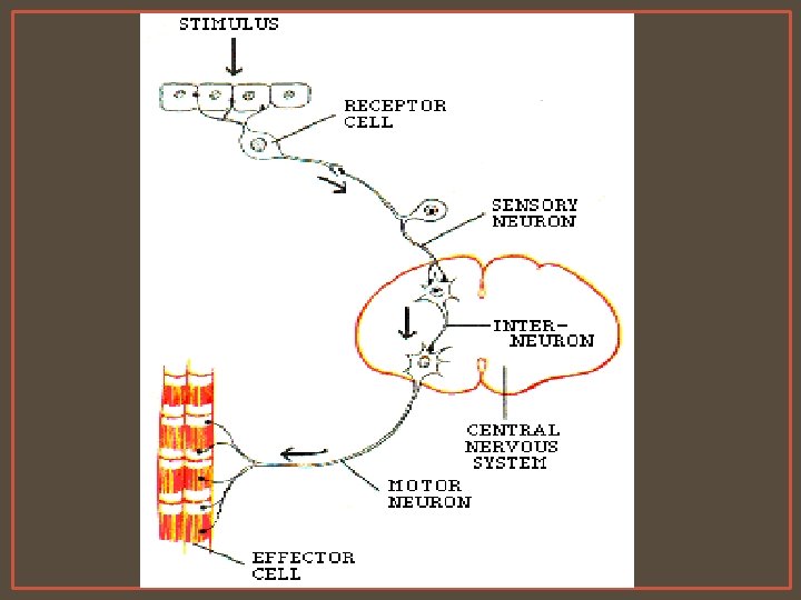
NEURONS Classified according to the DIRECTION OF IMPULSE -Afferent (sensory) – toward brain and spinal cord -Efferent (motor) – toward muscles/glands/organs -Interneurons (AKA Association) – CNS only, connect afferent and efferent fibers
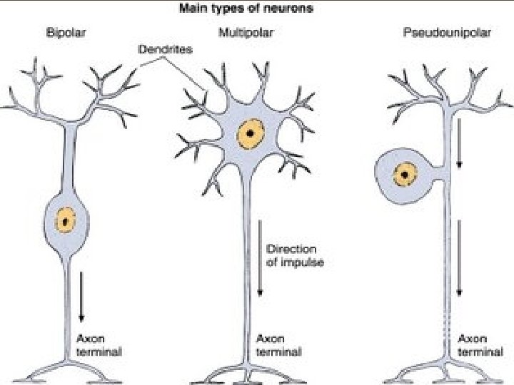
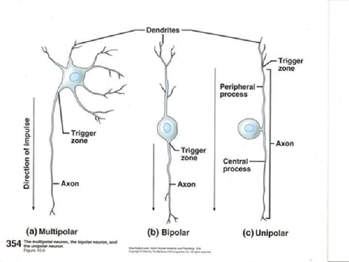
Classified according to STRUCTURE • -Multipolar – many dendrites, single axon (CNS to skel. muscle) • -Bipolar – single dendrite, single axon (special sensory areas) • -Unipolar – one fiber extending from the body, it splits into two branches (one to the spinal cord (axon); the other to the peripheral body (dendrite) (sensory from skin to sp. cord).
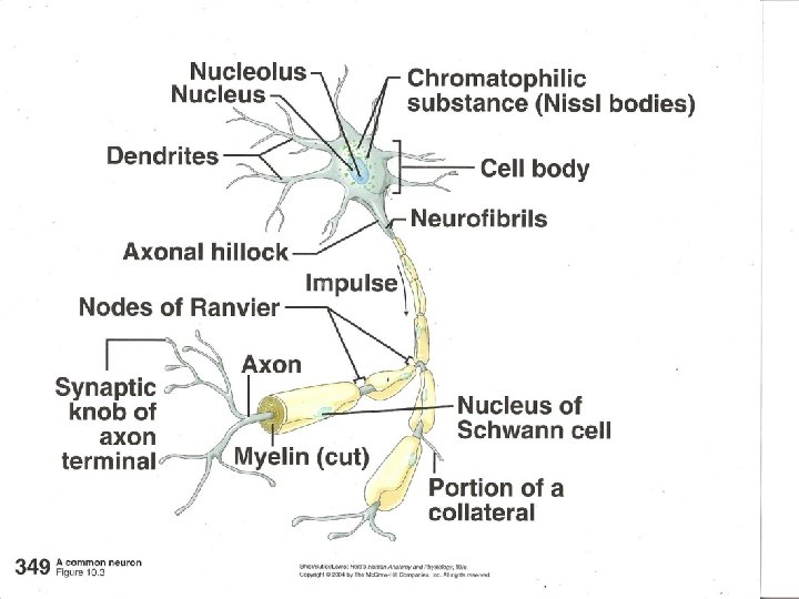
NEURON ANATOMY Cell body- has nucleus, cell membrane • At least 2 processes • Dendrites –receivers (impulses from other neurons – go to cell body) • Axon –senders (away from cell body) – 1 per neuron (may have branches, collaterals) • Nissl bodies – extra protein synthesis (Rough ER)
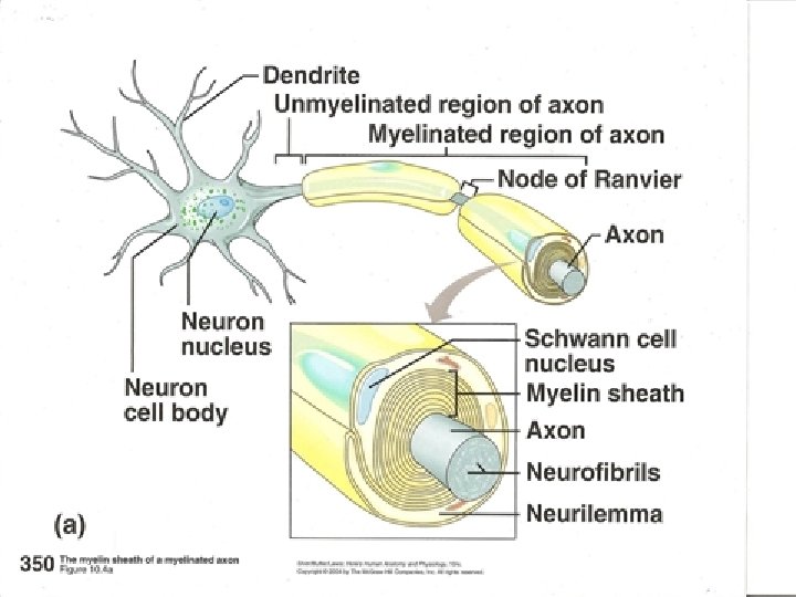
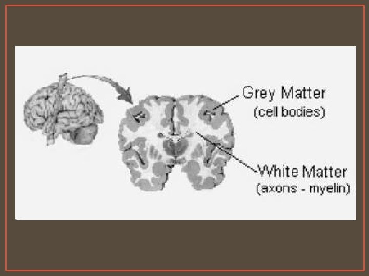
Myelin sheath – around axon (fat) insulation • – speeds up transmission (Schwann or oligodendroglia) • myelin – (white matter) inside – medulla • non-myelin (gray matter) outside – cortex Node of Ranvier- gaps in between that aid in impulse conduction Neurolemma – outer covering of axons [Schwann] • – helps in regenerating nerves • – none in brain and cord *no regeneration • Loses spindle fibers at about 3 yo – no longer able to mitotically divide