Divisions of the Human Nervous System Central Nervous


 · Paired (left and right) superior parts of the brain ·")


 · The surface is made of ridges (gyri) and grooves (sulci)")

 divide the cerebrum into lobes ·")


 Cerebrum divided into 4 lobes: Frontal: motor function, motivation, aggression,")
 The sulcus dividing frontal and parietal lobes is")


 Sum electrical activity can be read as waves with")
 3 types: Sensory ~ less than a second Short-term ~")
 Long term memories are divided into 2 types: Declarative memory")
 Deepest portion of cerebrum and brainstem associations called")
 Not really a “dominant” side Hemispheres communicate via the corpus")
- Slides: 31
Divisions of the Human Nervous System Central Nervous System-the brain and the spinal cord Peripheral Nervous System-the nerves outside the brain and spinal cord Two Division of the PNS Somatic Nervous System-the nerves that convey messages from the sense organs to the CNS and from the CNS to the muscles and glands Autonomic Nervous System-a set of neurons that control the heart, the intestines, and other organs
Figure 4. 1 The human nervous system Both the central nervous system and the peripheral nervous system have major subdivisions. The closeup of the brain shows the right hemisphere
The Brain: the big picture… The basic components of the CNS include the: • Cerebrum • Diencephalon • Cerebellum • Brain stem • Spinal cord
The Brain: development… Our central nervous system has humble origins early in our development (by 3 rd wk. ) A plate (neural plate) of cells develop crests (neural crests) that rise and eventually meet and fuse leaving the hollow dorsal nerve cord & and ventricles of our CNS
BI 201 Human Anatomy & Physiology The Brain: development… What happens if those crests DON’T meet and fuse? Failure of neural arch formation can result in Spina bifida. In mild cases results in a dimple or discolored spot (S. b. occulta), severe cases (S. b. cystica), meninges, CSF and nervous tissue may protrude.
The Brain: from the outside in… The brain and spinal cord are protected by meninges 3 layers: Dura mater ~ outermost, tough, continuous with periosteum Arachnoid mater ~ middle layer, spiderweb appearance Pia mater ~ innermost layer, not visible to naked eye
The Brain: from the outside in… The dura mater helps keep the brain in position and the cerebral spinal fluid (CSF) helps cushion/protect the brain Where does the fluid come from? Choroid plexuses on ventricle walls Ependymal cells allow fluids in from capillaries
The Brain: from the outside in… The CSF circulates throughout the ventricles, down the central canal of the spinal cord and throughout the subarachnoid space. CSF is reabsorbed back into blood primarily at the venous sinuses found within the dura mater.
Cerebral Hemispheres (Cerebrum) · Paired (left and right) superior parts of the brain · Include more than half of the brain mass Figure 7. 13 a Slide
Layers of the Cerebrum · Gray matter · Outer layer · Composed mostly of neuron cell bodies Figure 7. 13 a Slide
Layers of the Cerebrum · White matter · Fiber tracts inside the gray matter · Example: corpus callosum connects hemispheres Figure 7. 13 a Slide
Cerebral Hemispheres (Cerebrum) · The surface is made of ridges (gyri) and grooves (sulci) Figure 7. 13 a Slide
The Lobes The Occipital Lobe-posterior end of cortex Contains primary visual cortex The Parietal Lobe-between occipital love the central sulcus Contains the primary somatosensory cortex-receiving touch sensation, muscle-stretch information and joint position information The Temporal Lobe-lateral portion of each hemisphere, near the temples Contains targets for audition, essential for understanding spoken language, complex visual processes, emotional and motivational behaviors The Frontal Lobe-extends from the central sulcus to the anterior limit of the brain Contains Primary Motor Cortex-fine movements Contributes to shifting attention, planning of action, delayed response tasks as examples
Lobes of the Cerebrum · Fissures (deep grooves) divide the cerebrum into lobes · Surface lobes of the cerebrum · Frontal lobe · Parietal lobe · Occipital lobe · Temporal lobe Slide
Lobes of the Cerebrum Figure 7. 15 a Slide
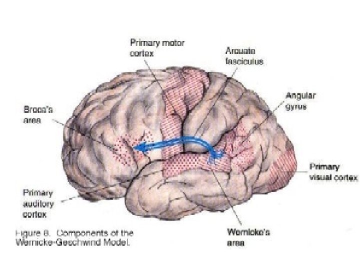
Specialized Areas of the Cerebrum · Somatic sensory area – receives impulses from the body’s sensory receptors · Primary motor area – sends impulses to skeletal muscles · Broca’s area – involved in our ability to speak Slide 7. 30
Sensory and Motor Areas of the Cerebral Cortex Figure 7. 14 Slide 7. 31
Specialized Area of the Cerebrum · Cerebral areas involved in special senses · Gustatory area (taste) · Visual area · Auditory area · Olfactory area Slide
Specialized Area of the Cerebrum · Interpretation areas of the cerebrum · Speech/language region · Language comprehension region · General interpretation area Copyright © 2003 Pearson Education, Inc. publishing as Benjamin Cummings Slide
Specialized Area of the Cerebrum Figure 7. 13 c Slide
The Brain: Cerebrum (lobes) Cerebrum divided into 4 lobes: Frontal: motor function, motivation, aggression, smell and mood Parietal: reception and evaluation of sensory info. Temporal: smell, hearing, memory and abstract thought Occipital: visual processing
The Brain: Cerebrum (bumps and grooves) The sulcus dividing frontal and parietal lobes is Central Sulcus. Ridges on either side are Pre & Post gyri Motor Pre CS ~ motor cortex Post CS ~ somatic sensory cortex Sensory
Layers of the Cerebrum · Basal nuclei – internal islands of gray matter · Regulates voluntary motor activities by modifying info sent to the motor cortex · Problems = ie unable to control muscles, spastic, jerky · Involved in Huntington’s and Parkinson’s Disease Figure 7. 13 a Slide
Figure 4. 20 Some major subdivisions of the human cerebral cortex The four lobes: occipital, parietal, temporal, and frontal.
The Brain: Cerebrum (makin’ waves) Sum electrical activity can be read as waves with EEG bio-feedback to control brain activity
The Brain: Cerebrum (memories) 3 types: Sensory ~ less than a second Short-term ~ seconds to minutes (about 7 - 12 bits) Long-term ~ minutes to life time associated with re-shaping neurons and formation of memory engrams (pattern of neurons and their connections) 1 9 2 1 3 6 0 3 7 8 2 4 3 1 1 3 5 7 9 2 4 6 8 0 1 3 5 7
The Brain: Cerebrum (memories) Long term memories are divided into 2 types: Declarative memory ~ ability to recall details, names etc. Procedural memory ~ ability to repeat behaviors or actions. Example: Pavlov and conditioning
The Brain: Cerebrum (memories in “limbo”) Deepest portion of cerebrum and brainstem associations called the Limbic system This is involved with mood, base emotions and interacts with hypothalamus to influence food/H 2 O acquisition. Major input is olfaction
The Brain: Cerebrum (hemispheres) Not really a “dominant” side Hemispheres communicate via the corpus callosum Structurally and functionally different Right brain -- usually Representational Left brain -- usually Categorical Anterior Smell R. Verb. Mem. Smell L. Shape mem. Musical Symbolic Language Intuitive Spatial Posterior