Multicellular life Evolution of multicellular life Animal tissue


 Campbell Fig 20. 5 A Holds other tissue in place")


 Campbell Fig 20. 5")


- Slides: 21
Multicellular life Evolution of multicellular life Animal tissue types
Campbell Fig 1. 1
Campbell Fig 20. 2
Animal tissue types • What is a tissue? • A cooperative unit of many very similar cells that perform a specific function. • Examples – Epithelial – Connective – Muscle – Nervous
Epithelial tissue • Covers and lines the body and its parts • One surface free, the other bound to basement membrane • Tissues are named by – Shape of cells – Number of layers of cells
Epithelial tissue Campbell Fig 20. 4 • • • Simple = single layer Stratified = multiple layers Squamous = flat (tiles) Cuboidal = like dice Columnar = like bricks
Simple Squamous Simple Cuboidal In the kidney tubules Campbell Fig 20. 4 Lines the lungs
Stratified Squamous Epithelium Campbell Fig 20. 4 Lines the esophagus
Ciliated columnar epithelium Campbell Fig 20. 4 Lines the air ways in the respiratory system
Connective tissue • Binds other tissues an provides support matrices • Few cells in a nonliving matrix • Three fiber types – Collagen fibers – Elastic fibers – Reticular fibers • Fibroblasts - cells that produce connective tissue
Loose connective tissue (Areolar) Campbell Fig 20. 5 A Holds other tissue in place A “binding” material
Other Connective tissues Campbell Fig 20. 5 Loose Fibrous connective Adipose Cartilage Blood Bone
Tendons Dense connective tissue that Attaches muscle to bone Like Campbell Fig 30. 7
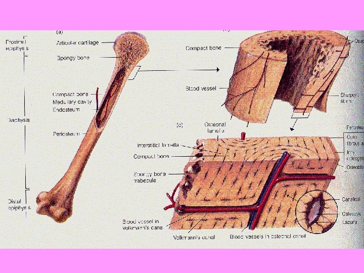
Bone Tissue • Osteocytes • Haversian canal • Lamelle (matrix) Campbell Fig 20. 5 D
Bone Development
Muscle tissue • Functions in movement • Bundles of long cells ( muscle fiber= muscle cell) • Skeletal muscle – Attached to bones by tendons, produces voluntary movement – Striated unbranched • Smooth muscle – Found in walls of digestive tract, produces involuntary movements – Unstriated, spindle shaped • Cardiac Muscle – Striated , branched, produces heartbeat
Muscle tissue Campbell 20. 6 Cardiac muscle Skeletal muscle Smooth muscle
Nervous Tissue • Responsible for coordinating body activties • Neurons are nerve cells • Motor neurons are nerves that activate muscles • Sensory neurons transmit information • Composed of cell body and dendrites • Supported by glial cells Campbell Fig 28. 3 A Modified
Nervous Tissue Campbell Fig 28. 2
Summary